. 2020 Apr;52(4):594-603.
doi: 10.1038/s12276-020-0410-4. Epub 2020 Apr 1.

PLCE1 regulates the migration, proliferation, and differentiation of podocytes

Affiliations

PLCE1 regulates the migration, proliferation, and differentiation of podocytes

Seyoung Yu et al. Exp Mol Med. 2020 Apr.

Abstract

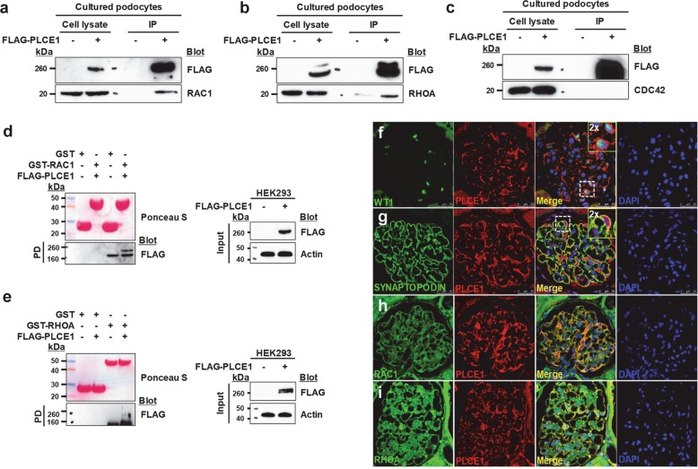
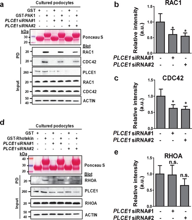
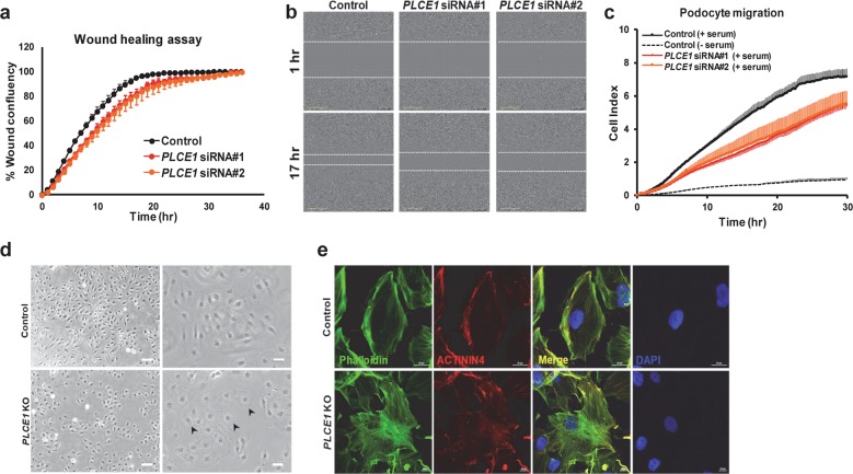
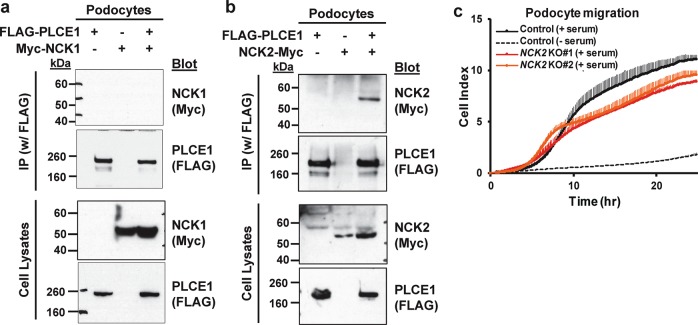
PLCE1 encodes phospholipase C epsilon, and its mutations cause recessive nephrotic syndrome. However, the mechanisms by which PLCE1 mutations result in defects associated with glomerular function are not clear. To address this, we investigated the function of PLCE1 in podocytes called glomerular epithelial cells, where the pathogenesis of nephrotic syndrome converges. PLCE1 colocalized with Rho GTPases in glomeruli. Further, it interacted with Rho GTPases through the pleckstrin homology domain and Ras GTP-binding domains 1/2. Knockdown or knockout of PLCE1 in podocytes resulted in decreased levels of GTP-bound Rac1 and Cdc42, but not those of RhoA, and caused a reduction in cell migration. PLCE1 interacted with NCK2 but not with NCK1. Similar to the PLCE1 knockout, NCK2 knockout resulted in decreased podocyte migration. Knockout of PLCE1 reduced the EGF-induced activation of ERK and cell proliferation in podocytes, whereas knockout of NCK2 did not affect proliferation. Further, the knockout of PLCE1 also resulted in decreased expression of podocyte markers, including NEPH1, NPHS1, WT1, and SYNPO, upon differentiation, but the knockout of NCK2 did not affect the expression of these markers. Therefore, our findings demonstrate that PLCE1 regulates Rho GTPase activity and cell migration through interacting with NCK2 and that PLCE1 also plays a role in the proliferation and differentiation of podocytes, regardless of the presence of NCK2.


Conflict of interest statement

The authors declare that they have no conflict of interest.

Figures

Fig. 1
Fig. 1. PLCE1 interacts with Rac1 and RhoA.
ac Immunoprecipitation of PLCE1 with Rho GTPase, Rac1 (a), RhoA (b), and Cdc42 (c) in cultured podocytes. Immunoblotting showed that PLCE1 interacts with Rac1 and RhoA but not with Cdc42. de FLAG-PLCE1 expressed in HEK293 cells was precipitated by GST- Rac1 (d) and GST-RhoA (e). Ponceau red staining at the top shows the GST proteins used. fi Costaining of PLCE1 with WT1 (f), SYNAPTOPODIN (g), Rac1 (h) and RhoA (i) using immunofluorescence in adult rat glomeruli. PLCE1 colocalized with Rac1 and RhoA in podocyte cell bodies. Immunoprecipitation (IP) and pulldown (PD) experiments are representative of more than three experiments.
Fig. 2
Fig. 2. The PH and RA1/2 domains of PLCE1 are required for the interaction with Rac1 and RhoA.
a Schematic representation of the PLCE1 domains. PLCE1 contains a RasGEF domain, a pleckstrin homology (PH) domain, two regions of sequence among PLCs (X and Y), a C2 domain, and two Ras-binding (RA) domains. The numbers represent the amino acid positions. The fragments of PLCE1 were fused to GFP at their N-terminus. b, c Interactions of PLCE1 domains with Rho GTPase Rac1 and RhoA. GFP-tagged PLCE1 fragments and Myc-tagged Rac1 (b) and RhoA (c) were transfected into podocytes, and immunoprecipitation was performed using anti-Myc agarose beads. Notably, both RAC1 and RhoA bind to PLCE1 through the PH and RA1/2 domains.
Fig. 3
Fig. 3. The effects of the PLCE1 knockdown on Rho GTPase activity.
a Active GTP-bound Rac1 and Cdc42 precipitated from podocytes transfected with PLCE1 siRNA using a GST-PAK1 (CRIB domain) pulldown assay. Ponceau red staining at the top shows the GST proteins used. Compared with control podocytes, podocytes transfected with PLCE1 siRNA exhibited a significant decrease in GTP-bound Rac1 and Cdc42 levels. b, c Quantification of Rac1 (b) and Cdc42 (c) in PLCE1 knockdown cells compared to that in control cells. d Active GTP-bound RhoA precipitated from podocytes transfected with PLCE1 siRNA using a GST-rhotekin (RBD domain) pulldown assay. Cells transfected with or without PLCE1 siRNA exhibited no significant differences in GTP-bound RhoA. e Quantification of RhoA in PLCE1 knockdown cells compared with that in control cells. Error bars indicate the standard deviations for more than three independent experiments. *P < 0.05; n.s. not significant.
Fig. 4
Fig. 4. Depletion of PLCE1 resulted in decreased podocyte migration and changed cell morphology.
a Wound closure was reduced in podocytes transfected with PLCE1 siRNAs (red and orange dots) compared to that in control podocytes (black dots). b Movement of cells into the wound is shown at 1 and 17 h after scratching. PLCE1 knockdown podocytes showed delayed wound closure. c The effect of PLCE1 knockdown on serum-induced podocyte migration. Podocytes transfected with PLCE1 siRNA (red and orange lines) exhibited decreased migration compared to that of the control podocytes (solid black line). Error bars are shown in only one direction for clarity and indicate standard deviations for more than three independent experiments. d, e The effect of PLCE1 knockout (KO) on podocyte morphology. PLCE1 KO resulted in the loss of terminal arborizations in undifferentiated podocytes (d) and an increased number of stress fibers in differentiated PLCE1 KO podocytes relative to that in control podocytes (e).
Fig. 5
Fig. 5. NCK2 interacts with PLCE1 and affects the migration in podocytes.
a, b Interaction of PLCE1 and NCKs. The plasmids containing FLAG-tagged PLCE1 and Myc-tagged NCKs were transfected into podocytes, and cell lysates were immunoprecipitated using anti-FLAG agarose beads. Notably, PLCE1 interacted with NCK2 (b) but not with NCK1 (a). The immunoblots are representative of more than three experiments. IP, immunoprecipitation. c The effect of NCK2 knockout (KO) on podocyte migration. NCK2-depleted podocytes exhibited decreased migration (red and orange lines) compared with that of the control podocytes (solid black line). Error bars are shown in only one direction for clarity and indicate standard deviations for more than three independent experiments.
Fig. 6
Fig. 6. PLCE1 regulates mitogen-activated protein kinases (MAPKs) in podocytes.
a MAPK phosphorylation was induced by 20 ng/µl epidermal growth factor (EGF) treatment for 45 or 120 min. EGF-induced phosphorylation of ERK and JNK was significantly reduced in PLCE1 knockout (KO) podocytes. b, c The effects of PLCE1 or NCK2 KO on podocyte proliferation. PLCE1 KO podocytes exhibited lower proliferation (b, red and orange lines), whereas NCK2 KO podocytes exhibited no significant differences in proliferation (c, red and orange lines) with respect to the control cells (black line). Error bars are shown in one direction only for clarity and indicate standard deviations from more than three independent experiments. d Expression analysis of podocyte markers. The expression of NEPH1, NPHS1, WT1, and SYNPO was measured by quantitative real-time PCR. The expression of podocyte markers was significantly reduced in PLCE1 KO podocytes, whereas no difference in the levels of podocyte markers was observed between NCK2 KO and control podocytes. Gene expression was normalized relative to that of actin. Data are presented as the mean ± standard deviation (n = 3). *P < 0.05.
Fig. 7
Fig. 7. Functions of PLCE1 in podocytes.
PLCE1 plays multiple roles in podocytes: (1) PLCE1 acts as a phospholipase and degrades phosphatidylinositol 4,5-bisphosphate (PIP2) into diacylglycerol (DAG) and inositol triphosphate (IP3). (2) PLCE1 can be activated by a G-protein and transduces mitogenic signals through its RasGEF activity to stimulate the Ras and MAPK pathways. (3) PLCE1 regulates the activity of Rho GTPases, which are the key regulators of actin dynamics and cell migration. Upon loss of PLCE1, the levels of active Rac1 and Cdc42 were reduced. (4) PLCE1 interacts with NCK2, which plays a pivotal role in stimulating N-WASP-Arp2/3-induced actin nucleation.

References

    1. Tryggvason K, Patrakka J, Wartiovaara J. Hereditary proteinuria syndromes and mechanisms of proteinuria. N. Engl. J. Med. 2006;354:1387–1401. doi: 10.1056/NEJMra052131. - DOI - PubMed
    1. Somlo S, Mundel P. Getting a foothold in nephrotic syndrome. Nat. Genet. 2000;24:333–335. doi: 10.1038/74139. - DOI - PubMed
    1. Hildebrandt F. Genetic kidney diseases. Lancet (Lond., Engl.) 2010;375:1287–1295. doi: 10.1016/S0140-6736(10)60236-X. - DOI - PMC - PubMed
    1. Sadowski CE, et al. A single-gene cause in 29.5% of cases of steroid-resistant nephrotic syndrome. J. Am. Soc. Nephrol. 2015;26:1279–1289. doi: 10.1681/ASN.2014050489. - DOI - PMC - PubMed
    1. Rao J, et al. Advillin acts upstream of phospholipase C 1 in steroid-resistant nephrotic syndrome. J. Clin. Investig. 2017;127:4257–4269. doi: 10.1172/JCI94138. - DOI - PMC - PubMed

Publication types

MeSH terms

LinkOut - more resources