. 2020 Sep;41(9):1208-1222.
doi: 10.1038/s41401-020-0359-9. Epub 2020 Apr 1.

Cinnamaldehyde protects against rat intestinal ischemia/reperfusion injuries by synergistic inhibition of NF-κB and p53

Affiliations

Cinnamaldehyde protects against rat intestinal ischemia/reperfusion injuries by synergistic inhibition of NF-κB and p53

Marwan Almoiliqy et al. Acta Pharmacol Sin. 2020 Sep.

Abstract

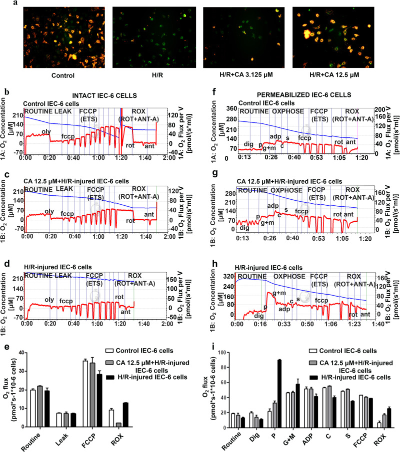
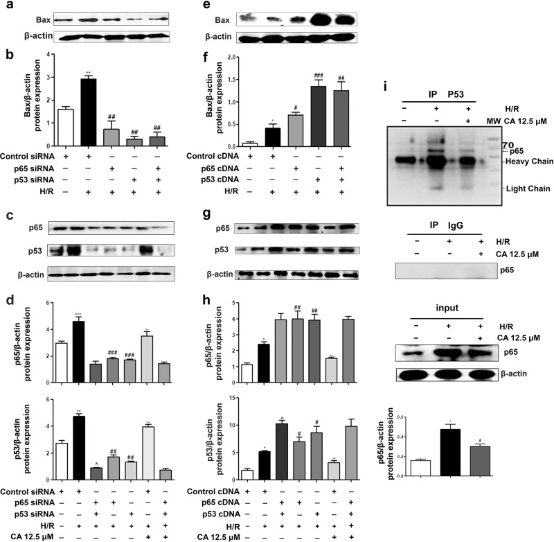
Our preliminary study shows that cinnamaldehyde (CA) could protect against intestinal ischemia/reperfusion (I/R) injuries, in which p53 and NF-κB p65 play a synergistic role. In this study, we conducted in vivo and in vitro experiments to verify this proposal. SD rats were pretreated with CA (10 or 40 mg · kg-1 · d-1, ig) for 3 days, then subjected to 1 h mesenteric ischemia followed by 2 h reperfusion. CA pretreatment dose-dependently ameliorated morphological damage and reduced inflammation evidenced by decreased TNF-α, IL-1β, and IL-6 levels and MPO activity in I/R-treated intestinal tissues. CA pretreatment also attenuated oxidative stress through restoring SOD, GSH, LDH, and MDA levels in I/R-treated intestinal tissues. Furthermore, CA pretreatment significantly reduced the expression of inflammation/apoptosis-related NF-κB p65, IKKβ, IK-α, and NF-κB p50, and downregulated apoptotic protein expression including p53, Bax, caspase-9 and caspase-3, and restoring Bcl-2, in I/R-treated intestinal tissues. We pretreated IEC-6 cells in vitro with CA for 24 h, followed by 4 h hypoxia and 3 h reoxygenation (H/R) incubation. Pretreatment with CA (3.125, 6.25, and 12.5 μmol · L-1) significantly reversed H/R-induced reduction of IEC-6 cell viability. CA pretreatment significantly suppressed oxidative stress, NF-κB activation and apoptosis in H/R-treated IEC-6 cells. Moreover, CA pretreatment significantly reversed mitochondrial dysfunction in H/R-treated IEC-6 cells. CA pretreatment inhibited the nuclear translocation of p53 and NF-κB p65 in H/R-treated IEC-6 cells. Double knockdown or overexpression of p53 and NF-κB p65 caused a synergistic reduction or elevation of p53 compared with knockdown or overexpression of p53 or NF-κB p65 alone. In H/R-treated IEC-6 cells with double knockdown or overexpression of NF-κB p65 and p53, CA pretreatment caused neither further decrease nor increase of NF-κB p65 or p53 expression, suggesting that CA-induced synergistic inhibition on both NF-κB and p53 played a key role in ameliorating intestinal I/R injuries. Finally, we used immunoprecipitation assay to demonstrate an interaction between p53 and NF-κB p65, showing the basis for CA-induced synergistic inhibition. Our results provide valuable information for further studies.

Keywords: NF-κB; apoptosis; cinnamaldehyde; inflammation; mesenteric ischemia/reperfusion injury; mitochondria; oxidative stress; p53.


Conflict of interest statement

The authors declare no competing interests.

Figures

Fig. 1
Fig. 1
Cinnamaldehyde protected against intestinal I/R-induced morphological damage and inflammation. a The chemical structure of cinnamaldehyde. b Representative images of intestinal histology (H&E staining, original magnification ×100). c Chiu’s score of the intestine after intestinal I/R. d Tissue levels of TNF-α, IL-1β, IL-6 and MPO. Data are expressed as the mean ± SD (n = 5), ***P < 0.001, **P < 0.01, and *P < 0.05 vs sham group; ###P < 0.001, ##P < 0.01, and #P < 0.05 vs I/R group
Fig. 2
Fig. 2
Cinnamaldehyde protected against intestinal I/R- and H/R-induced oxidative stress. a Tissue levels of SOD, GSH, LDH, and MDA. b The levels of SOD protein expression in intestinal tissues from three individual samples of randomly selected rats from each group. c Quantification analysis of SOD in intestinal tissues is performed for three independent experiments. d The levels of SOD protein expression in normal/H/R-injured IEC-6 cells from three individual samples. e Quantification of SOD in normal/H/R-injured IEC-6 cells from three independent experiments. f ROS assays were performed on the IEC-6 cell line. g The effect of CA (1.56, 3.125, 6.25, 12.5, 25, and 50 μmol · L−1) on IEC-6 cell viability for 24 h under normal conditions. h The effect of CA (1.56, 3.125, 6.25, 12.5, 25, and 50 μmol · L−1) on IEC-6 cell viability for 24 h in H/R conditions. Data are expressed as the mean ± SD (n = 3), ***P < 0.001, **P < 0.01, and *P < 0.05 vs sham group; ###P < 0.001, ##P < 0.01, and #P < 0.05 vs I/R group
Fig. 3
Fig. 3
Cinnamaldehyde improved mitochondrial respiration function and MMP (ΔΨm) in H/R-injured IEC-6 cells. a Mitochondrial membrane potential (MMP ΔΨm) assays were performed by using JC-1 staining in normal/H/R-injured IEC-6 cells. b Mitochondrial respiration of intact IEC-6 cells is shown; the plot of oxygen consumption flux per 1 × 106 cells in DMEM at 37 °C: control IEC-6 cells, (c) CA 12.5 μmol · L−1 + H/R-injured IEC-6 cells, and (d) H/R-injured IEC-6 cells. e Shown are the average basal respiration (ROUTINE), Oligomycin/Oly (LEAK), FCCP (maximal electron transport system ETS) and rotenone/Rot and Antimycin-A/ Ant-A (ROX). f Mitochondrial respiration of permeabilized IEC-6 is shown; the graph represents the plot of oxygen consumption flux per 1 × 106 cells in DMEM at 37 °C: control IEC-6 cells, (g) CA 12.5 μmol · L−1 + H/R-injured IEC-6 cells, and (h) H/R-injured IEC-6 cells. i Average values are shown for digitonin (Dig), pyruvate (P), glutamate (G) and malate (M), ADP, cytochrome-c (C), succinate (S), FFCP, rotenone (Rot) and antimycin-A (Ant-A)
Fig. 4
Fig. 4
Cinnamaldehyde ameliorated apoptosis in intestinal I/R-injuries and H/R injuries. a Apoptotic protein levels of Bax, Bcl-2, caspase-9, caspase-3, and p53 in sham-operated/I/R-injured intestinal tissues from three individual samples of randomly selected rats from each group. b Quantification analysis of apoptotic protein levels in intestinal tissues of three independent experiments. c The protein levels of Bax, Bcl-2, caspase-9, caspase-3, and p53 in 3 individual samples of normal/H/R-injured IEC-6. d Quantification analysis of apoptotic protein levels in three independent experiments for normal/H/R-injured IEC-6 cells. e p53 mRNA levels in intestinal tissues. f p53 mRNA levels in normal/H/R-injured IEC-6 cells. g TUNEL staining in intestinal I/R-injured tissues (All images original magnification ×200). Data are expressed as the mean ± SD (n = 3), ***P < 0.001, **P < 0.01, and *P < 0.05 vs control group; ###P < 0.001, ##P < 0.01, and #P < 0.05 vs I/R group
Fig. 5
Fig. 5
Cinnamaldehyde protected against intestinal I/R injuries through inhibition of NF-κB in rats. a Protein levels of IKKβ, IK-α, NF-κB p50, and NF-κB p65 in the intestinal tissues from three individual samples of randomly selected rats from each group. b Quantification analysis of NF-κB-related proteins in the intestinal tissues from three independent experiments. c NF-κB p65 mRNA levels in intestinal tissues. d Immunohistochemical staining of NF-κB p65 in I/R-injured rats (original image magnification ×200). Data are expressed as the mean ± SD (n = 3), ***P < 0.001, **P < 0.01, and *P < 0.05 vs sham group; ###P < 0.001, ##P < 0.01, and #P < 0.05 vs I/R group
Fig. 6
Fig. 6
Cinnamaldehyde protected against H/R injuries through inhibition of NF-κB in IEC-6 cells. a Protein levels of IKKβ, IK-α, NF-κB p50, and NF-κB p65 in three individual samples of normal/H/R-injured IEC-6 cells. b Quantification analysis of NF-κB-related proteins in three independent samples of normal/H/R-injured IEC-6 cells. c NF-κB p65 mRNA levels in normal/H/R-injured IEC-6 cells. Data are expressed as the mean ± SD (n = 3), ***P < 0.001, **P < 0.01, and *P < 0.05 vs sham group; ###P < 0.001, ##P < 0.01, and #P < 0.05 vs H/R group
Fig. 7
Fig. 7
Cinnamaldehyde inhibited NF-κB and p53 nuclear translocation in H/R-injured IEC-6 cells. a Immunofluorescence assay of NF-κB p65 in normal/H/R-injured IEC-6 cells. b Immunofluorescence assay of p53 in normal/H/R-injured IEC-6 cells
Fig. 8
Fig. 8
Effects of cinnamaldehyde in NF-κB p65 + p53 siRNA and cDNA cotransfected IEC-6 cells. a Protein level of Bax in control siRNA-transfected IEC-6 cells, in control siRNA-transfected H/R-injured IEC-6 cells, in NF-κB p65 siRNA-transfected H/R-injured IEC-6 cells, in p53 siRNA-transfected H/R-injured IEC-6 cells, and in NF-κB p65 + p53 siRNA cotransfected H/R-injured IEC-6 cells. b Quantification analysis of the parameters in (a). c Protein levels of NF-κB p65 and p53 in control siRNA-transfected IEC-6 cells, in control siRNA-transfected H/R-injured IEC-6 cells, in NF-κB p65 siRNA-transfected H/R-injured IEC-6 cells, in p53 siRNA-transfected H/R-injured IEC-6 cells, in NF-κB p65 + p53 siRNA cotransfected H/R-injured IEC-6 cells, in control siRNA transfected and CA pretreated H/R-injured IEC-6 cells, and in CA pretreated and NF-κB p65 + p53 siRNA cotransfected H/R-injured IEC-6 cells. d Quantification analysis of the parameters in (c). e Protein level of Bax in control cDNA-transfected IEC-6 cells, in control cDNA-transfected H/R-injured IEC-6 cells, in NF-κB p65 cDNA-transfected H/R-injured IEC-6 cells, in p53 cDNA-transfected H/R-injured IEC-6 cells, and in NF-κB p65 + p53 cDNA cotransfected H/R-injured IEC-6 cells. f Quantification analysis of the parameters in (e). g Protein levels of NF-κB p65 and p53 in control cDNA-transfected IEC-6 cells, in control cDNA-transfected H/R-injured IEC-6 cells, in NF-κB p65 cDNA-transfected H/R-injured IEC-6 cells, in p53 cDNA-transfected H/R-injured IEC-6 cells, in NF-κB p65 + p53 cDNA cotransfected H/R-injured IEC-6 cells, in control cDNA-transfected and CA pretreated H/R-injured IEC-6 cells, and in CA pretreated and NF-κB p65 + p53 cDNA cotransfected H/R-injured IEC-6 cells. h Quantification analysis of the parameters in (g). i A p53 antibody was immunoprecipitated after incubation with extract from control IEC-6 cells, H/R-injured IEC-6 cells, and CA pretreated and H/R-injured IEC-6 cells; NF-κB p65 was used for immunoblotting. Data are expressed as the mean ± SD (n = 3), ***P < 0.001, **P < 0.01, and *P < 0.05 vs control siRNA or cDNA IEC-6 cells; ###P < 0.001, ##P < 0.01, and #P < 0.05 vs H/R + siRNA or cDNA IEC-6 cells; ~P < 0.05 vs protein expression before H/R in transfection with NF-κB p65 and p53; ^P < 0.05 vs single transfection

References

    1. Ou-Yang L, Liu Y, Wang BY, Cao P, Zhang JJ, Huang YY, et al. Carnosine suppresses oxygen-glucose deprivation/recovery-induced proliferation and migration of reactive astrocytes of rats in vitro. Acta Pharmacol Sin. 2018;39:24–34. - PMC - PubMed
    1. Liu S, Yang Y, Song YQ, Geng J, Chen QL. Protective effects of N(2)LalanylLglutamine mediated by the JAK2/STAT3 signaling pathway on myocardial ischemia reperfusion. Mol Med Rep. 2018;17:5102–8. - PMC - PubMed
    1. Gao S, Zhu Y, Li H, Xia Z, Wu Q, Yao S, et al. Remote ischemic postconditioning protects against renal ischemia/reperfusion injury by activation of T-LAK-cell-originated protein kinase (TOPK)/PTEN/Akt signaling pathway mediated anti-oxidation and anti-inflammation. Int Immunopharmacol. 2016;38:395–401. - PubMed
    1. Gonul Y, Ozsoy M, Kocak A, Ozkececi ZT, Karavelioglu A, Bozkurt MF, et al. Antioxidant, antiapoptotic and inflammatory effects of interleukin-18 binding protein on kidney damage induced by hepatic ischemia reperfusion. Am J Med Sci. 2016;351:607–15. - PubMed
    1. Li Y, Feng D, Wang Z, Zhao Y, Sun R, Tian D, et al. Ischemia-induced ACSL4 activation contributes to ferroptosis-mediated tissue injury in intestinal ischemia/reperfusion. Cell Death Differ. 2019;26:2284–99. - PMC - PubMed

MeSH terms