. 2021 Jan 4;113(1):80-87.
doi: 10.1093/jnci/djaa047.

Germline MBD4 Mutations and Predisposition to Uveal Melanoma

Affiliations

Germline MBD4 Mutations and Predisposition to Uveal Melanoma

Anne-Céline Derrien et al. J Natl Cancer Inst. .

Abstract

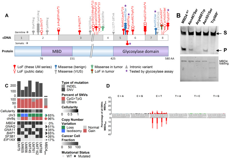
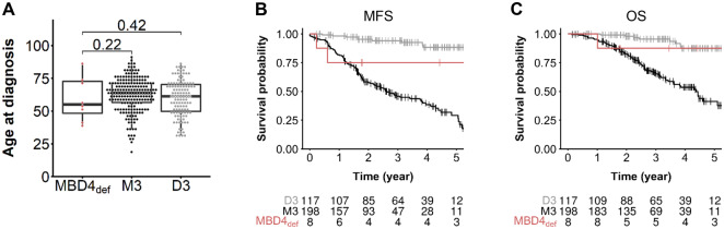
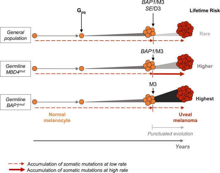
Background: Uveal melanoma (UM) arises from malignant transformation of melanocytes in the uveal tract of the eye. This rare tumor has a poor outcome with frequent chemo-resistant liver metastases. BAP1 is the only known predisposing gene for UM. UMs are generally characterized by low tumor mutation burden, but some UMs display a high level of CpG>TpG mutations associated with MBD4 inactivation. Here, we explored the incidence of germline MBD4 variants in a consecutive series of 1093 primary UM case patients and a series of 192 UM tumors with monosomy 3 (M3).

Methods: We performed MBD4 targeted sequencing on pooled germline (n = 1093) and tumor (n = 192) DNA samples of UM patients. MBD4 variants (n = 28) were validated by Sanger sequencing. We performed whole-exome sequencing on available tumor samples harboring MBD4 variants (n = 9). Variants of unknown pathogenicity were further functionally assessed.

Results: We identified 8 deleterious MBD4 mutations in the consecutive UM series, a 9.15-fold (95% confidence interval = 4.24-fold to 19.73-fold) increased incidence compared with the general population (Fisher exact test, P = 2.00 × 10⁻⁵, 2-sided), and 4 additional deleterious MBD4 mutations in the M3 cohort, including 3 germline and 1 somatic mutations. Tumors carrying deleterious MBD4 mutations were all associated with high tumor mutation burden and a CpG>TpG hypermutator phenotype.

Conclusions: We demonstrate that MBD4 is a new predisposing gene for UM associated with hypermutated M3 tumors. The tumor spectrum of this predisposing condition will likely expand with the addition of MBD4 to diagnostic panels. Tumors arising in such a context should be recognized because they may respond to immunotherapy.


Figures

Figure 1.
Figure 1.
Functional consequences and phenotype associated with germline and somatic MBD4 deleterious variants. A) Schematic representation of MBD4 cDNA (top) and protein (bottom) sequences. Functional methyl-binding domain (MBD) and glycosylase domain are indicated. The position of all MBD4 variants identified in the 2 uveal melanoma (UM) series (consecutive germline UM series and tumor monosomy 3 [M3] series) is highlighted, with germline and somatic variants above and below the cDNA sequence, respectively, and the 2 variants from the tumor M3 cohort with unknown somatic or germline origin circled in green. These MBD4 variants include loss-of-function (LoF, in red), missense (either benign, in blue-filled circles, or of unknown biological significance [VUS] in gray-filled circles) and intronic (gray triangles) variants. Each circle represents 1 patient harboring the variant. Other MBD4 germline deleterious variants mined on public data are also shown (empty red circles). B) Top: Glycosylase activity assay of recombinant wild-type MBD4 (MBD4WT) and mutant proteins resulting from missense variants and 1 stop gain variant (purple star in 1A) residing in the MBD4 glycosylase domain. Substrate = S; cleaved product = P. Bottom: loading blot for MBD4 wild-type and mutant recombinant proteins corresponding to the glycosylase assay. C) Tumor characteristics of MBD4-deficient (MBD4def) patients compared with that of MBD4-proficient UM patients (MBD4pro) (18). MBD4def patients include UM75, UM605, and UM656 from the consecutive germline series and UMT45, UMT61, UMT162, and UMT88 from the M3 UM tumor series. All patients harbor germline MBD4 variants, except for UMT88 with a somatic MBD4 variant. Top: tumor mutation burden estimated by number of variants (single nucleotide variants [SNVs] in dark gray, and insertions-deletions [INDELs] in light gray) in the exome; middle: proportion of CpG>TpG transitions (red) relative to all SNVs (gray); bottom: copy number alterations in chromosomes 3 and 8q, and mutational status of MBD4, GNAQ, GNA11, BAP1, SF3B1, and EIF1AX, represented as percentage for the MBD4pro series (18). The clonality or subclonality of these key mutational events is indicated by their cancer cell fraction in black-gray gradation, taking into account the variant allele frequency (VAF), copy number change, and cellularity. A plot of the VAF distribution of all variants in the 7 exomes is available in Supplementary Figure 4 (available online). For each exome in the MBD4def group, tumor cellularity is indicated by black-gray shading (and quantified in Supplementary Table 2, available online). D) Mutational patterns of the MBD4def (top) and MBD4pro (bottom) groups based on the relative proportion (y-axis) of each of the 96 types of trinucleotide substitution (x-axis). Dark or bright colors correspond to sense or antisense strands. Individual mutational pattern for all tumor exomes assessed are available in Supplementary Figure 3 (available online).
Figure 2.
Figure 2.
Uveal melanoma (UM) clinical characteristics in an MBD4-deficient (MBD4def) context. A) Age of UM onset of MBD4def patients (n = 8) in the germline consecutive UM series compared with disomy 3 (D3, n = 117) and monosomy 3 (M3, n = 198) MBD4-proficient (MBD4pro) UMs. Wilcoxon test, 1-sided (testing early UM onset in MBD4def patients): MBD4def vs M3: P = .22, MBD4def vs D3: P = .42; no age difference found between D3 and M3 groups, Wilcoxon test, 2-sided P = .087; – not shown). B and C) Metastasis-free survival (MFS, B) and overall survival (OS, C) of MBD4def UM patients (n = 8) and MBD4pro UM patients with M3 or D3. Time zero refers to time at primary UM diagnosis. MFS was defined as the interval between the date of primary UM diagnosis and the date of distant metastasis (first imaging) or death from any cause. The number of patients in each group at each time point (year) is indicated. Survival distributions were estimated by the Kaplan-Meier method and compared using the log-rank test: log-rank test, 2-sided, M3 vs D3: P = 1.98 × 10–9 (OS), P = 1.11 × 10–16 (MFS); M3 vs MBD4def: P = .11 (OS), P = .06 (MFS); D3 vs MBD4def: P = .62 (OS), P = .10 (MFS).
Figure 3.
Figure 3.
Working model for uveal melanoma (UM) malignant transformation process throughout time in different genetic backgrounds. Germline MBD4mut/BAP1mut: population with MBD4/BAP1 germline mutation. Following the Gαq activating mutation, secondary mutational events in each genetic background are indicated, along with their association with either disomy 3 (D3) or monosomy 3 (M3). SE = SF3B1 or EIF1AX mutation. Relative lifetime risk of UM is represented by the expansion size and color from normal melanocytes to UM. Dashed and full red arrows indicate the rate of accumulation of somatic mutations throughout time (low and high, respectively).

References

    1. Aronow ME, Topham AK, Singh AD.. Uveal melanoma: 5-year update on incidence, treatment, and survival (SEER 1973-2013). Ocul Oncol Pathol. 2018;4(3):145–151. - PMC - PubMed
    1. Khoja L, Atenafu EG, Suciu S, et al. Meta-analysis in metastatic uveal melanoma to determine progression free and overall survival benchmarks: an international rare cancers initiative (IRCI) ocular melanoma study. Ann Oncol. 2019;30(8):1370–1380. - PubMed
    1. Carvajal RD, Schwartz GK, Tezel T, et al. Metastatic disease from uveal melanoma: treatment options and future prospects. Br J Ophthalmol. 2017;101(1):38–44. - PMC - PubMed
    1. Harbour JW, Onken MD, Roberson EDO, . et al. Frequent mutation of BAP1 in metastasizing uveal melanomas. Science. 2010;330(6009):1410–1413. - PMC - PubMed
    1. Wiesner T, Murali R, Fried I, et al. A distinct subset of atypical Spitz tumors is characterized by BRAF mutation and loss of BAP1 expression. Am J Surg Pathol. 2012;36(6):818–830. - PMC - PubMed

Publication types