

Association of Long-Duration Spaceflight With Anterior and Posterior Ocular Structure Changes in Astronauts and Their Recovery
- PMID: 32239198
- PMCID: PMC7118682
- DOI: 10.1001/jamaophthalmol.2020.0673
Association of Long-Duration Spaceflight With Anterior and Posterior Ocular Structure Changes in Astronauts and Their Recovery
Abstract
Importance: During long-duration spaceflights, nearly all astronauts exhibit some change in ocular structure within the spectrum of spaceflight-associated neuro-ocular syndrome.
Objective: To quantitatively determine in a prospective study whether changes in ocular structures hypothesized to be associated with the development of spaceflight-associated neuro-ocular syndrome occur during 6-month missions on board the International Space Station (ISS).
Design, setting, and participants: The Ocular Health ISS Study of astronauts is a longitudinal prospective cohort study that uses objective quantitative imaging modalities. The present cohort study investigated the ocular structure of 11 astronauts before, during, and after a 6-month mission on board the ISS.
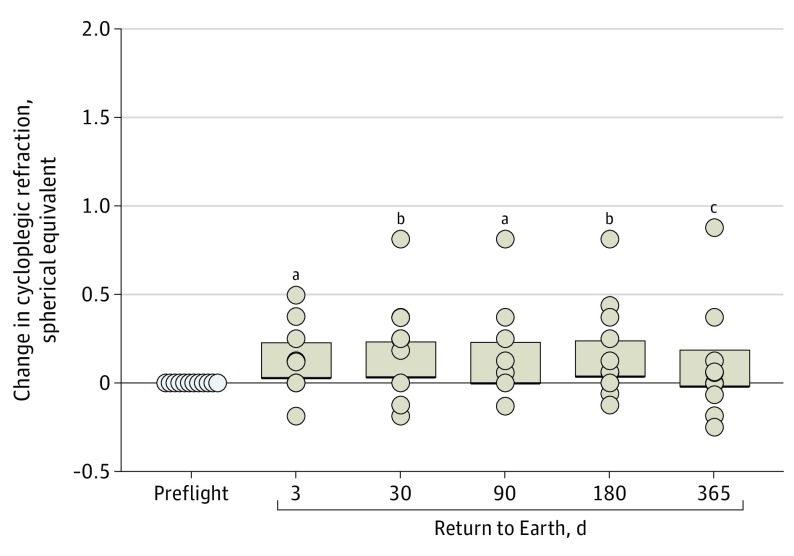
Main outcomes and measures: Changes in ocular structure (peripapillary edema, axial length, anterior chamber depth, and refraction) hypothesized to be associated with the development of spaceflight-associated neuro-ocular syndrome during 6-month missions on board the ISS were assessed. Statistical analyses were conducted from August 2018 to January 2019.
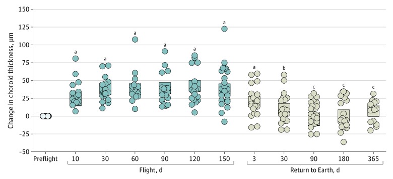
Results: Before launch, the 11 astronauts were a mean (SD) age of 45 (5) years, a mean (SD) height of 1.76 (0.05) m, and a mean (SD) weight of 75.3 (7.1) kg. Six astronauts did not have prior spaceflight experience, 3 had completed short-duration missions on board the Space Shuttle, and 2 had previous long-duration spaceflight missions on board the ISS. Their mean (SD) duration on board the ISS in the present study was 170 (19) days. Optic nerve head rim tissue and peripapillary choroidal thickness increased from preflight values during early spaceflight, with maximal change typically near the end of the mission (mean change in optic nerve head rim tissue thickness on flight day 150: 35.7 μm; 95% CI, 28.5-42.9 μm; P < .001; mean choroidal thickness change on flight day 150: 43 μm; 95% CI, 35-46 μm; P < .001). The mean postflight axial length of the eye decreased by 0.08 mm (95% CI, 0.10-0.07 mm; P < .001) compared with preflight measures, and this change persisted through the last examination (1 year after spaceflight: 0.05 mm; 95% CI, 0.07-0.03 mm; P < .001).
Conclusions and relevance: This study found that spaceflight-associated peripapillary optic disc edema and choroid thickening were observed bilaterally and occurred in both sexes. In addition, this study documented substantial peripapillary choroid thickening during spaceflight, which has never been reported in a prospective study cohort population and which may be a contributing factor in spaceflight-associated neuro-ocular syndrome. Data collection on spaceflight missions longer than 6 months will help determine whether the duration of the mission is associated with exacerbating these observed changes in ocular structure or visual function.
Conflict of interest statement
Figures


References
-
- Stenger MB, Tarver WJ, Brunstetter T, et al. NASA Human Research Program evidence report: human health countermeasures element: risk of spaceflight associated neuro-ocular syndrome (SANS). Published November 30, 2017. Accessed February 15, 2018. https://humanresearchroadmap.nasa.gov/evidence/reports/SANS.pdf?rnd=0.43...
