Review
. 2020 Apr 2;14(4):e0007951.
doi: 10.1371/journal.pntd.0007951. eCollection 2020 Apr.

Schistosome migration in the definitive host

Affiliations
Review

Schistosome migration in the definitive host

Catherine S Nation et al. PLoS Negl Trop Dis. .

Abstract

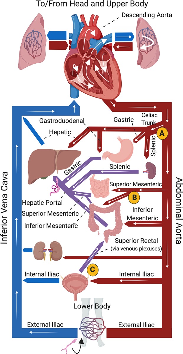
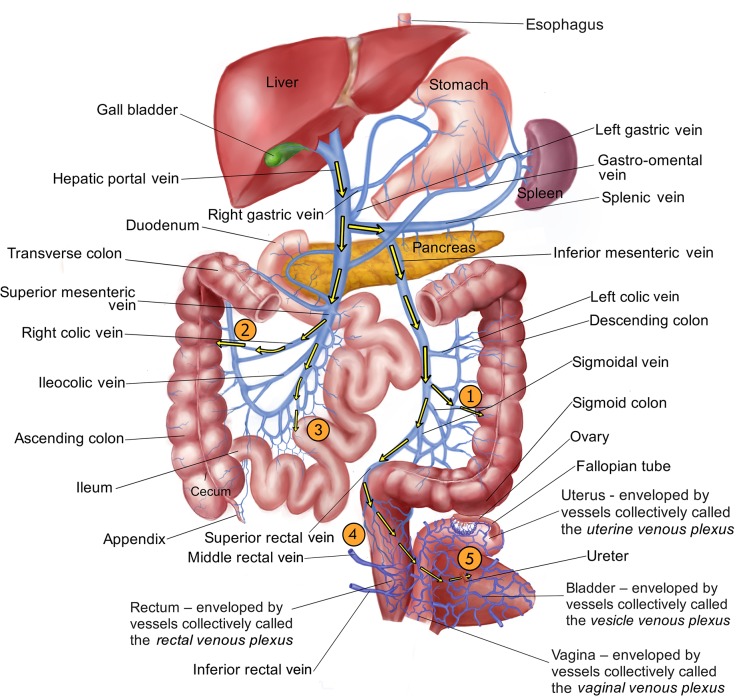
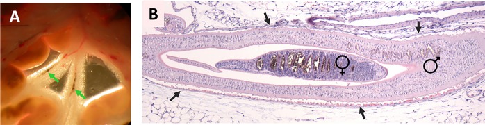
Schistosomes are parasitic blood flukes that infect >200 million people around the world. Free-swimming larval stages penetrate the skin, invade a blood vessel, and migrate through the heart and lungs to the vasculature of the liver, where maturation and mating occurs. From here, the parasite couples migrate to their preferred egg laying sites. Here, we compare and contrast what is known about the migration patterns within the definitive host of the three major species of human schistosome: Schistosoma mansoni, S. japonicum, and S. haematobium. We conclude that intravascular schistosomes are inexorable colonizers whose migration and egg laying strategy is profligate; all three species (and their eggs) can be found throughout the mesenteric venules, the rectal venous plexus, and, to a greater or lesser extent, the urogenital venous plexuses. In addition, it is common for parasite eggs to be deposited in locations that lack easy access to the exterior, further demonstrating the relentless exploratory nature of these intravascular worms.


Conflict of interest statement

The authors have declared that no competing interests exist.

Figures

Fig 1
Fig 1. Diagrammatical representation of core mammalian vasculature showing (white arrows) possible routes of migration of a schistosome juvenile to the portal vasculature, where the parasites mature.
A cercaria (depicted at bottom) has penetrated the skin of the lower body and invaded the vasculature, as suggested by the black arrow. White arrows track the parasite travelling via the inferior vena cava to the heart, then to the left lung before returning to the heart and entering the abdominal aorta. From the aorta, pathway “A” indicates parasites entering the celiac trunk to the gastroduodenal, hepatic, or splenic arteries to the liver. Pathway “B” indicates parasites entering the superior or inferior mesenteric arteries and moving through the hepatic portal vessels to the liver. Pathway “C” depicts parasites entering the internal iliac artery to the vessels of the bladder, from where they could reach the liver via the superior rectal and hepatic portal vasculature. Arteries are depicted in red, most veins in blue; hepatic portal veins in purple. Fig 1 was created with Biorender.com.
Fig 2
Fig 2. Diagrammatical representation of the hepato-portal and associated vasculature showing possible routes of migration (yellow arrows) of schistosomes from the blood vessels of the liver to their egg laying sites.
From the hepatic portal vein, pathway “1” indicates parasites entering the splenic vein, then the inferior mesenteric vein to the venules of the descending colon. Pathway “2” indicates parasites entering the superior mesenteric vein to right colic vein and the venules of the ascending colon. Pathway “3” indicates parasites leaving the superior mesenteric vein via the jejunal and ileal veins to the venules draining the small intestine (jejunum and ilium). Pathway “4” indicates parasites traveling via the inferior mesenteric vein into the superior rectal (hemorrhoidal) vein to the venules of the rectum. Finally, pathway “5” indicates parasites migrating beyond the vasculature of the rectum via the rectal venous plexus through the uterine and vaginal venous plexuses (in females, as illustrated, or the prostatic venous plexus in males, not shown) into the vesicle venous plexus draining the bladder. Image credit: Thanks to Taoli Shen for illustrating Fig 2.
Fig 3
Fig 3
(A) Exposed mesenteries of a 7-week infected female Swiss Webster mouse showing intravascular adult S. mansoni worms (indicated by arrows). (B) Hematoxylin and eosin–stained, formalin-fixed section of a blood vessel of a 7-week infected female Balb/c mouse containing an adult S. mansoni couple. The male worm and his female partner (within his gynaecophoric canal) are both indicated. The vascular endothelium bounding the worm couple is indicated by arrows.

Similar articles

Cited by

References

    1. Stirewalt MA. Schistosoma mansoni: cercaria to schistosomule. Adv Parasitol. 1974;12: 115–182. 10.1016/s0065-308x(08)60388-7 - DOI - PubMed
    1. Miller P, Wilson RA. Migration of the schistosomula of Schistosoma mansoni from the lungs to the hepatic portal system. Parasitology. 1980;80: 267–288. 10.1017/s0031182000000743 - DOI - PubMed
    1. Crabtree JE, Wilson RA. Schistosoma mansoni: an ultrastructural examination of skin migration in the hamster cheek pouch. Parasitology. 1985;91: 111–120. 10.1017/s0031182000056559 - DOI - PubMed
    1. Wheater PR, Wilson RA. Schistosoma mansoni: a histological study of migration in the laboratory mouse. Parasitology. 1979;79: 49–62. 10.1017/s0031182000051970 - DOI - PubMed
    1. He YX, Chen L. Schistosoma mansoni, S. haematobium, and S. japonicum: early events associated with penetration and migration of schistosomula through human skin. Exp Parasitol. 2002;102: 99–108. 10.1016/s0014-4894(03)00024-9 - DOI - PubMed

Publication types

MeSH terms