

Reproducibility, and repeatability of corneal topography measured by Revo NX, Galilei G6 and Casia 2 in normal eyes
- PMID: 32240192
- PMCID: PMC7117679
- DOI: 10.1371/journal.pone.0230589
Reproducibility, and repeatability of corneal topography measured by Revo NX, Galilei G6 and Casia 2 in normal eyes
Abstract
Purpose: To test the repeatability and reproducibility of the topography module in posterior segment spectral domain optical coherence tomography with Revo NX (new device) and to compare keratometry values obtained by a Scheimpflug tomography (Galilei G6) and a swept source OCT (Casia 2).
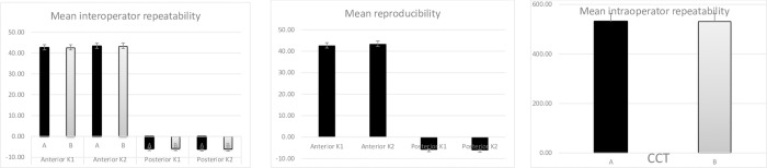
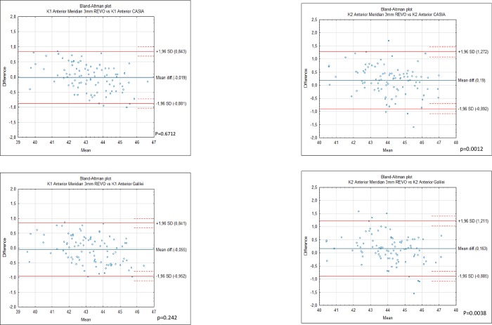
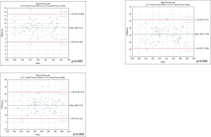
Methods: In this prospective study, healthy subjects with nonoperated eyes had their central corneal thickness (CCT), anterior and posterior K1/K2 corneal power measured with the new device. Two operators made 6 measurements on the new device to check intraobserver repeatability and reproducibility, and measurement on Casia 2 and Galilei G6. Bland-Altman plots were used to assess the agreement between the devices for each analyzed variable.
Results: 94 eyes (94 patients) were studied. All devices produced significantly different mean CCT, the highest for Galilei 569.13±37.58 μm followed by Casia 545.00 ±36.15 μm and Revo 537.39±35.92 μm. The mean anterior K1 was 43.21 ± 1.37 for Casia 2 43.21 ± 1.55 for Revo NX and 43.19 ± 1.39 for Galilei G6, and the differences were insignificant p = 0.617. The posterior K1 for Revo NX was -5.77 ± 0.25 whereas for Casia 2 it was -5.98±0.22 and for Galilei G6-6.09±0.28 D p< 0.0001. The Revo NX showed intraclass correlation coefficient ranging from 0.975 for the posterior K2 surface, and 0.994 for anterior K1 and 0.998 for CCT.
Conclusions: Revo NX is independent of the user and offers a high level of repeatability for the anterior and posterior cornea. The wide range of differences between the devices suggests they should not be used interchangeably.
Conflict of interest statement
Optopol Technology Ltd. provided the Revo NX equipment with corneal topography module used in this study. AW received a speaker's honorarium form Carl Zeiss and works as a consultant for Carl Zeiss Meditec. Aw has a patent pending for the OCT angiography algorithm. Polish patent office P.418979. This does not alter our adherence to PLOS ONE policies on sharing data and materials.
Figures

References
MeSH terms
LinkOut - more resources
Full Text Sources
Research Materials
Miscellaneous
