. 2020 Apr 2;15(4):e0230711.
doi: 10.1371/journal.pone.0230711. eCollection 2020.

Replication-inducible vaccinia virus vectors with enhanced safety in vivo

Affiliations

Replication-inducible vaccinia virus vectors with enhanced safety in vivo

Caitlin M O'Connell et al. PLoS One. .

Abstract

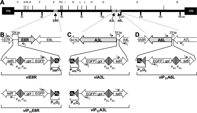
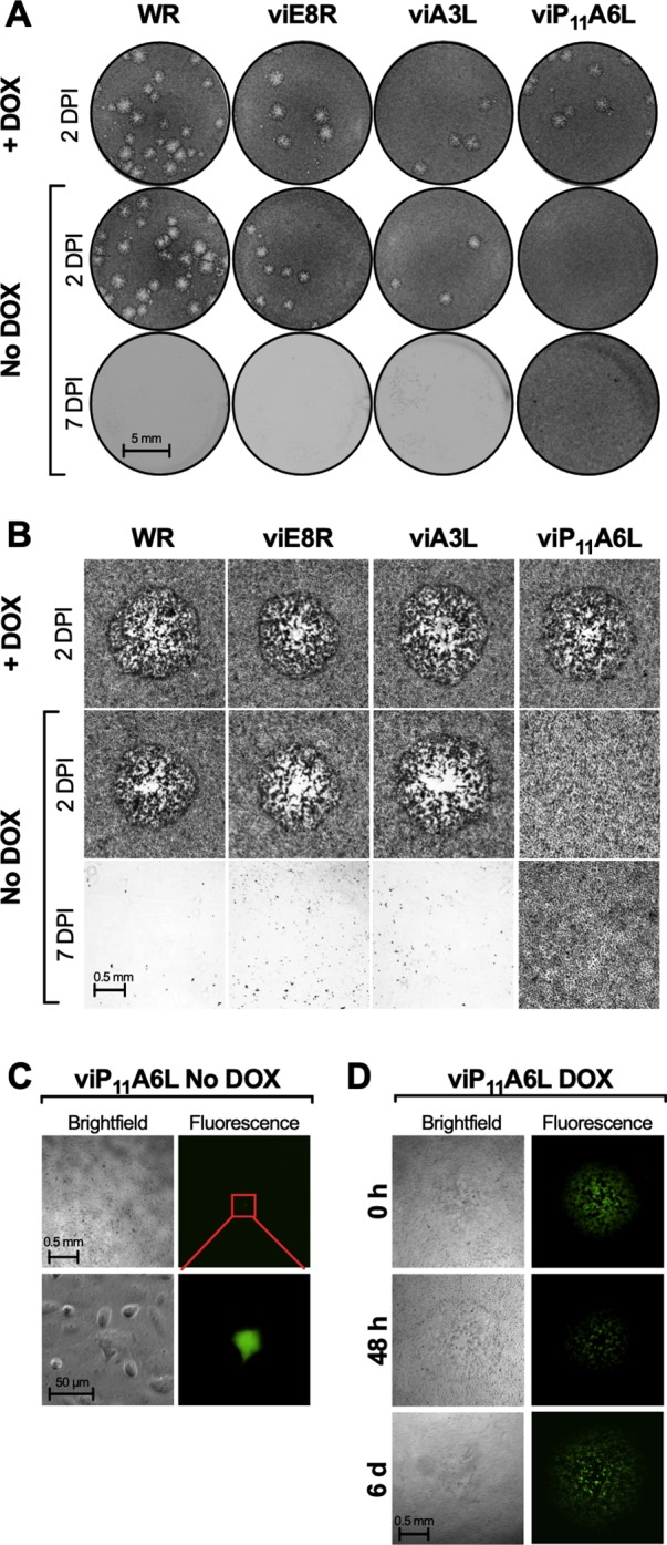
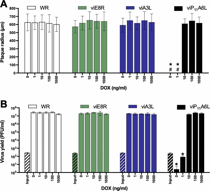
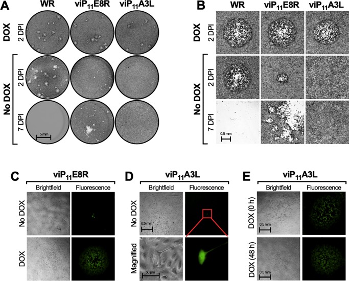
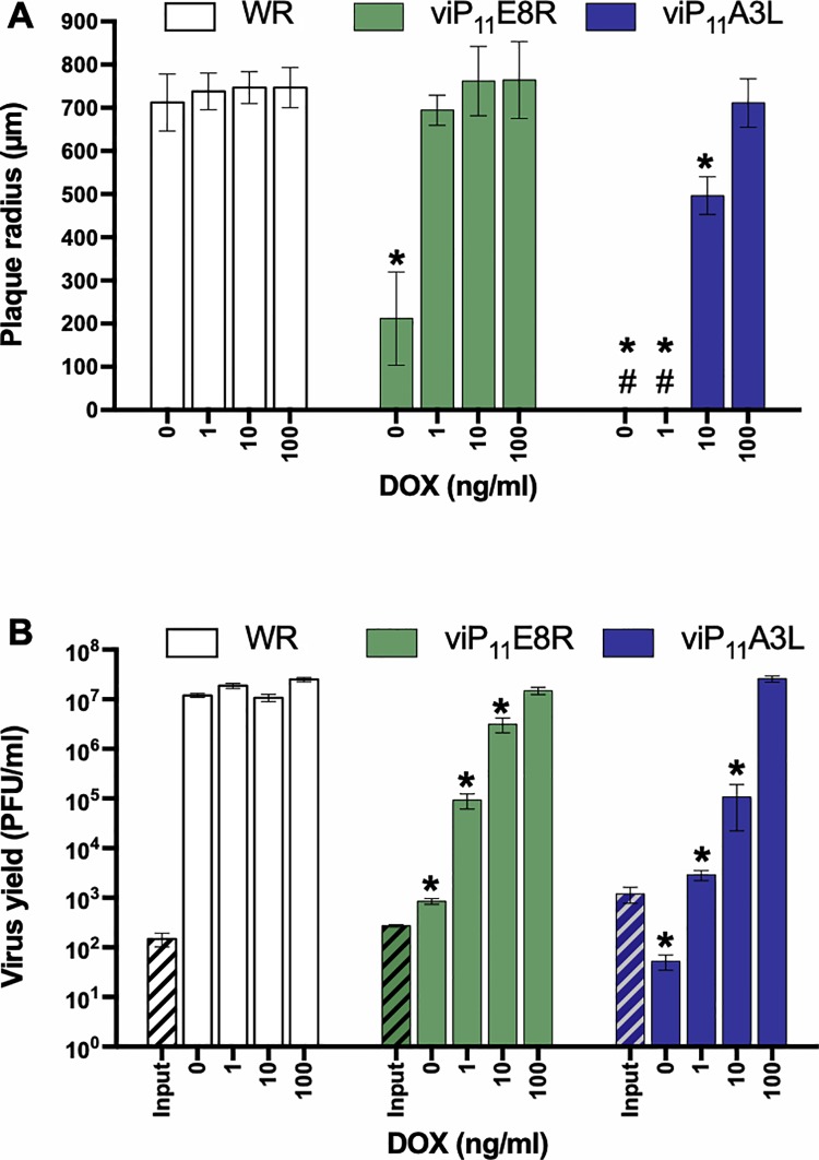
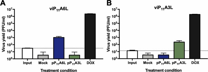
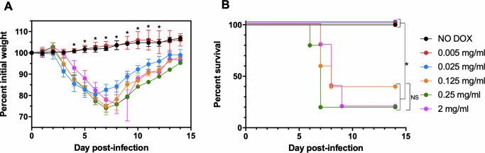
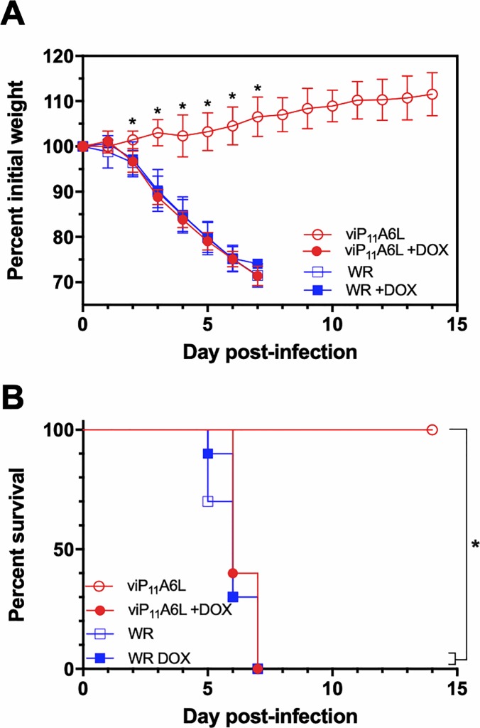
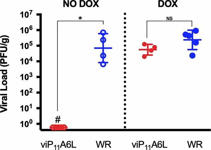
Vaccinia virus (VACV) has been used extensively as the vaccine against smallpox and as a viral vector for the development of recombinant vaccines and cancer therapies. Replication-competent, non-attenuated VACVs induce strong, long-lived humoral and cell-mediated immune responses and can be effective oncolytic vectors. However, complications from uncontrolled VACV replication in vaccinees and their close contacts can be severe, particularly in individuals with predisposing conditions. In an effort to develop replication-competent VACV vectors with improved safety, we placed VACV late genes encoding core or virion morphogenesis proteins under the control of tet operon elements to regulate their expression with tetracycline antibiotics. These replication-inducible VACVs would only express the selected genes in the presence of tetracyclines. VACVs inducibly expressing the A3L or A6L genes replicated indistinguishably from wild-type VACV in the presence of tetracyclines, whereas there was no evidence of replication in the absence of antibiotics. These outcomes were reflected in mice, where the VACV inducibly expressing the A6L gene caused weight loss and mortality equivalent to wild-type VACV in the presence of tetracyclines. In the absence of tetracyclines, mice were protected from weight loss and mortality, and viral replication was not detected. These findings indicate that replication-inducible VACVs based on the conditional expression of the A3L or A6L genes can be used for the development of safer, next-generation live VACV vectors and vaccines. The design allows for administration of replication-inducible VACV in the absence of tetracyclines (as a replication-defective vector) or in the presence of tetracyclines (as a replication-competent vector) with enhanced safety.


Conflict of interest statement

The authors have declared that no competing interests exist.

Figures

Fig 1
Fig 1. Genomic organization of the VACVs inducibly expressing the E8R, A3L, or A6L genes.
(A) Genome of the WR strain of VACV showing HindIII restriction fragments A through P and the location of the E8R, A3L, and A6L genes. Cassettes containing the putative E8R (PE8R) or A3L (PA3L) promoters, or the P11 promoter, followed by the tet operator (O2) were inserted upstream of the E8R, A3L, or A6L genes to generate the recombinant VACVs viE8R (B), viA3L (C), and viP11A6L (D), respectively. Replacement of PE8R and PA3L promoters with P11 resulted in viP11E8R (B, lower panel) and viP11A3L (C, lower panel). The cassettes also contain the tetR gene and the gpt-EGFP fusion gene under back-to-back synthetic early/late VACV promoters (PE/L). Arrows with numbers indicate primers (Table 2) used to amplify specific genomic regions for characterization of the viruses. ITR, inverted terminal repeat. Panels B-D are not drawn to scale.
Fig 2
Fig 2. viP11A6L forms plaques only in the presence of DOX.
BS-C-1 cell monolayers were infected with the indicated VACVs at approximately 5–20 PFU/well in the absence or presence of 1 μg/ml DOX and cells were stained with crystal violet 2 or 7 DPI (A and B) or imaged by brightfield (phase) and fluorescence microscopy (C and D). (A) Image of representative wells showing the plaque phenotypes. (B) Representative brightfield microscopic images of stained cells showing plaques, when present. WR refers to VACV WR (parental strain). In the absence of DOX only single EGFP+ cells were observed 2 DPI for viP11A6L (C), and under higher magnification, EGFP expression was contained to single cells and was the only indication of infection (red inset), suggesting abortive infections. When DOX was added at the time of infection (0 h), 48 h, or 6 days after infection (D), plaques were visible 2 days later (2, 4, or 8 DPI, respectively). Data is representative of two separate experiments.
Fig 3
Fig 3. viP11A6L replicates indistinguishably from WR in the presence of DOX.
(A) The effect of DOX on plaque size was examined by infecting BS-C-1 cell monolayers with the VACVs in the absence or presence of multiple concentrations of DOX. At 36 hpi, cells were stained with crystal violet and the size (radius) of approximately 20 representative isolated plaques was measured (# indicates absence of plaques). (B) The effect of DOX on virus replication was examined by infecting BS-C-1 cell monolayers with the indicated VACVs at an MOI of 0.01. Cells were collected immediately to determine input titer (hatched bars) or after 48 h in the absence or presence of multiple concentrations of DOX to determine virus yield (solid bars). Titers were determined on BS-C-1 cells in the presence of 1 μg/ml DOX. The data shown represent the mean viral yields from triplicate samples assayed in duplicate. Error bars indicate standard deviation. An asterisk indicates statistically significant differences (p < 0.05 by two-way ANOVA followed by Tukey’s multiple comparisons test) between WR and the inducible viruses at a given DOX concentration. Data is representative of two separate experiments.
Fig 4
Fig 4. viP11A3L does not form plaques and causes abortive infections in the absence of DOX.
BS-C-1 cell monolayers were infected with the indicated VACVs at approximately 5–20 PFU/well in the absence or presence of 1 μg/ml DOX and cells were stained with crystal violet 2 or 7 DPI (A and B) or imaged by brightfield (phase) and fluorescence microscopy (C, D, and E). (A) Image of representative wells showing the plaque phenotypes. (B) Representative brightfield microscopic images of stained cells showing plaques, when present. (C) In the absence of DOX smaller plaques formed 2 DPI with viP11E8R. (D) In the absence of DOX, EGFP expression was contained to single viP11A3L-infected cells and was the only indication of infection. (E) When DOX was added at the time of infection or 48 h after infection, plaques were visible 2 and 4 days later, respectively. Data is representative of two separate experiments.
Fig 5
Fig 5. viP11A3L replicates indistinguishably from wild-type VACV in the presence of DOX.
(A) The effect of DOX on plaque size was examined by infecting BS-C-1 cell monolayers with the VACVs in the absence or presence of multiple concentrations of DOX. At 36 hpi, cells were stained with crystal violet and the size (radius) of approximately 20 representative isolated plaques was measured (# indicates absence of plaques). (B) The effect of DOX on virus replication was examined by infecting BS-C-1 cell monolayers with the indicated VACVs at an MOI of 0.01. Cells were collected immediately to determine input titer (hatched bars) or after 48 h in the absence or presence of multiple concentrations of DOX to determine virus yield (solid bars). Titers were determined on BS-C-1 cells in the presence of 1 μg/ml DOX. The data shown represent the mean viral yields from triplicate samples assayed in duplicate. Error bars indicate standard deviation. An asterisk indicates statistically significant differences (p < 0.05 by two-way ANOVA followed by Tukey’s multiple comparisons test) between WR and the inducible viruses at a given DOX concentration.
Fig 6
Fig 6. Transient complementation allows viP11A6L and viP11A3L replication in the absence of DOX.
BS-C-1 cell monolayers were infected with viP11A6L (A) or viP11A3L (B) at an MOI of 0.01 in the absence of DOX and transfected with plasmids expressing the A6L (pP11A6L) or A3L (pP11A3L) genes under the constitutive VACV P11 promoter, or no plasmid (mock). Infections were also performed in the presence of 1 μg/ml DOX (DOX). Cells were collected immediately after infection (input, dotted line) or 2 DPI. Virus yield was determined by plaque assay on BS-C-1 cells in the presence of 1 μg/ml DOX. The data shown represent the mean viral yields from triplicate samples assayed in duplicate. Error bars indicate standard deviation. Data are representative of two separate experiments.
Fig 7
Fig 7. viP11A6L causes weight loss in mice in the presence of DOX.
Groups of female CB6F1/J mice (n = 5) were inoculated intranasally with ~5 × 10⁴ PFU viP11A6L in the absence or presence of different concentrations of DOX in drinking water. Weight and mortality were assessed daily. Animals were euthanized if weight loss was ≥ 25%. (A) Mean group weights are displayed as a percentage of group weight on day 0. An asterisk represents statistically significant differences (p < 0.01) determined using one-way ANOVA followed by Dunnett’s multiple comparisons test comparing NO DOX to all other groups at each day post-infection. Error bars indicate standard deviation. (B) Percent survival is shown. An asterisk represents statistically significant differences (p < 0.05) by log-rank (Mantel-Cox) test for differences in survival adjusted for multiple comparisons using the Bonferroni post-hoc test. NS = not significant.
Fig 8
Fig 8. viP11A6L causes weight loss and mortality similar to WR in the presence of DOX.
Groups of female CB6F1/J mice (n = 10) were inoculated intranasally with ~2 × 10⁶ PFU viP11A6L or WR in the absence or presence of DOX. Weight and mortality were assessed daily. Animals were euthanized if weight loss was ≥ 25%. Mean group weights as a percentage of group weight on Day 0 (A), or percent survival (B) are shown. Asterisks indicate statistical significance (p < 0.01) by one-way ANOVA followed by Sidak’s multiple comparisons test (A), or by log-rank (Mantel-Cox) test between indicated groups adjusted for multiple comparisons using the Bonferroni post-hoc test (B). Error bars indicate standard deviation.
Fig 9
Fig 9. viP11A6L replicates indistinguishably from wild-type VACV in mice treated with DOX.
Groups of five female CB6F1/J were inoculated intraperitoneally with ~2 × 10⁶ PFU viP11A6L or WR in the presence or absence of DOX in drinking water. Mice were euthanized 6 DPI, and ovaries collected and processed. Ovarian homogenates were added to BS-C-1 cells in the presence of 1 μg/ml DOX to determine viral loads (# indicates absence of plaques via plaque assay). Asterisk indicates statistically significant differences (p < 0.01) by Mann-Whitney test between groups in each DOX treatment. NS = not significant.

References

    1. Wehrle PF. A reality in our time—certification of the global eradication of smallpox. J Infect Dis. 1980;142(4):636–8. Epub 1980/10/01. 10.1093/infdis/142.4.636 . - DOI - PubMed
    1. Prevention CfDCa. Update: multistate outbreak of monkeypox—Illinois, Indiana, Kansas, Missouri, Ohio, and Wisconsin, 2003. MMWR Morb Mortal Wkly Rep. 2003;52(27):642–6. Epub 2003/07/12. . - PubMed
    1. Reynolds MG, Damon IK. Outbreaks of human monkeypox after cessation of smallpox vaccination. Trends Microbiol. 2012;20(2):80–7. Epub 2012/01/14. 10.1016/j.tim.2011.12.001 . - DOI - PubMed
    1. Durski KN, McCollum AM, Nakazawa Y, Petersen BW, Reynolds MG, Briand S, et al. Emergence of Monkeypox—West and Central Africa, 1970–2017. MMWR Morb Mortal Wkly Rep. 2018;67(10):306–10. Epub 2018/03/16. 10.15585/mmwr.mm6710a5 . - DOI - PMC - PubMed
    1. Fenner F. Smallpox and its eradication. Geneva: World Health Organization; 1988. xvi, 1460 p. p.

Publication types