. 2020 Jul 1;202(1):100-111.
doi: 10.1164/rccm.201906-1232OC.

Nanoparticle Delivery of Proangiogenic Transcription Factors into the Neonatal Circulation Inhibits Alveolar Simplification Caused by Hyperoxia

Affiliations

Nanoparticle Delivery of Proangiogenic Transcription Factors into the Neonatal Circulation Inhibits Alveolar Simplification Caused by Hyperoxia

Craig Bolte et al. Am J Respir Crit Care Med. .

Abstract

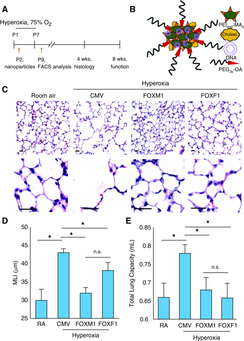
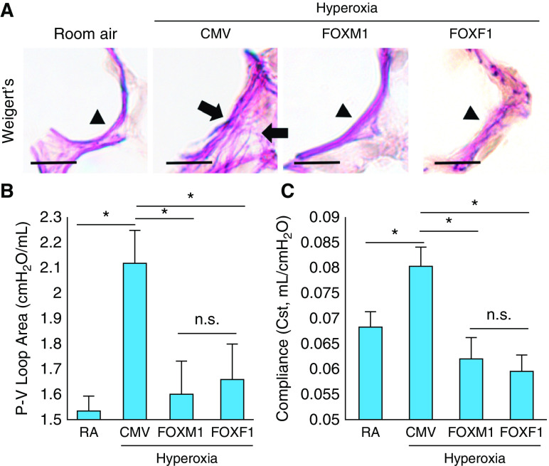
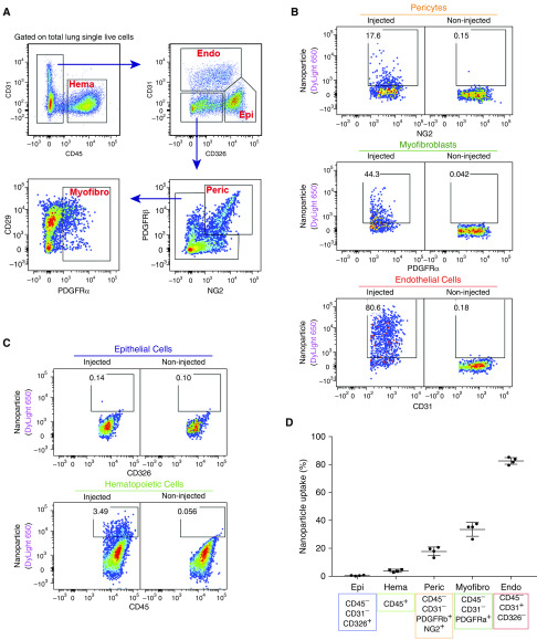
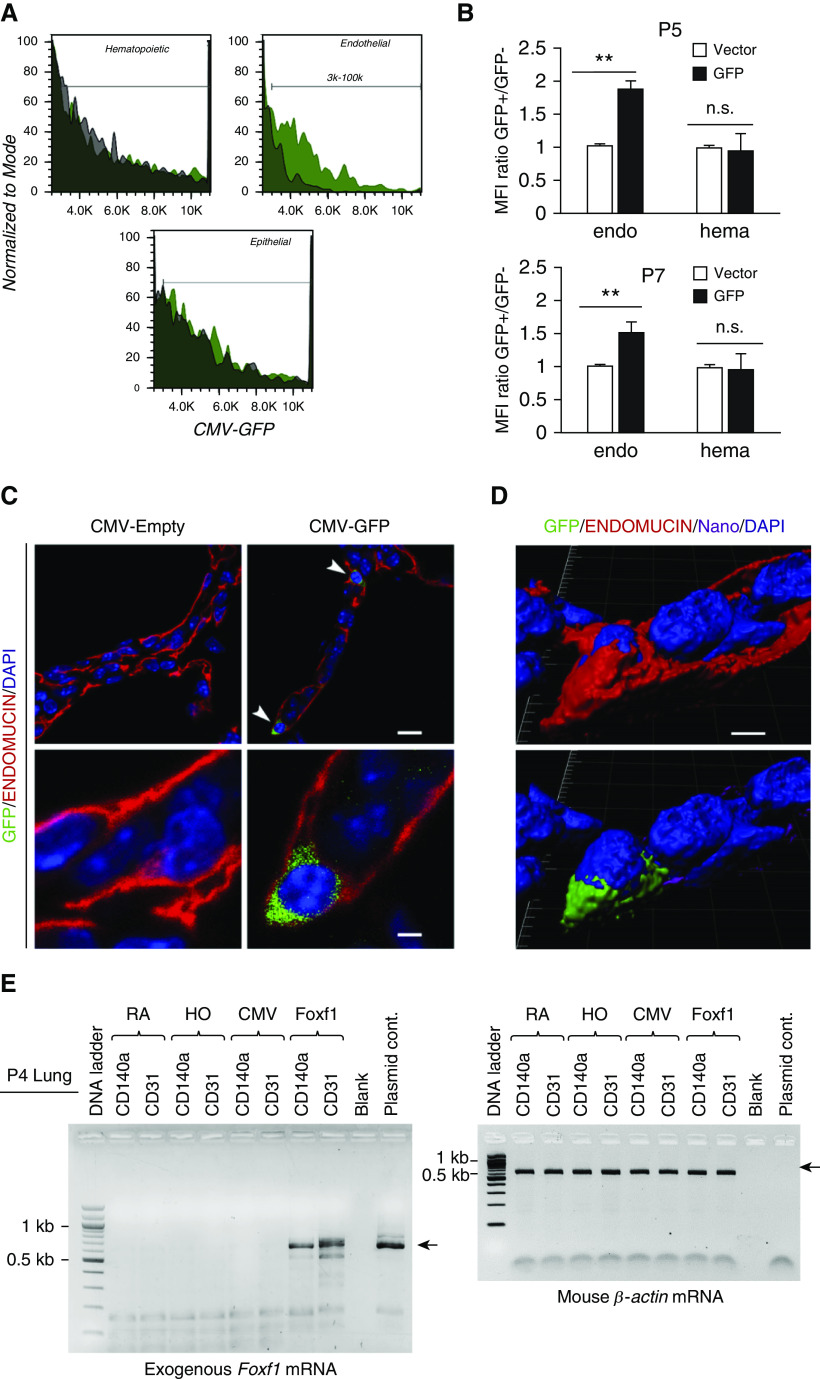
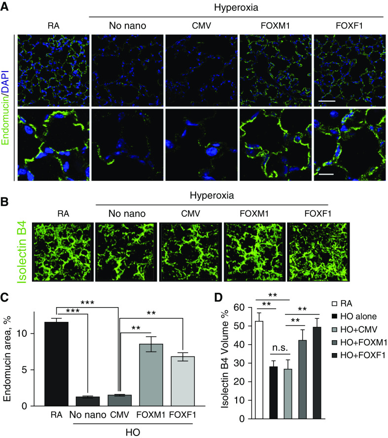
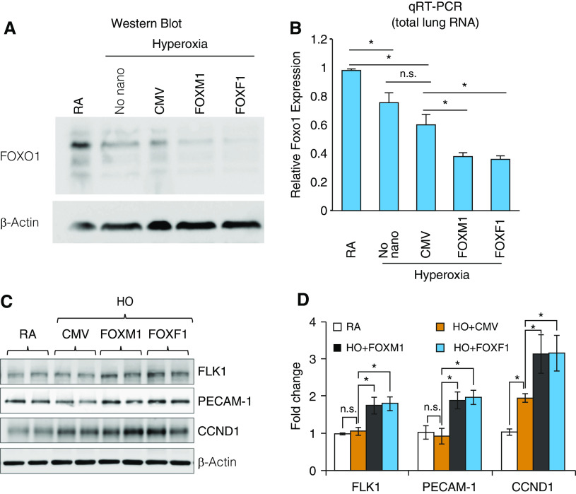
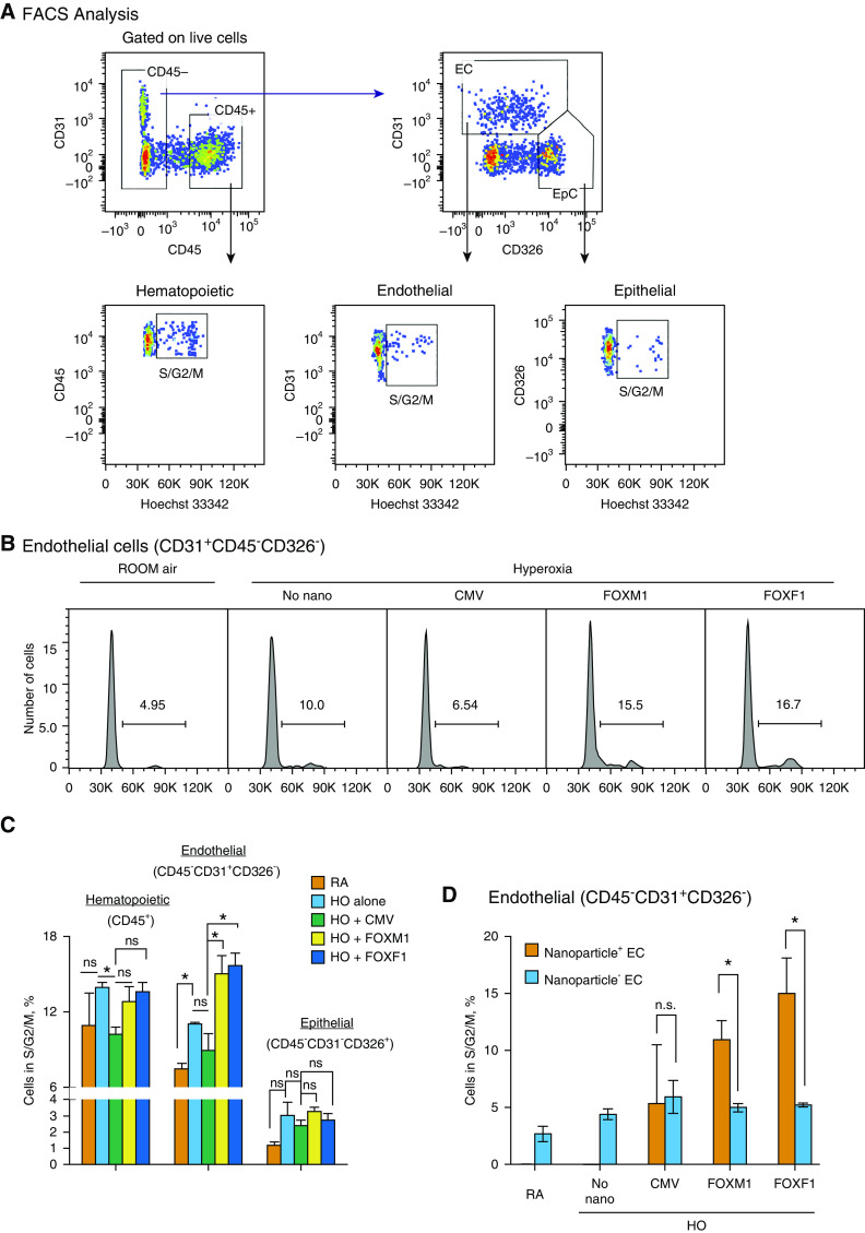
Rationale: Advances in neonatal critical care have greatly improved the survival of preterm infants, but the long-term complications of prematurity, including bronchopulmonary dysplasia (BPD), cause mortality and morbidity later in life. Although VEGF (vascular endothelial growth factor) improves lung structure and function in rodent BPD models, severe side effects of VEGF therapy prevent its use in patients with BPD.Objectives: To test whether nanoparticle delivery of proangiogenic transcription factor FOXM1 (forkhead box M1) or FOXF1 (forkhead box F1), both downstream targets of VEGF, can improve lung structure and function after neonatal hyperoxic injury.Methods: Newborn mice were exposed to 75% O2 for the first 7 days of life before being returned to a room air environment. On Postnatal Day 2, polyethylenimine-(5) myristic acid/polyethylene glycol-oleic acid/cholesterol nanoparticles containing nonintegrating expression plasmids with Foxm1 or Foxf1 cDNAs were injected intravenously. The effects of the nanoparticles on lung structure and function were evaluated using confocal microscopy, flow cytometry, and the flexiVent small-animal ventilator.Measurements and Main Results: The nanoparticles efficiently targeted endothelial cells and myofibroblasts in the alveolar region. Nanoparticle delivery of either FOXM1 or FOXF1 did not protect endothelial cells from apoptosis caused by hyperoxia but increased endothelial proliferation and lung angiogenesis after the injury. FOXM1 and FOXF1 improved elastin fiber organization, decreased alveolar simplification, and preserved lung function in mice reaching adulthood.Conclusions: Nanoparticle delivery of FOXM1 or FOXF1 stimulates lung angiogenesis and alveolarization during recovery from neonatal hyperoxic injury. Delivery of proangiogenic transcription factors has promise as a therapy for BPD in preterm infants.

Keywords: FOX transcription factors; VEGF signaling; bronchopulmonary dysplasia; nanoparticle gene delivery systems; neonatal hyperoxic lung injury.


Figures

Figure 1.
Figure 1.
Nanoparticle delivery of FOXM1 (forkhead box M1) or FOXF1 (forkhead box F1) decreases alveolar simplification caused by neonatal hyperoxia. (A) Schematic representation of 7-day hyperoxia treatment of wild-type newborn mice. Control mice were exposed to room air (RA). Polyethylenimine-(5) myristic acid/polyethylene glycol oleic acid/cholesterol (PEI600-MA5/PEG-OA/Cho) nanoparticles were delivered at Postnatal Day 2 (P2) via the facial vein. (B) Structure of PEI600-MA5/PEG-OA/Cho nanoparticles containing plasmid DNA, PEI600-MA5, and polyethylene glycol oleic acid (PEG2k-OA). (C) Hematoxylin and eosin (H&E) staining of paraffin-embedded lung sections shows alveolar simplification in hyperoxia-treated mice. Mice were exposed to hyperoxia or RA from P1 to P7, followed by RA exposure until lung harvest at P28. Nanoparticle delivery was performed at P2. Delivery of either FOXM1 or FOXF1 expression vectors improves lung structure in hyperoxia-treated mice compared with cytomegalovirus (CMV)-empty control. Scale bars, 50 μm. (D) Nanoparticle delivery of FOXM1 or FOXF1 decreases alveolar simplification. Mean linear intercept (MLI) was calculated using 15 random H&E–stained lung fields (n = 4–6 mice per group). (E) Nanoparticle delivery of FOXM1 or FOXF1 normalizes TLC in hyperoxia-treated mice. TLC was measured using the flexiVent ventilator when mice were 8 weeks of age (n = 4–6 mice per group). Error bars are mean ± SE. *P < 0.05. FACS = fluorescence-activated cell sorter; n.s. = not significant.
Figure 2.
Figure 2.
Nanoparticle delivery of FOXM1 (forkhead box M1) or FOXF1 (forkhead box F1) improves lung function in hyperoxia-treated lungs. (A) Elastin staining of paraffin-embedded lung sections shows disorganization of elastin fibers in hyperoxia-treated lungs (arrows). Mice were exposed to hyperoxia or room air (RA) from Postnatal Day 1 (P1) to P7, followed by RA exposure until lung harvest at P28. Nanoparticle delivery was performed at P2. Delivery of either FOXM1 or FOXF1 expression vectors improves elastin fiber deposition in alveolar septa of hyperoxia-treated mice. Arrowheads show normal elastic fibers. Scale bars, 50 μm. (B and C) Nanoparticle delivery of FOXM1 or FOXF1 improves lung function in hyperoxia-treated mice. Pressure–volume (P-V) loop area and lung compliance were measured using the flexiVent ventilator when mice were 8 weeks of age (n = 4–6 mice per group). Error bars are mean ± SE. *P < 0.05. CMV = cytomegalovirus; n.s. = not significant.
Figure 3.
Figure 3.
Polyethylenimine-(5) myristic acid/polyethylene glycol oleic acid/cholesterol (PEI600-MA5/PEG-OA/Cho) nanoparticles efficiently target endothelial cells in the neonatal lung. (A) Fluorescence-activated cell sorter (FACS) gating strategy to identify hematopoietic cells (Hema; CD45+CD31), endothelial cells (Endo; CD31+CD45CD326), epithelial cells (Epi; CD326+CD45CD31), pericytes (Peric; NG2+PDGFRb+CD45CD31CD326), and myofibroblasts (Myofibro; PDGFRa+CD45CD31CD326). DyLight 650–labeled nanoparticles were delivered at Postnatal Day 2 (P2). FACS analysis of enzymatically digested lung tissue was performed at P5. (B and C) Dot plots show the presence of nanoparticles in different populations of pulmonary cells. Noninjected mice were used as control animals to identify cells containing nanoparticles. (D) Percentage of nanoparticle-targeted cells is shown among pericytes; myofibroblasts; and epithelial, endothelial, and hematopoietic cells (n = 4 mice per group). Error bars are mean ± SE. NG2 = melanoma-associated chondroitin sulfate proteoglycan 4; PDGFRα = platelet-derived growth factor receptor-α; PDGFRβ = platelet-derived growth factor receptor-β.
Figure 4.
Figure 4.
Polyethylenimine-(5) myristic acid/polyethylene glycol oleic acid/cholesterol (PEI600-MA5/PEG-OA/Cho) nanoparticles deliver cytomegalovirus (CMV)-GFP (green fluorescent protein) reporter plasmid to pulmonary endothelial cells. (A and B) GFP fluorescence is detected in pulmonary endothelial cells after treatment with PEI600-MA5/PEG-OA/Cho nanoparticles. CMV-GFP plasmid was encapsulated into nanoparticles, and the nanoparticle–DNA complexes were injected at Postnatal Day 2 (P2). Lungs were harvested at P5 and P7 and used for fluorescence-activated cell sorter (FACS) analysis. Histograms in A show GFP fluorescence in different cell types. GFP (green area) is detected in endothelial cells (CD31+CD45CD326). Autofluorescence is shown as the black area. Data were quantitated in B by comparing GFP mean fluorescence intensity (MFI) with autofluorescence in each cell type (n = 3–4 mice per group). Error bars are mean ± SE. **P < 0.01. (C) Confocal images show that GFP is present in perinuclear regions of endothelial cells stained with endomucin (arrowheads). Lungs of mice treated with nanoparticles containing CMV–empty plasmid were used as controls. Scale bars: top panels, 10 μm; bottom panels, 2 μm. (D) High-magnification confocal images show the presence of GFP (green) and DyLight 650 quantum dots (purple) in cytoplasm of microvascular endothelial cells stained with endomucin. Cell nuclei were stained with DAPI (blue). Cell surface endomucin was removed using a deconvolution option in Imaris software (imaris.oxinst.com) (bottom image), indicating the presence of GFP and DyLight 650 inside the cell. Scale bars, 2 μm. (E) RT-PCR shows the presence of exogenous, plasmid-derived Foxf1 (forkhead box F1) mRNA in endothelial cells (CD31+CD45CD326) and myofibroblasts (CD140a+CD31CD45CD326) that were FACS sorted from P4 lungs. β-Actin was used as a loading control. endo = endothelial cells; hema = hematopoietic cells; HO = hyperoxia; n.s. = not significant; RA = room air.
Figure 5.
Figure 5.
Nanoparticle delivery of FOXM1 (forkhead box M1) or FOXF1 (forkhead box F1) improves alveolar microvascular network in hyperoxia (HO)-treated mice. (A) Immunostaining for endomucin shows alveolar microvascular networks (green) in P28 lungs. Mice were exposed to HO or room air (RA) from Postnatal Day 1 (P1) to P7, followed by RA exposure until lung harvest at P28. Nanoparticles containing cytomegalovirus (CMV)-Foxm1, CMV-Foxf1, or CMV-empty (control) were delivered at P2. DAPI was used to stain cell nuclei. The alveolar microvascular network is improved after nanoparticle delivery of FOXM1 or FOXF1. Scale bars: top panels, 50 μm; bottom panels, 10 μm. (B) Nanoparticle delivery of FOXM1 or FOXF1 increases capillary density in HO-injured lungs. Isolectin B4 was injected i.v. 1 hour before harvesting the mice at P14 (n = 3–4 mice per each group). (C and D) Endomucin-stained area (C) and isolectin B4–labeled volume (D) were quantified using 10 random lung images (n = 3–6 mice per group). Error bars are mean ± SD. **P < 0.01 and ***P < 0.001. n.s. = not significant.
Figure 6.
Figure 6.
Nanoparticle delivery of FOXM1 (forkhead box M1) or FOXF1 (forkhead box F1) alters expression of cell cycle regulatory genes in hyperoxia (HO)-treated lungs. (A) Immunoblots show the amounts of FOXO1 (forkhead box O1) and β-actin proteins in lung extracts after nanoparticle delivery of FOXM1 or FOXF1. Mice were exposed to HO or room air (RA) from Postnatal Day 1 (P1) to P7, followed by RA exposure. Nanoparticle–DNA complexes were injected at P2. Cytomegalovirus (CMV)–empty plasmid was used as a control. (B) qRT-PCR shows the expression of Foxo1 mRNA in whole-lung RNA after nanoparticle delivery of FOXM1 or FOXF1 (n = 3 mice per group). Foxo1 mRNA was normalized to β-actin mRNA. (C and D) Immunoblots show expression of FLK1 (vascular endothelial growth factor receptor 2), PECAM-1 (platelet endothelial cell adhesion molecule 1), and CCND1 (cyclin D1) in lung protein extracts after nanoparticle delivery of FOXM1 or FOXF1. Images were quantified using densitometry (n = 3–4 mice per group). Error bars are mean ± SE. *P < 0.05. n.s. = not significant.
Figure 7.
Figure 7.
Nanoparticle delivery of FOXM1 (forkhead box M1) or FOXF1 (forkhead box F1) increases endothelial cell proliferation during the recovery period after neonatal hyperoxia (HO). (A) Fluorescence-activated cell sorter (FACS) gating strategy to identify hematopoietic (CD45+CD31), epithelial (EpC; CD326+CD45CD31), and endothelial cells (EC; CD31+CD45CD326) in mouse lung tissue. Mice were exposed to HO or room air (RA) from Postnatal Day 1 (P1) to P7, followed by RA exposure. FACS analysis of enzymatically digested lung tissue was performed 2 days after injury at P9. Dot plots show FACS analysis of cells obtained from HO-treated lungs. Hoechst 33342 dye was used to identify cells undergoing S, G2, and M phases of the cell cycle. (B and C) Histograms in B show the percentage of EC in S, G2, and M phases of the cell cycle after nanoparticle delivery of FOXM1 or FOXF1 compared with cytomegalovirus (CMV)-empty control. Data were quantitated in C and compared between different pulmonary cell types (n = 3–4 mice per group). Nanoparticle delivery of FOXM1 or FOXF1 increases the percentage of proliferating EC in HO-treated lungs. (D) Comparison of EC with and without nanoparticles. Cell proliferation is higher in EC containing nanoparticles with FOXM1 or FOXF1 (Nanoparticle+ EC) than in EC without nanoparticles (Nanoparticle EC) (n = 3–4 mice per group). Error bars are mean ± SE. *P < 0.05. n.s. = not significant.

Comment in

Similar articles

Cited by

References

    1. Ohtani S, Iwamaru A, Deng W, Ueda K, Wu G, Jayachandran G, et al. Tumor suppressor 101F6 and ascorbate synergistically and selectively inhibit non–small cell lung cancer growth by caspase-independent apoptosis and autophagy. Cancer Res. 2007;67:6293–6303. - PubMed
    1. Horiguchi M, Kojima H, Sakai H, Kubo H, Yamashita C. Pulmonary administration of integrin-nanoparticles regenerates collapsed alveoli. J Control Release. 2014;187:167–174. - PubMed
    1. Naderi N, Karponis D, Mosahebi A, Seifalian AM. Nanoparticles in wound healing; from hope to promise, from promise to routine. Front Biosci. 2018;23:1038–1059. - PubMed
    1. Jiang X, Malkovskiy AV, Tian W, Sung YK, Sun W, Hsu JL, et al. Promotion of airway anastomotic microvascular regeneration and alleviation of airway ischemia by deferoxamine nanoparticles. Biomaterials. 2014;35:803–813. - PMC - PubMed
    1. Abman SH. Impaired vascular endothelial growth factor signaling in the pathogenesis of neonatal pulmonary vascular disease. Adv Exp Med Biol. 2010;661:323–335. - PubMed

Publication types

MeSH terms