

High-resolution computed tomography manifestations of COVID-19 infections in patients of different ages
- PMID: 32240913
- PMCID: PMC7102649
- DOI: 10.1016/j.ejrad.2020.108972
High-resolution computed tomography manifestations of COVID-19 infections in patients of different ages
Abstract
Purpose: We aimed to compare chest HRCT lung signs identified in scans of differently aged patients with COVID-19 infections.
Methods: Case data of patients diagnosed with COVID-19 infection in Hangzhou City, Zhejiang Province in China were collected, and chest HRCT signs of infected patients in four age groups (<18 years, 18-44 years, 45-59 years, ≥60 years) were compared.
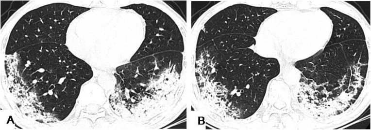
Results: Small patchy, ground-glass opacity (GGO), and consolidations were the main HRCT signs in 98 patients with confirmed COVID-19 infections. Patients aged 45-59 years and aged ≥60 years had more bilateral lung, lung lobe, and lung field involvement, and greater lesion numbers than patients <18 years. GGO accompanied with the interlobular septa thickening or a crazy-paving pattern, consolidation, and air bronchogram sign were more common in patients aged 45-59 years, and ≥60 years, than in those aged <18 years, and aged 18-44 years.
Conclusions: Chest HRCT manifestations in patients with COVID-19 are related to patient's age, and HRCT signs may be milder in younger patients.
Keywords: COVID-19; Ground-glass opacity; High-resolution computed tomography; Pure ground-glass opacity.
Copyright © 2020 Elsevier B.V. All rights reserved.
Conflict of interest statement
Declaration of Competing Interest No conflict of interest needs to be disclosed.
Figures
Comment in
-
Chest CT in coronavirus pandemic. Are there really age based radiological and clinical differences?Eur J Radiol. 2020 Aug;129:109107. doi: 10.1016/j.ejrad.2020.109107. Epub 2020 Jun 1. Eur J Radiol. 2020. PMID: 32512503 Free PMC article. No abstract available.
References
-
- Wuhan Municipal Health Commission; Wuhan, China: 2019. Report of Clustering Pneumonia of Unknown Etiology in Wuhan City. Google Scholar.
-
- WHO . 2020. Novel Coronavirus – China. Jan 12.http://www.who.int/csr/don/12-january-2020-novel-coronavirus-china/en/ (Accessed Jan 19, 2020)
-
- World Health Organization . 2020. Novel Coronavirus (2019-nCoV) Situation Report -11.https://www.who.int/docs/default-source/coronaviruse/situationreports/20... Sfvrsn = de7c0f7_4.
-
- World Health Organization . 2020. Coronavirus Disease 2019 (COVID-19) Situation Report – 54.https://www.who.int/docs/default-source/coronaviruse/situation-reports/2...
MeSH terms
LinkOut - more resources
Full Text Sources
