Review
. 2020 Apr 2;21(1):85.
doi: 10.1186/s13059-020-02003-5.

Lamina-associated domains: peripheral matters and internal affairs

Affiliations
Review

Lamina-associated domains: peripheral matters and internal affairs

Nolwenn Briand et al. Genome Biol. .

Abstract

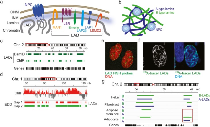
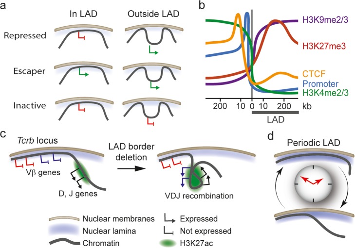
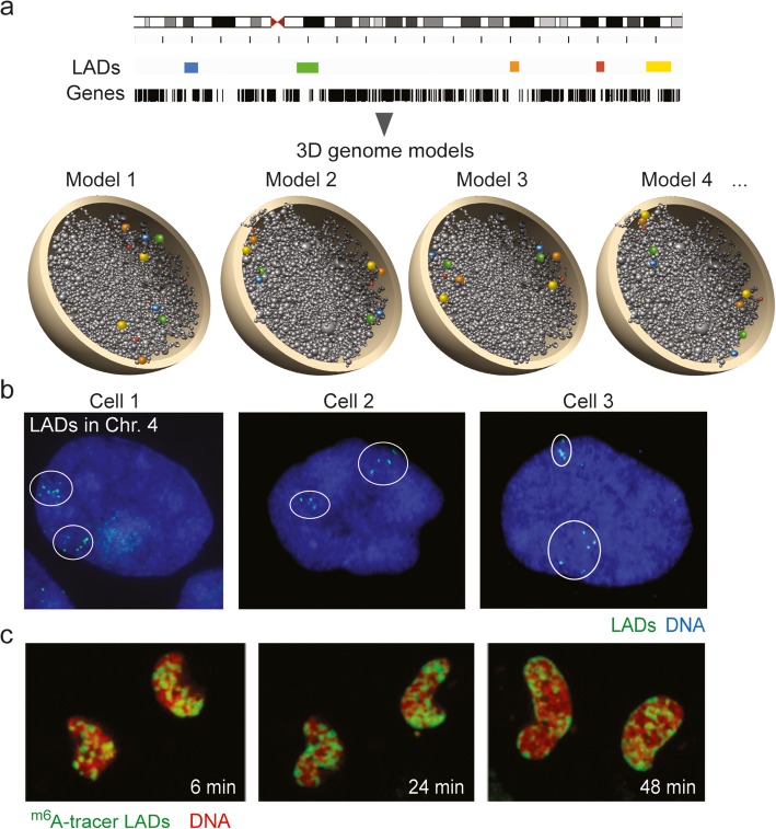
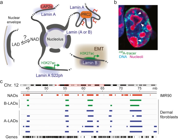
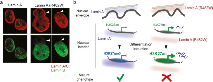
At the nuclear periphery, associations of chromatin with the nuclear lamina through lamina-associated domains (LADs) aid functional organization of the genome. We review the organization of LADs and provide evidence of LAD heterogeneity from cell ensemble and single-cell data. LADs are typically repressive environments in the genome; nonetheless, we discuss findings of lamin interactions with regulatory elements of active genes, and the role lamins may play in genome regulation. We address the relationship between LADs and other genome organizers, and the involvement of LADs in laminopathies. The current data lay the basis for future studies on the significance of lamin-chromatin interactions in health and disease.

Keywords: 3D genome; Chromatin; LAD; Lamin A mutation; Nuclear envelope; Nuclear lamin; Radial positioning.


Conflict of interest statement

The authors declare that they have no competing interests.

Figures

Fig. 1
Fig. 1
Associations of chromatin with the nuclear envelope. a Association of chromatin with the nuclear envelope via inner nuclear membrane proteins, lamina interactions, and interactions with proteins of the NPC. A sample of INM proteins interacting with the nuclear lamina and chromatin is depicted. Chromatin interacting with the NPC is loose and euchromatic, in contrast to the compact and heterochromatic nature of domains interacting with the lamina and INM proteins (LADs). b A- and B-type lamins form distinct filaments in the nuclear lamina. c Browser view of LADs identified by lamin B DamID-seq and lamin B ChIP-seq in a region of human chromosome 2. DamID data (HT1080 cells) are from [13]; ChIP data (dermal fibroblasts) are from [14]. d Detection of LAD using the Enriched Domain Detector (EDD) algorithm is using a tunable gap penalty parameter (Gap). “Gap 1” (here, an arbitrary calling corresponding to EDD’s default gap penalty with this dataset) is more stringent than “Gap 2.” ChIP data are from [14]. e FISH visualization of 25 LADs in two nuclei: note the dispersion of LADs around the nuclear periphery and in the nucleoplasm. Reproduced from [15] with permission. fm6A tracer visualization of LADs. The m6A tracer approach is a DamID variation enabling GFP labeling of Dam-methylated lamin-interacting sites in living cells [15]. Reproduced from [15] with permission. g Lamin A LAD (A-LAD) and lamin B LAD (B-LAD) detection by ChIP-seq analysis of lamin A/C and lamin B1 using the EDD algorithm [16], in a region of human chromosome 2. Boxed area, a variable A/B-LAD absent in adipose stem cells but detectable in adipocytes. LAD data are compiled from [8, 14, 16, 17]
Fig. 2
Fig. 2
LADs are dynamic and heterogeneous domains. a Three classes of promoters identified in LADs: “repressed promoters” become active when experimentally removed from their LAD context, “escaper promoters” are active even when in a LAD, and “inactive promoters” are inactive regardless of their LAD context [80]. b Enrichment profiles of indicated genomic features in LAD borders. c Deletion of a LAD border within the Tcrb locus spatially reorganizes the locus [70]. When inactive, the locus contains a LAD border that separates repressed Vβ genes from the H3K27 acetylated recombination center containing active D and J genes. Deletion of the LAD border elicits spreading of H3K27ac into the LAD, activation of a set of Vβ genes, and looping of the active Vβ genes and the recombination center, favoring VDJ recombination. d A subset of LADs in mouse liver displays periodic patterns of association with the nuclear lamina [78]. LAD borders, rather than entire LADs, are most often affected
Fig. 3
Fig. 3
Heterogeneous spatial distribution of LADs in individual cells. a Schematic representation of five LADs in a fictive ensemble ChIP-seq experiment (browser view) and in four fictive 3D genome models recapitulating the heterogeneity of LAD positioning in individual cells in a population [14]. b FISH image of five labeled LADs (within circles) in both homologous chromosomes 4 in human primary adipose stem cells. Note the variation between cells in the position of these LADs relative to each other and to the nuclear edge, and the heterogeneity of relative LAD positions between two homologous chromosomes (our unpublished data). cm6A tracer visualization of LAD reassembly post-mitosis in daughter nuclei. LAD targeting to the nuclear periphery occurs during G1 phase [15]. Reproduced from [15] with permission
Fig. 4
Fig. 4
Associations of lamins with chromatin in the nuclear interior. a Lamin associations with chromatin in various intranuclear contexts. A-type lamins interact with euchromatic LADs (via LAP2α) [31, 32] and with promoters and enhancers bound by Polycomb (PRC2) [–112]. Phosphorylated lamin A (lamin A S22ph) also interacts with H3K27ac-rich and active enhancers [113]. Lamins have been shown at the periphery of nucleoli [114, 115], but it remains unclear whether they are implicated in tethering NADs. Lamin B has also been reported to interact with acetylated enhancers and expressed genes during EMT [116] (boxed area). bm6A tracer visualization of lamin B LADs at the nuclear envelope and in the vicinity of nucleoli. Taken from [15] with permission. c Genome browser view of NADs and LADs in a segment of human chromosome 12. Note the overlap between NADs (from [117]) and LADs (from [14])
Fig. 5
Fig. 5
Lamin A mutations causing laminopathies alter peripheral and internal nuclear architecture. a Nuclear envelope deformations in the nuclei of patients bearing the FLPD2-causing lamin A (R482W) mutation. Note the nuclear blebs essentially devoid of lamin B (arrows); reproduced from [20] with permission. b The lamin A (R482W) mutant elicits chromatin rearrangements at the nuclear periphery and in the nuclear interior in patient cells and in stem cell models of FPLD2 [14, 111, 112]. Whereas peripheral A-LADs are moderately affected at the nuclear periphery, punctual (non-LAD) lamin A interactions with promoters and enhancers in the nuclear interior are impaired in cells expressing lamin A (R482W). In wild-type cells (left), a weakly H3K27-acetylated developmentally regulated promoter bound by lamin A is inactivated during differentiation, in association with increased lamin A binding and H3K27 methylation. In mutant cells (right), defective binding of lamin A (R482W) coincides with expression of the gene in the undifferentiated state, and unscheduled overexpression and H3K27 acetylation after induction of differentiation. Differentiation is, however, abortive [111, 112]

References

    1. Rowley MJ, Corces VG. Organizational principles of 3D genome architecture. Nat Rev Genet. 2018;19:789–800. - PMC - PubMed
    1. Zheng H, Xie W. The role of 3D genome organization in development and cell differentiation. Nat Rev Mol Cell Biol. 2019;20:535–550. - PubMed
    1. Lieberman-Aiden E, van Berkum NL, Williams L, Imakaev M, Ragoczy T, Telling A, Amit I, Lajoie BR, Sabo PJ, Dorschner MO, et al. Comprehensive mapping of long-range interactions reveals folding principles of the human genome. Science. 2009;326:289–293. - PMC - PubMed
    1. Dekker J, Marti-Renom MA, Mirny LA. Exploring the three-dimensional organization of genomes: interpreting chromatin interaction data. Nat Rev Genet. 2013;14:390–403. - PMC - PubMed
    1. Rao SS, Huntley MH, Durand NC, Stamenova EK, Bochkov ID, Robinson JT, Sanborn AL, Machol I, Omer AD, Lander ES, Aiden EL. A 3D map of the human genome at kilobase resolution reveals principles of chromatin looping. Cell. 2014;159:1665–1680. - PMC - PubMed

Publication types

LinkOut - more resources