. 2021 May 1;106(5):1290-1302.
doi: 10.3324/haematol.2020.246603.

Donor sex, age and ethnicity impact stored red blood cell antioxidant metabolism through mechanisms in part explained by glucose 6-phosphate dehydrogenase levels and activity

Affiliations

Donor sex, age and ethnicity impact stored red blood cell antioxidant metabolism through mechanisms in part explained by glucose 6-phosphate dehydrogenase levels and activity

Angelo D'Alessandro et al. Haematologica. .

Abstract

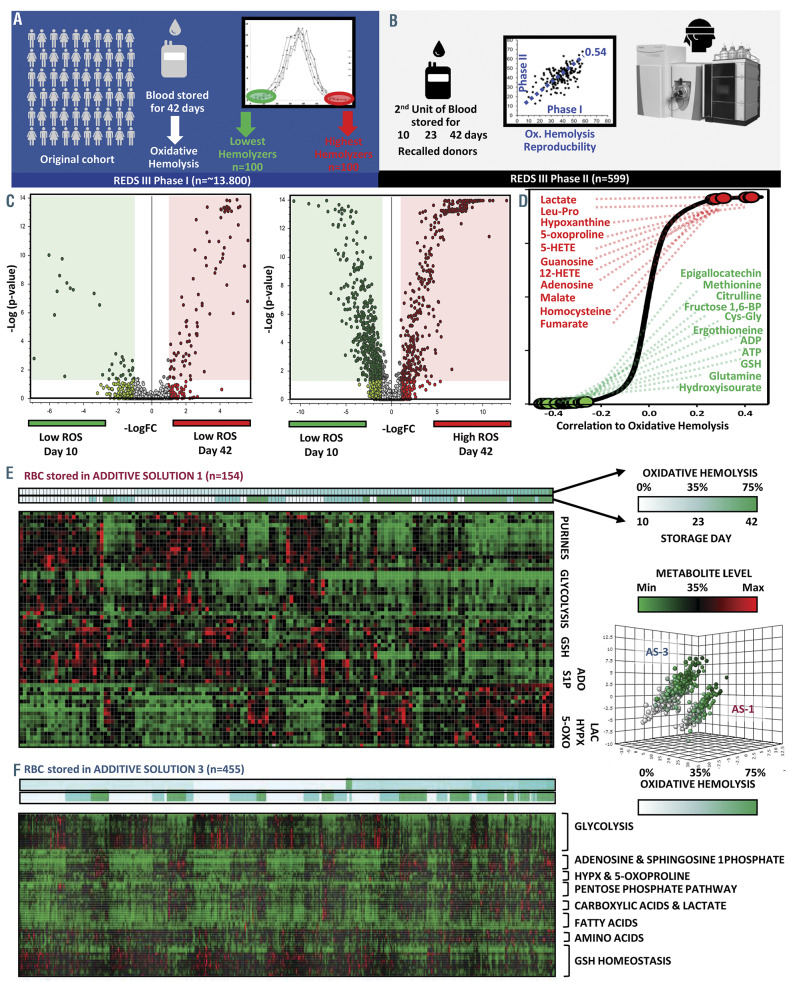
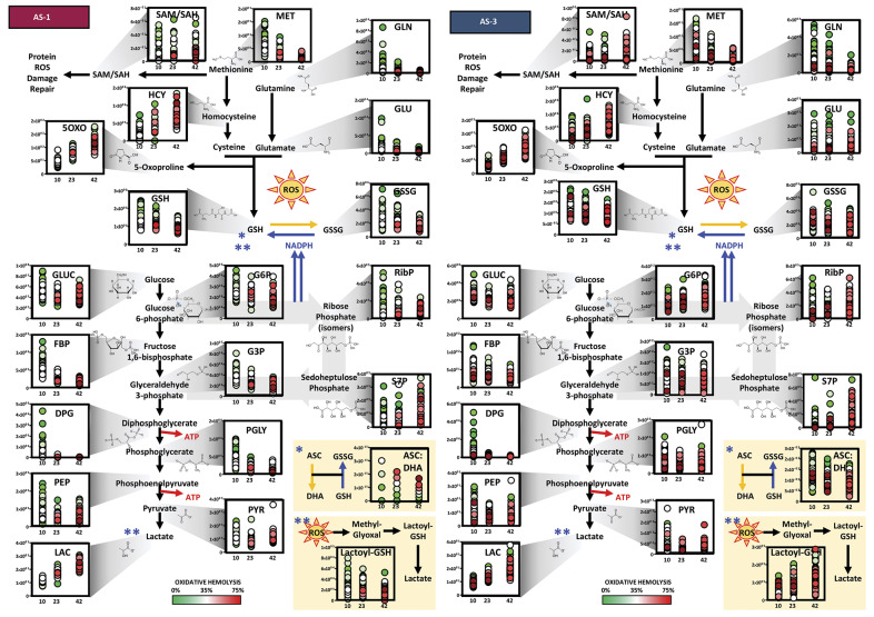
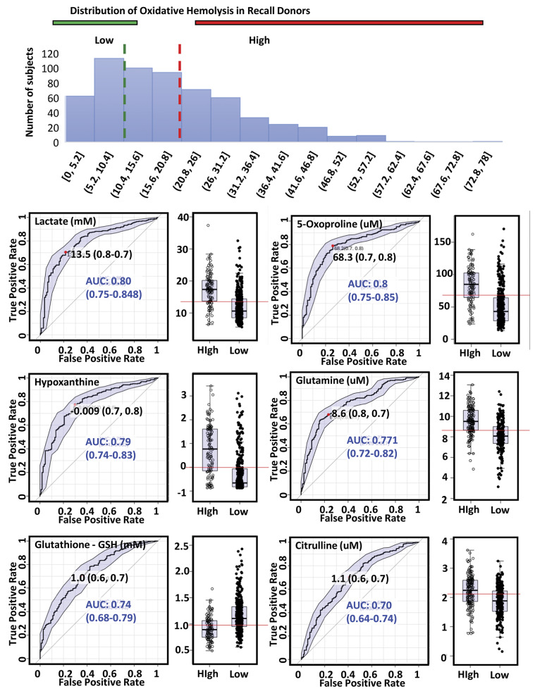
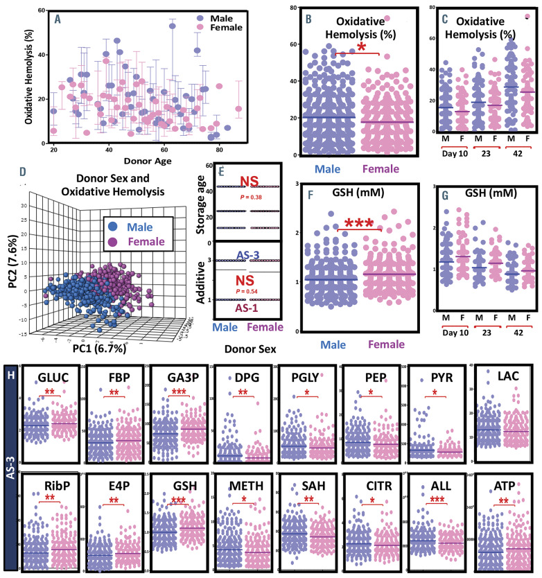
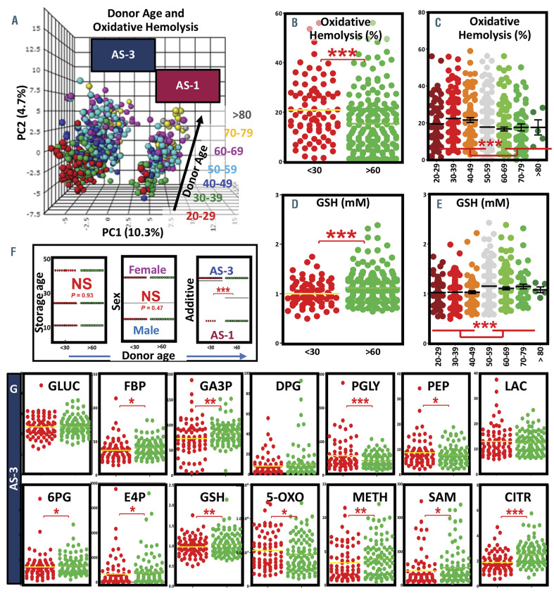
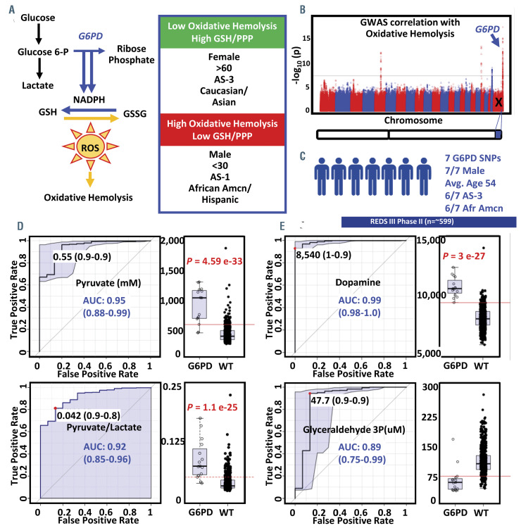
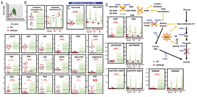
Red blood cell storage in the blood bank promotes the progressive accumulation of metabolic alterations that may ultimately impact the erythrocyte capacity to cope with oxidant stressors. However, the metabolic underpinnings of the capacity of RBCs to resist oxidant stress and the potential impact of donor biology on this phenotype are not known. Within the framework of the REDS-III RBC-Omics study, RBCs from 8,502 healthy blood donors were stored for 42 days and tested for their propensity to hemolyze following oxidant stress. A subset of extreme hemolyzers donated a second unit of blood, which was stored for 10, 23, and 42 days and profiled again for oxidative hemolysis and metabolomics (599 samples). Alterations of RBC energy and redox homeostasis were noted in donors with high oxidative hemolysis. RBCs from females, donors over 60 years old, donors of Asian/South Asian race-ethnicity, and RBCs stored in additive solution-3 were each independently characterized by improved antioxidant metabolism compared to, respectively, males, donors under 30 years old, Hispanic and African American race ethnicity donors, and RBCs stored in additive solution-1. Merging metabolomics data with results from an independent GWAS study on the same cohort, we identified metabolic markers of hemolysis and G6PD-deficiency, which were associated with extremes in oxidative hemolysis and dysregulation in NADPH and glutathione-dependent detoxification pathways of oxidized lipids. Donor sex, age, ethnicity, additive solution and G6PD status impact the metabolism of the stored erythrocyte and its susceptibility to hemolysis following oxidative insults.


Figures

Figure 1.
Figure 1.
Within the framework of the REDS-III Omics study, 13,800 healthy donor volunteers were enrolled and donated a unit of blood, which was stored for 42 days prior to measuring red blood cells (RBC) susceptibility to hemolysis following oxidative insult with AAPH (A). The 5th and 95th extreme hemolyzers (n = 200) were recalled and donated a second unit of blood, which was stored for 10, 23 and 42 days prior to testing of oxidative hemolysis (AAPH) and metabolomics (B). Volcano plot representation of metabolic changes at day 42 vs day 10 clearly indicate minimal storage-induced metabolic derangements in RBC from low oxidative hemolyzers (C), while thousands of significant changes were observed in RBC from high oxidative hemolyzers (D). Top metabolic changes positively (red) and negatively (green) correlating (Spearman correlation) with oxidative hemolysis (D) were determined and graphed as heat maps, as a function of oxidative hemolysis and storage day (white to green), storage additives AS-1 (E) vs. AS-3 (F) – a key variable identified by principal component analysis – box on the right), relative abundance (low to high = green to red). Abbreviations: 5-OXO: 5-oxoproline; ADO: adenosine; GSH: reduced glutathione; HYPX: hypoxanthine; LAC: lactate; LogFC: -log(2) fold-change; ROS: reactive oxygen species; S1P: sphingosine 1-phosphate; AAPH: 2,2'-azobis-2-methyl-propanimidamide dihydrochloride.
Figure 2.
Figure 2.
Metabolic changes in glycolysis, pentose phosphate pathway and glutathione homeostasis as a function of storage additives (AS-1 – left; AS-3 – right). On the x axis of each graph, storage day 10, 23 and 42 are represented. Each dot represents an independent measurement and colors are proportional to the oxidative hemolysis measurement for the same sample (green to red = low to high oxidative hemolysis). 5-OXO: 5-oxoproline; ASC:DHA: ascorbate to dehydroascorbate ratios; DPG: 2,3-diphosphoglycerate; FBP: fructose bisphosphate; HCY: homocysteine; G3P: glyceraldehyde 3-phosphate; G6P: glucose 6-phosphate; GLN: glutamine; GLU: glutamate; GLUC: glucose; GSH: reduced glutathione; GSSG: oxidized glutathione; LAC: lactate; LAC-GSH: lactoyl-glutathione; MET: methionine; PEP: phosphoenolpyruvate; PGLY: phosphoglycerate; PYR: pyruvate; RibP: ribosome phosphate (isomers); SAM/SAH: S-adenosylmethionine to S-adenosylhomocysteine ratios; S7P: sedoheptulose phosphate.
Figure 3.
Figure 3.
Receiver Operating Characteristic (ROC) curves of metabolic predictors of low and high (33rd and 66th percentile) oxidative hemolysis across the recalled donor population. Specificity, sensitivity and absolute quantitative threshold for each metabolite are noted. AUC: area under the curve.
Figure 4.
Figure 4.
Oxidative hemolysis as a function of donor sex (male vs. female = blue vs. pink) and age (A). Oxidative hemolysis was higher in male donors at any given storage day (B) or storage day 10, 23 and 42 (C). Vice versa, reduced glutathione (GSH) was lower in male donors at any given storage day (F) and in the longitudinal analysis at day 10, 23 and 42 (G). Partial least squares discriminant analysis (PLS-DA) discriminated between male and female donors (D), in the absence of significant enrichment for AS-3 or AS-1 sample in either group (E). Red blood cells (RBC) from male donors have altered glycolysis, lower activation of the pentose phosphate pathway, altered methionine and citrulline metabolism (samples are plotted with no distinction of storage days, only samples stored in AS-3 are shown in this panel - H). ALL: allantoin; ATP: adenosine triphosphate; CITR: citrate; DPG: 2,3-diphosphoglycerate; E4P: erythrose phosphate; FBP: fructose bisphosphate; GA3P: glyceraldehyde 3-phosphate; GLUC: glucose; GSH: reduced glutathione; LAC: lactate; METH: methionine; PEP: phosphoenolpyruvate; PGLY: phosphoglycerate; PYR: pyruvate; RibP: ribose phosphate (isomers); SAH: S-adenosylhomocysteine.
Figure 5.
Figure 5.
Oxidative hemolysis as a function of donor age (buckets in principal components analysis [PCA] indicate increments of age by decade across second principal component [PC2]). (A). Oxidative hemolysis and reduced glutathione (GSH - donor under 30 years of age vs. over 60 – B-D, respectively; breakdown by decades – C-E) are higher and lower, respectively in red blood cells (RBS) from donors younger than 30 at any given storage day. In F, RBC from donors over 60 vs. under 30 were not significantly different with respect to storage age or donor sex, though the number of RBC from older donors that were stored in AS-1 was significantly higher. Even when controlling for storage additive (AS-3 only - G), RBC from young donors have altered glycolysis, lower levels of pentose phosphate pathway metabolites, altered methionine and citrulline metabolism (samples are plotted with no distinction of storage days). 5-OXO: 5-oxoproline; 6PG: 6-phosphogluconate; CITR: citrate; DPG: 2,3-diphosphoglycerate; E4P: erythrose phosphate; FBP: fructose bisphosphate; GA3P: glyceraldehyde 3-phosphate; GLUC: glucose; GSH: reduced glutathione; LAC: lactate; METH: methionine; NS: not-significant; PEP: phosphoenolpyruvate; PGLY: phosphoglycerate; SAM: S-adenosylmethionine.
Figure 6.
Figure 6.
Glucose 6-phosphate dehydrogenase (G6PD) deficiency (A) is a critical factor influencing oxidative hemolsysis. Genome wide association study analysis (GWAS) (B) in a subset of the recalled donor population in the REDS-III study (C). Biomarker analysis of metabolomics data identifies pyruvate and pyruvate/lactate ratios (D), dopamine and glyceraldehyde 3-phosphate (E) as a potential predictor of G6PD deficiency. G6PD: glucose 6-phosphate dehydrogenase; GSH: reduced glutathione; GSSG: oxidized glutathione; NADPH: nicotinamide adenine dinucleotide phosphate, reduced; WT: non-G6PD deficient. AUC: area under the curve.
Figure 7.
Figure 7.
Glucose 6-phosphate dehydrogenase (G6PD) deficient donor from the REDS-III Omics study (A) had higher oxidative hemolysis (B), lower glutathione and lower pentose phosphate pathway activation than G6PD sufficient donors (C). By limiting the availability of reduced glutathione (GSH) and NADPH, G6PD deficiency negatively impact recycling of peroxidized lipids through the glyoxalase pathway and aldehyde dehydrogenase 1 (ALDH1)-dependent steps (D). 4HNE: 4-hydroxynonenal; 6PG: 6-phosphogluconate; ALDH1: aldehyde dehydrogenase 1; ATP: adenosine triphosphate; DPG: 2,3-diphosphoglycerate; E4P: erythrose phosphate; FBP: fructose bisphosphate; G3P: glyceraldehyde 3-phosphate; GLN: glutamine; GLUC: glucose; GSDHN: glutathionyl dihydroxynonenal; GSH: reduced glutathione; HNA: hydroxynonenoic acid; HPX: hypoxanthine; LAC: lactate; LACTALDH: lactaldehyde; METH: methionine; PEP: phosphoenolpyruvate; PGLY: phosphoglycerate; RibP: ribose phosphate (isomers); SAM/SAH: S-adenosylmethionine to S-Adenosylhomocysteine ratios;

References

    1. D’Alessandro A, Kriebardis AG, Rinalducci S, et al. An update on red blood cell storage lesions, as gleaned through biochemistry and omics technologies. Transfusion. 2015; 55(1):205-219. - PubMed
    1. Bardyn M, Rappaz B, Jaferzadeh K, et al. Red blood cells ageing markers: a multiparametric analysis. Blood Transfus. 2017;15(3):239-248. - PMC - PubMed
    1. Blasi B, D’Alessandro A, Ramundo N, Zolla L. Red blood cell storage and cell morphology. Transfus. Med Oxf Engl. 2012;22(2):90-96. - PubMed
    1. Roussel C, Monnier S, Dussiot M, et al. Fluorescence exclusion: a simple method to assess projected surface, volume and morphology of red blood cells stored in blood bank. Front Med. 2018;5:164. - PMC - PubMed
    1. Yoshida T, Prudent M, D’alessandro A. Red blood cell storage lesion: causes and potential clinical consequences. Blood Transfus. 2019;17(1):27-52. - PMC - PubMed

Publication types