. 2020 Apr 21;117(16):8948-8957.
doi: 10.1073/pnas.1917303117. Epub 2020 Apr 2.

Exceptional diversity of opsin expression patterns in Neogonodactylus oerstedii (Stomatopoda) retinas

Affiliations

Exceptional diversity of opsin expression patterns in Neogonodactylus oerstedii (Stomatopoda) retinas

Megan L Porter et al. Proc Natl Acad Sci U S A. .

Abstract

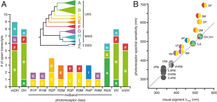
Stomatopod crustaceans possess some of the most complex animal visual systems, including at least 16 spectrally distinct types of photoreceptive units (e.g., assemblages of photoreceptor cells). Here we fully characterize the set of opsin genes expressed in retinal tissues and determine expression patterns of each in the stomatopod Neogonodactylus oerstedii Using a combination of transcriptome and RACE sequencing, we identified 33 opsin transcripts expressed in each N. oerstedii eye, which are predicted to form 20 long-wavelength-sensitive, 10 middle-wavelength-sensitive, and three UV-sensitive visual pigments. Observed expression patterns of these 33 transcripts were highly unusual in five respects: 1) All long-wavelength and short/middle-wavelength photoreceptive units expressed multiple opsins, while UV photoreceptor cells expressed single opsins; 2) most of the long-wavelength photoreceptive units expressed at least one middle-wavelength-sensitive opsin transcript; 3) the photoreceptors involved in spatial, motion, and polarization vision expressed more transcripts than those involved in color vision; 4) there is a unique opsin transcript that is expressed in all eight of the photoreceptive units devoted to color vision; and 5) expression patterns in the peripheral hemispheres of the eyes suggest visual specializations not previously recognized in stomatopods. Elucidating the expression patterns of all opsin transcripts expressed in the N. oerstedii retina reveals the potential for previously undocumented functional diversity in the already complex stomatopod eye and is a first step toward understanding the functional significance of the unusual abundance of opsins found in many arthropod species' visual systems.

Keywords: Stomatopoda; evolution; in situ hybridization; opsin; retinal expression.


Conflict of interest statement

The authors declare no competing interest.

Figures

Fig. 1.
Fig. 1.
The complex photoreceptor arrangement and spectral absorbance in the N. oerstedii compound eye. (A) Image of N. oerstedii. (Scale bar: 5 mm.) (B) The external division of the ommatidia into five regions: The marginal dorsal hemisphere (mDH), dorsal hemisphere (DH), MB composed of six rows of ommatidia, ventral hemisphere (VH), and marginal ventral hemisphere (mVH). (C) A schematic of the arrangement of the dorsal hemisphere, MB (rows 1 to 6), and ventral hemisphere ommatidia, including the R8 cells that sit atop R1–7 cells that form either one photoreceptive unit (hemispheres and MB rows 5 and 6) or two photoreceptive units (MB rows 1 to 4). Photoreceptors are colored to match their spectral absorbance curves (9) as in D.
Fig. 2.
Fig. 2.
Maximum-likelihood phylogeny of crustacean opsins, including the 33 opsins characterized from N. oerstedii eyes. N. oerstedii opsins are in bold, and divided into subgroups based on spectral clade (LWS, subgroups A to E; MWS, subgroups G to I; SWS/UVS, subgroups J to K). Clades of opsins from related taxonomic groups have been collapsed for clarity. For stomatopod opsins, clades of opsins from a single species have been collapsed, and the number of opsins indicated in parentheses behind the species name. Maximum-likelihood bootstrap support and Bayesian posterior probabilities are mapped to the visible branches.
Fig. 3.
Fig. 3.
ISH tissue expression patterns for 33 N. oerstedii opsin transcripts. Each 10- to 14-μm retinal tissue section is oriented as in Fig. 1C and has been mapped onto a phylogeny of only N. oerstedii opsins. A schematic showing the expression patterns in the retina for each transcript or set of transcripts is placed side by side with representative ISH images for clarity. (A) LWS opsin clade ISH; (B) MWS opsin clade ISH; (C) SWS/UVS opsin clade ISH. N. oerstedii opsin subgroups are labeled A to K and are colored as in Fig. 2. A condensed tree is shown to depict clade and subgroup relationships. Numbers on branches represent bootstrap support values. mDH = marginal dorsal hemisphere; DH = dorsal hemisphere; MB = midband (rows 1-6); VH = ventral hemisphere; mVH = marginal ventral hemisphere; * = no detected ISH labeling, data not shown. Data for SWS subgroups J and K from ref. . (Scale bars in all panels: 200 μm.)
Fig. 4.
Fig. 4.
(A) Summary of the number of opsin transcripts expressed in each of the LWS and MWS photoreceptor types found in the N. oerstedii retina. Bars are colored by subgroup and labeled A through G as in Fig. 3, and as indicated in the inset collapsed phylogeny of all N. oerstedii opsins. (B) Plot of photoreceptor spectral sensitivity, measured spectral absorbance (visual pigment λmax), and subgroups of opsin transcripts expressed. Each circle represents a photoreceptor, containing a pie chart depicting the number of opsins expressed, colored by subgroup as in A. Photoreceptors specialized for polarization sensitivity are indicated by circles surrounded by tick marks. Photoreceptors in both panels are abbreviated as follows: marginal dorsal hemisphere, mDH; dorsal hemisphere, DH; MB row 1 proximal tier photoreceptor, 1P; MB row 1 middle tier photoreceptor, 1M; MB row 2 proximal tier photoreceptor, 2P; MB row 2 middle tier photoreceptor, 2M; MB row 3 proximal tier photoreceptor, 3P; MB row 3 middle tier photoreceptor, 3M; MB row 4 proximal tier photoreceptor, 4P; MB row 4 middle tier photoreceptor, 4M; MB rows 5 and 6–5/6; ventral hemisphere, VH; marginal ventral hemisphere, mVH; retinula cell 8 photoreceptor, R8. The dashed line represents the margin where the photoreceptor spectral sensitivity and visual pigment spectral absorbance peaks are equivalent. Spectral sensitivity maxima deviating from this line are influenced by tiering and by photostable optical filters.

Similar articles

Cited by

References

    1. Schram F. R., et al. , “Subclass Hoplocarida Calman, 1904: Order Stomatopoda Latreille, 1817” in Treatise on Zoology—Anatomy, Taxonomy, Biology; The Crustacea; Revised and Updated, as Well as Extended from the Traité de Zoologie, Vaupel Klein J. C., Charmantier-Daures M., Schram F. R., Eds. (Brill, Leiden-Boston, 2013), Vol. 4, pp. 179–356.
    1. Marshall N. J., A unique colour and polarization vision system in mantis shrimps. Nature 333, 557–560 (1988). - PubMed
    1. Marshall N. J., Land M. F., King C. A., Cronin T. W., The compound eyes of mantis shrimps (Crustacea, Hoplocarida, Stomatopoda). I. Compound eye structure: The detection of polarized light. Philos. Trans. R. Soc. Lond. B Biol. Sci. 334, 33–56 (1991).
    1. Marshall N. J., Land M. F., King C. A., Cronin T. W., The compound eyes of mantis shrimps (Crustacea, Hoplocarida, Stomatopoda). II. Colour pigments in the eyes of stomatopod crustaceans: Polychromatic vision by serial and lateral filtering. Philos. Trans. R. Soc. Lond. B Biol. Sci. 334, 57–84 (1991).
    1. Marshall J., Cronin T. W., Kleinlogel S., Stomatopod eye structure and function: A review. Arthropod Struct. Dev. 36, 420–448 (2007). - PubMed

Publication types

Associated data

LinkOut - more resources