Review
. 2020 May 15;295(20):6888-6925.
doi: 10.1074/jbc.REV120.006194. Epub 2020 Apr 2.

Biosynthesis of the modified tetrapyrroles-the pigments of life

Affiliations
Review

Biosynthesis of the modified tetrapyrroles-the pigments of life

Donald A Bryant et al. J Biol Chem. .

Abstract

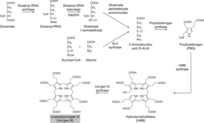
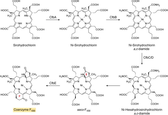
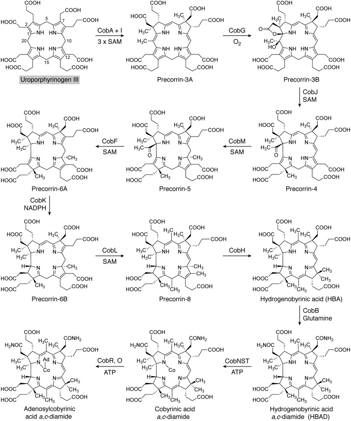
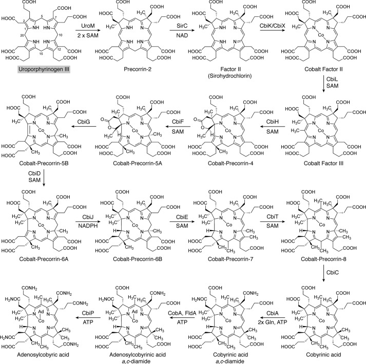
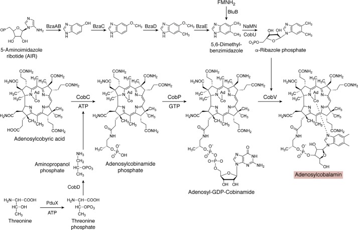
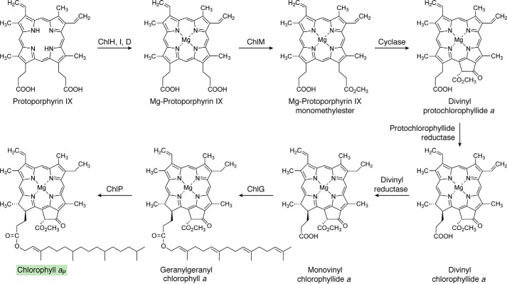
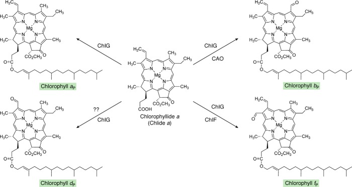
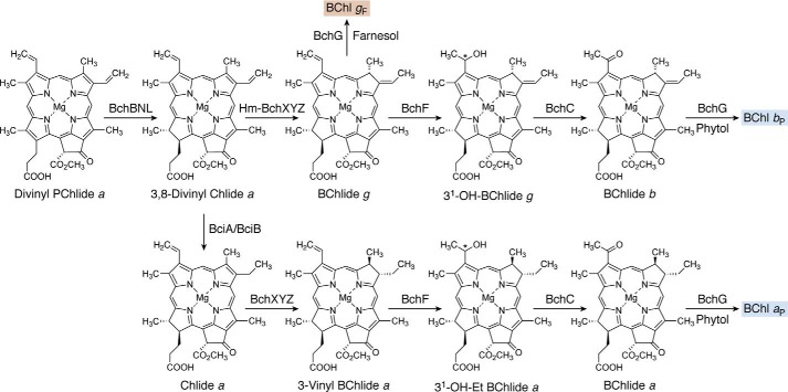
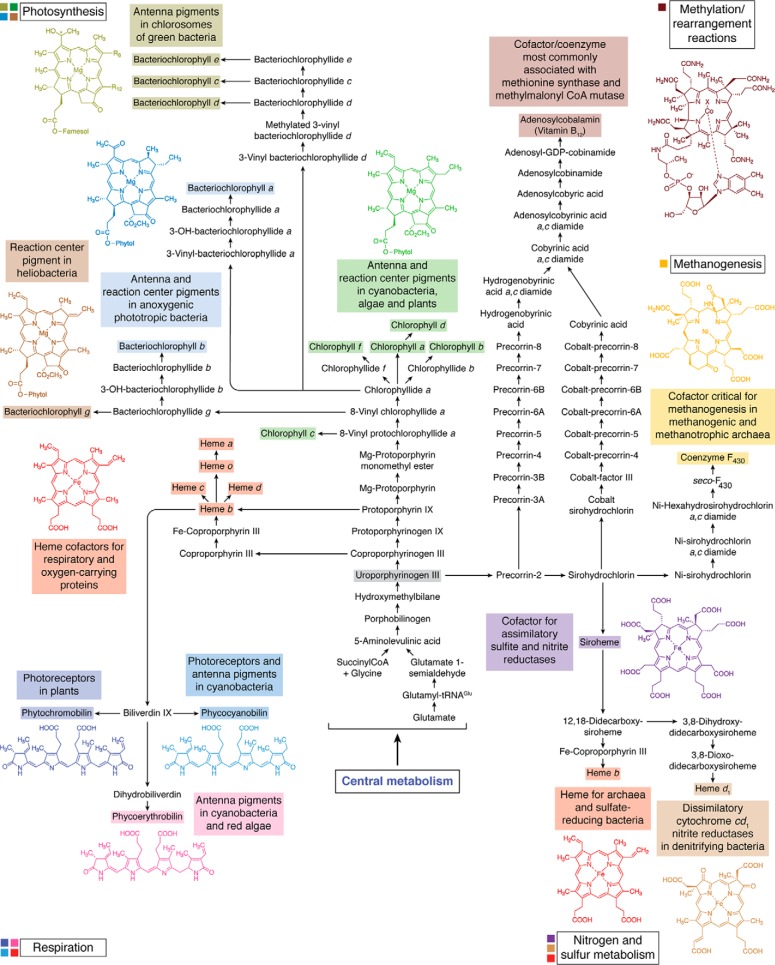
Modified tetrapyrroles are large macrocyclic compounds, consisting of diverse conjugation and metal chelation systems and imparting an array of colors to the biological structures that contain them. Tetrapyrroles represent some of the most complex small molecules synthesized by cells and are involved in many essential processes that are fundamental to life on Earth, including photosynthesis, respiration, and catalysis. These molecules are all derived from a common template through a series of enzyme-mediated transformations that alter the oxidation state of the macrocycle and also modify its size, its side-chain composition, and the nature of the centrally chelated metal ion. The different modified tetrapyrroles include chlorophylls, hemes, siroheme, corrins (including vitamin B12), coenzyme F430, heme d1, and bilins. After nearly a century of study, almost all of the more than 90 different enzymes that synthesize this family of compounds are now known, and expression of reconstructed operons in heterologous hosts has confirmed that most pathways are complete. Aside from the highly diverse nature of the chemical reactions catalyzed, an interesting aspect of comparative biochemistry is to see how different enzymes and even entire pathways have evolved to perform alternative chemical reactions to produce the same end products in the presence and absence of oxygen. Although there is still much to learn, our current understanding of tetrapyrrole biogenesis represents a remarkable biochemical milestone that is summarized in this review.

Keywords: 5-aminolevulinic acid; adenosylcobalamin (AdoCbl); bacteriochlorophyll; bilin; biosynthesis; chlorophyll; cobalamin; coenzyme F430; heme; heme d1; photosynthesis; precorrin; tetrapyrrole; uroporphyrinogen III; vitamin B12.


Conflict of interest statement

The authors declare that they have no conflicts of interest with the contents of this article.

Figures

Figure 1.
Figure 1.
Structures of the major modified tetrapyrroles outlined in this review and their structural relationship to the first macrocyclic primogenitor, uroporphyrinogen III. The major modified tetrapyrroles shown surrounding the central uroporphyrinogen III include chlorophyll aP, coenzyme F430, siroheme, cobalamin, biliverdin IXα, heme d1, and heme b. The asymmetrically arranged pyrrole rings in uroporphyrinogen III are named A–D, with the D ring inverted with respect to the other rings. The numbering scheme for the macrocycle is shown for uroporphyrinogen, where positions 1, 2, 5, 7, 10, 12, 15, and 20 are highlighted. In Chls there is a fifth ring, termed ring E, and similarly in F430, there are two extra rings that are termed E and F as shown. For cobalamin (vitamin B12), the side chains are designated (a–f), and these are labeled. The X above the cobalt is a cyanide group in vitamin B12; this position is occupied by either a methyl or adenosyl group in the major biological forms of cobalamin. The shaded boxes surrounding the names of these end-product compounds coordinate with the colors in other pathway figures and in the summary pathway depicted in Fig. 14.
Figure 2.
Figure 2.
The two routes for the biosynthesis of 5-ALA and the subsequent biosynthesis of uroporphyrinogen III. The Shemin, or C4, route involves the condensation of glycine and succinyl-CoA and is mediated by the enzyme 5-aminolevulinic acid synthase. The C5 pathway acquires the intact carbon skeleton from glutamate and utilizes glutamyl-tRNA as an intermediate. The glutamyl-tRNA undergoes a reduction by glutamyl-tRNA reductase to give GSA. The final step involves the enzyme GsaM, which rearranges the GSA into 5-ALA. Then two molecules of 5-ALA are condensed into PBG by the action of the enzyme porphobilinogen synthase. Next, four molecules of PBG are deaminated and linked together to give a linear bilane called HMB in a reaction catalyzed by HMB synthase. The final step involves the cyclization and inversion of the terminal D ring to give uroporphyrinogen III. The gray box for uroporphyrinogen III also identifies this central intermediate in Figs. 3 and 14.
Figure 3.
Figure 3.
The biosynthesis of siroheme from uroporphyrinogen III. Initially, uroporphyrinogen is methylated at positions C2 and C7 to give precorrin-2 and then undergoes dehydrogenation to give sirohydrochlorin and finally ferrochelation to yield siroheme. The reactions are either mediated by three independent enzymes, such as SirA, -C, and -B, or by two enzymes, such as a uroporphyrinogen methyltransferase (CobA or Met1p)) and a bifunctional dehydrogenase/chelatase (Met8p), or by a single multifunctional enzyme, CysG. The shaded boxes surrounding the names of compounds coordinate with other pathway figures and the summary depiction in Fig. 14.
Figure 4.
Figure 4.
The transformation of sirohydrochlorin into coenzyme F430. The steps involved in the biosynthesis of F430 from sirohydrochlorin are outlined. Initially, sirohydrochlorin is chelated with nickel by the enzyme CfbA to give nickel sirohydrochlorin. Next, the two acetic acid side chains on rings A and B, the a and c side chains, are amidated in a reaction catalyzed by CfbB that also requires glutamine and ATP as substrates. This generates nickel sirohydrochlorin a,c-diamide, which acts as the substrate for the reductase system that is catalyzed by CfbC and -D. The reductase removes three double bonds from the macrocycle, which also spontaneously results in the formation of the lactam ring E, thereby generating seco-F430. The final step, mediated by CfbE, results in the formation of the cyclic hexanone ring F in another ATP-requiring process. The shaded box for coenzyme F430 coordinates with other pathway figures and the summary depiction in Fig. 14.
Figure 5.
Figure 5.
The aerobic biosynthesis of adenosylcobyrinic acid a,c-diamide from uroporphyrinogen III. The individual steps along the aerobic route for cobalamin synthesis are shown. Initially, uroporphyrinogen III undergoes three methylation steps at C2, C7, and C20, before hydroxylation at the C20 position generates precorrin-3B, a masked pinacol that is primed for ring contraction through rearrangement. The contraction is mediated by CobJ, which also methylates at C17. More methylations, a decarboxylation, and a mutase reaction generate the orange-colored hydrogenobyrinic acid (HBA) intermediate. Cobalt insertion followed by adenosylation and amidation of the side chains generates adenosylcobyrinic acid a,c-diamide, the point where the aerobic and anaerobic (see Figs. 6 and 14) pathways rejoin. The gray shading surrounding Uroporphyrinogen III coordinates with other pathway figures and the summary in Fig. 14.
Figure 6.
Figure 6.
The anaerobic biosynthesis of adenosylcobyric acid from uroporphyrinogen III. The aerobic pathway starts with the synthesis of sirohydrochlorin, which is sometimes also referred to as Factor II. Metal insertion at this stage generates cobalt-sirohydrochlorin, which then undergoes a further methylation at C20 to give cobalt-factor III. Ring contraction is mediated by CbiH, which forms a δ-lactone in the generation of cobalt-precorrin-4. Further methylations coupled with lactone ring opening and rearrangement give rise to cobyrinic acid. Amidations together with adenosylation ultimately give rise to the formation of adenosylcobyric acid. The shaded box surrounding uroporphyrinogen III coordinates with other pathway figures and the summary in Fig. 14.
Figure 7.
Figure 7.
The final stages of cobalamin biosynthesis. Adenosylcobyric acid is converted into adenosylcobalamin through the action of three further enzymes. Initially, an aminopropanol phosphate linker is attached to the propionate side chain found on ring D to give adenosylcobinamide phosphate. Aminopropanol is itself derived from threonine. A GDP moiety is attached to the aminopropanol phosphate linker to give adenosyl-GDP-cobinamide. Finally, the GDP moiety is replaced with another nucleotide called α-ribazole, itself made from the ligation of dimethylbenzimidazole with the ribose portion of nicotinamide mononucleotide (NaMN). This results in the formation of adenosylcobalamin. The shaded box surrounding adenosylcobalamin coordinates with other pathway figures and the summary in Fig. 14.
Figure 8.
Figure 8.
The three routes to heme from uroporphyrinogen III. The protoporphyrin route (gray arrow) involves the formation of protoporphyrin IX via coproporphyrinogen and protoporphyrinogen with the final step involving insertion of iron into protoporphyrin IX. There are aerobic and anaerobic forms of the enzymes associated with the formation of protoporphyrinogen and protoporphyrin, where the asterisks next to the enzyme (for CgdH, PgdH1, and PgdH2) indicate that these enzymes are found largely under anaerobic conditions. The siroheme route (pale blue arrow) involves the decarboxylation of siroheme to give didecarboxysiroheme, followed by the removal of the acetic acid side chains on rings A and B to give Fe-coproporphyrin before the final step, which involves the decarboxylation of the propionate side chains on rings A and B to produce heme. The coproporphyrin pathway (dusty rose arrow) is a hybrid between the first two routes: coproporphyrinogen is oxidized to give coproporphyrin, which is chelated with iron to give Fe-coproporphyrin. The final step is then the formation of the vinyl side chains through the decarboxylation of the propionate side chains on rings A and B. The conversion of Fe-coproporphyrin into heme is catalyzed by the same enzyme in both the siroheme and coproporphyrin pathways, although it has different names. The shaded boxes surrounding the names of some compounds coordinate with other pathway figures and the summary in Fig. 14.
Figure 9.
Figure 9.
The biosynthesis of chlorophyll aP from protoporphyrin IX. Magnesium insertion into ProtoIX directs the intermediate toward Chl synthesis by generating magnesium ProtoIX. This acts as the substrate for a methyltransferase (ChlM), which, together with SAM, gives rise to magnesium ProtoIX monomethyl ester. In the following reaction, the cyclase forms ring E of PChlide a, the C17=C18 double bond of which is then reduced, forming divinyl Chlide a. After reduction of one of the vinyl side chains, geranylgeraniol is attached to the propionate on ring D to form geranylgeranyl Chl a. Subsequent reduction of the geranylgeranyl group to phytol (P) gives rise to Chl aP. The shaded box surrounding Chl aP coordinates with other pathway figures and the summary depiction in Fig. 14.
Figure 10.
Figure 10.
Pathway to show the transformation of chlorophyllide a into other chlorophylls. The addition of the esterifying phytol moieties to the C17 propionates is presumably catalyzed by ChlG in all cases. CAO hydroxylates the C7 methyl group twice, producing a geminal diol that spontaneously dehydrates to form the formyl group of Chl b. The enzyme leading to Chl d in A. marina is unknown. Chl d is also found in some terrestrial cyanobacteria that can photoacclimate to utilize far-red light for oxygenic photosynthesis. There is evidence suggesting that thiol compounds and/or proteins, including cysteine-rich allophycocyanins produced in far-red light, and oxygen may catalyze the formation of Chl d. Note that conversion of the C3 vinyl group of Chl a to the C3 formyl group of Chl d requires the loss of one carbon. The C2 formyl group of Chl f is introduced by a photooxidoreductase, ChlF, which is an enzyme containing Chl a and pheophytin a and which is structurally related to the D1 subunit of photosystem II. The shaded boxes surrounding the names of Chls coordinate with other pathway figures and the summary in Fig. 14.
Figure 11.
Figure 11.
Synthesis of bacteriochlorophylls a, g, and b from divinyl-protochlorophyllide a. There are two types of Chlide a oxidoreductases. The type found in R. sphaeroides and most other anoxygenic phototrophs converts Chlide a into 3-vinyl BChlide a. However, organisms such as Heliobacterium modesticaldum (Hm) that produce BChl gF or Blastochloris viridis that produce BChl b have an enzyme that converts 3,8-divinyl Chlide into BChlide g, which has an ethylidene side chain at the C8 position. BChl a and b are usually esterified with phytol by the BChl synthases (BchG) that occur in those organisms. However, BChl g is esterified with farnesol by the Bchl g synthases that occur in heliobacteria. Note that divinyl PChlide is also the precursor for the synthesis of the family of pigments known as Chl c. For additional details, see section “Extension of the pathway beyond Chlide a/Chl a.” The shaded boxes surrounding the names of some compounds coordinate with other pathway figures and the summary in Fig. 14.
Figure 12.
Figure 12.
Synthesis of bacteriochlorophylls c, d, e, and f from chlorophyllide a. Purified BciD is active with both BChlide d and BChlide c as substrates. The conversion of the C7 methyl group to a formyl group proceeds via a geminal diol intermediate that spontaneously dehydrates to produce the formyl group. Esterifying alcohols are added by BchK in all cases. BChls c and d are characteristically found in green-colored green bacteria, whereas BChls e and f are produced by brown-colored green sulfur bacteria. Note that BChl f has not been observed in nature; however, it has been generated by mutation of bchU in C. limnaeum and can still produce functional chlorosomes (see section “Chlorobium Chls: BChls c, d, e, and f” for more details). In green sulfur bacteria, the esterifying alcohols of BChls c, d, e, and f are usually farnesol (F) (see inset, showing a farnesol group attached to ring D of a partial macrocycle). However, in members of the Chloroflexi and C. thermophilum, the esterifying alcohols are often highly variable and are frequently straight-chain alcohols or geranylgeraniol and its reduction products. All alcohols are added by BchK enzymes that are specific to individual organisms. In green sulfur bacteria and some other green bacteria, the ethyl side chain at C8 can be methylated by BchQ to produce propyl, isobutyl, and neopentyl side chains (see inset). Similarly, the methyl group at C12 can be methylated by BchR to produce an ethyl side chain. The asterisk near the hydroxyl group at C31 indicates that this is a chiral center that is mostly R but is S when the side chains at C8 and C12 are more extensively methylated. The shaded boxes surrounding the names of some compounds coordinate with other pathway figures and the summary in Fig. 14.
Figure 13.
Figure 13.
Synthesis of bilins from biliverdin, which is formed from heme b. Biliverdin is produced by the oxygen-dependent cleavage of heme b by the enzyme heme oxygenase. Regio-specific ferredoxin-dependent bilin reductases lead to phytochromobilin, phycocyanobilin, and phycoerythrobilin, and biliverdin reductase produces bilirubin. Note that phycoerythrobilin can be synthesized by one enzyme (PebS) or in two steps by PebA and PebB via a 17,18-dihydrobiliverdin intermediate. Phycoviolobilin and phycourobilin are produced from phycocyanobilin and phycoerythrobilin, respectively, by isomerization, which occurs during the attachment of these bilins to proteins by isomerizing bilin lyases. BVR, biliverdin reductase; HY2, phytochromobilin synthase. See section “Bilins: Chromophores of phycobiliproteins, phytochromes, and cyanobacteriochromes” for additional details. The shaded boxes surrounding the names of some compounds coordinate with the summary in Fig. 14.
Figure 14.
Figure 14.
Summary overview of the entire tetrapyrrole biosynthetic network. Those aspects of life on Earth that depend on tetrapyrroles are shown at the periphery in boxes with black outlines, adjacent to color-coded squares that correspond to various tetrapyrroles. Color-coded boxes specify the functions of nearby tetrapyrroles. The colors used in this figure coordinate with the colored boxes associated with the names of end-product compounds in other figures in this article. The link to central metabolism is indicated, which provides the starting point for the entire network. Note that all compounds produced in this pathway ultimately are derived from uroporphyrinogen III. Note also that heme b arises as a respiratory cofactor and as a precursor of hemes c, d, and o and biliverdin IX. Heme b also arises separately in archaea and sulfate-reducing bacteria as a cofactor for nitrogen and sulfur metabolism.

Similar articles

Cited by

References

    1. Battersby A. R. (2000) Tetrapyrroles: the pigments of life. Nat. Prod. Rep. 17, 507–526 10.1039/b002635m - DOI - PubMed
    1. Allen J. W., Barker P. D., Daltrop O., Stevens J. M., Tomlinson E. J., Sinha N., Sambongi Y., and Ferguson S. J. (2005) Why isn't “standard” heme good enough for c-type and d1-type cytochromes? Dalton Trans. 3410–3418 10.1039/b508139b - DOI - PubMed
    1. Hamza I., and Dailey H. A. (2012) One ring to rule them all: trafficking of heme and heme synthesis intermediates in the metazoans. Biochim. Biophys. Acta 1823, 1617–1632 10.1016/j.bbamcr.2012.04.009 - DOI - PMC - PubMed
    1. Barber J. (2008) Photosynthetic generation of oxygen. Philos. Trans. R. Soc. Lond. B Biol. Sci. 363, 2665–2674 10.1098/rstb.2008.0047 - DOI - PMC - PubMed
    1. Dailey H. A., Dailey T. A., Gerdes S., Jahn D., Jahn M., O'Brian M. R., and Warren M. J. (2017) Prokaryotic heme biosynthesis: multiple pathways to a common essential product. Microbiol. Mol. Biol. Rev. 81, e00048–16 10.1128/MMBR.00048-16 - DOI - PMC - PubMed

Publication types

MeSH terms