. 2020 May 15;295(20):6823-6830.
doi: 10.1074/jbc.RA120.013338. Epub 2020 Apr 2.

The C-terminal tails of the mitochondrial transcription factors Mtf1 and TFB2M are part of an autoinhibitory mechanism that regulates DNA binding

Affiliations

The C-terminal tails of the mitochondrial transcription factors Mtf1 and TFB2M are part of an autoinhibitory mechanism that regulates DNA binding

Urmimala Basu et al. J Biol Chem. .

Abstract

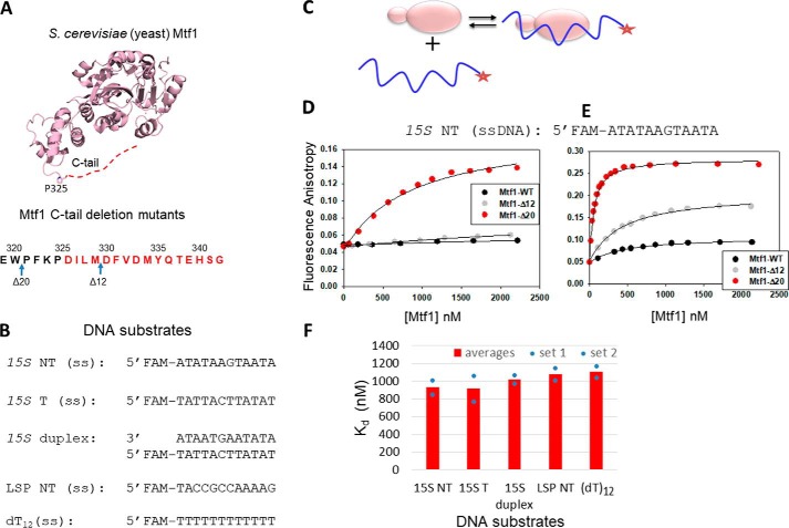
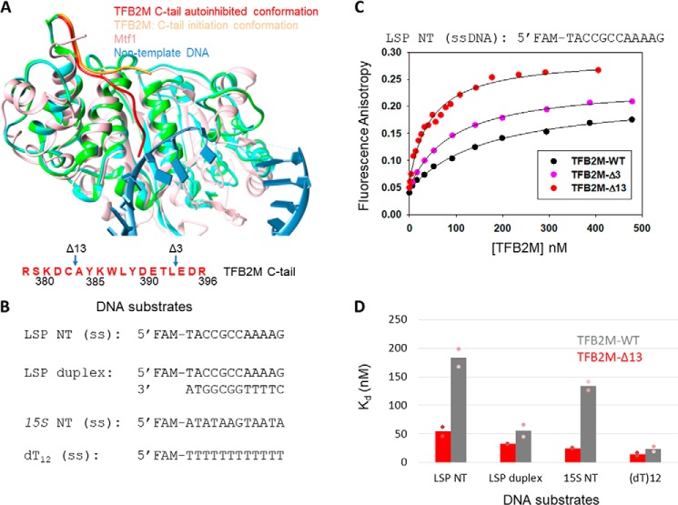
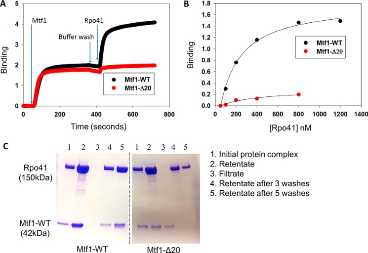
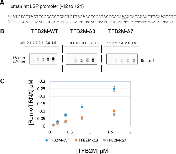
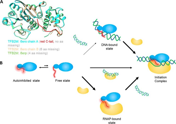
The structurally homologous Mtf1 and TFB2M proteins serve as transcription initiation factors of mitochondrial RNA polymerases in Saccharomyces cerevisiae and humans, respectively. These transcription factors directly interact with the nontemplate strand of the transcription bubble to drive promoter melting. Given the key roles of Mtf1 and TFB2M in promoter-specific transcription initiation, it can be expected that the DNA binding activity of the mitochondrial transcription factors is regulated to prevent DNA binding at inappropriate times. However, little information is available on how mitochondrial DNA transcription is regulated. While studying C-terminal (C-tail) deletion mutants of Mtf1 and TFB2M, we stumbled upon a finding that suggested that the flexible C-tail region of these factors autoregulates their DNA binding activity. Quantitative DNA binding studies with fluorescence anisotropy-based titrations revealed that Mtf1 with an intact C-tail has no affinity for DNA but deletion of the C-tail greatly increases Mtf1's DNA binding affinity. Similar observations were made with TFB2M, although autoinhibition by the C-tail of TFB2M was not as complete as in Mtf1. Analysis of available TFB2M structures disclosed that the C-tail engages in intramolecular interactions with the DNA binding groove in the free factor, which, we propose, inhibits its DNA binding activity. Further experiments showed that RNA polymerase relieves this autoinhibition by interacting with the C-tail and engaging it in complex formation. In conclusion, our biochemical and structural analyses reveal autoinhibitory and activation mechanisms of mitochondrial transcription factors that regulate their DNA binding activities and aid in specific assembly of transcription initiation complexes.

Keywords: Mtf1; RNA polymerase; TFB2M; autoinhibition; fluorescence anisotropy; mitochondria; mitochondrial RNA polymerase; mitochondrial transcription factors; protein–DNA interaction; transcriptional coactivator.


Conflict of interest statement

The authors declare that they have no conflicts of interest with the contents of this article. The content is solely the responsibility of the authors and does not necessarily represent the official views of the National Institutes of Health

Figures

Figure 1.
Figure 1.
The C-tail of Mtf1 drastically autoinhibits the DNA binding activity of Mtf1. A, structure of the yeast mitochondrial transcription factor Mtf1 (rose, PDB code 1I4W). The missing 16 aa of the C-tail of Mtf1 in the crystal structure are shown as a red dotted line and are also marked in red in the amino acid sequence of the C-tail of Mtf1. B, DNA sequences of the substrates used for the Mtf1 DNA binding studies. C, cartoon showing the basic scheme of the fluorescence anisotropy assays to monitor protein–DNA binding. D, representative binding curves showing the fluorescence anisotropy changes resulting from titration of the 15S NT DNA with Mtf1. 15S NT DNA (5 nm) was titrated with Mtf1-WT (black circles), Mtf1-Δ12 (gray circles), and Mtf1-Δ20 (red circles) in buffer A (see “Experimental procedures”). E, 15S NT (5 nm) was titrated with Mtf1-WT (black circles), Mtf1-Δ12 (gray circles), and Mtf1-Δ20 (red circles) in buffer A without potassium glutamate. The solid lines represent fit to the hyperbolic Equation 1 with Kd values as follows: Mtf1-WT = 447 ± 60 nm (amplitude, 0.059), Mtf1-Δ12 = 426 ± 33 nm (amplitude, 0.156), Mtf1-Δ20 = 51 ± 2.8 nm (amplitude: 0.24). F, the average DNA Kd values of Mtf1-Δ20 are shown for the DNA substrates in B. The blue dots are the individual values for set 1 and set 2 titration data, which are shown in Fig. S1.
Figure 2.
Figure 2.
The C-tail of TFB2M mildly autoinhibits the DNA binding activity of TFB2M. A, the aligned structures of free human TFB2M (PDB code 6ERO, cyan), TFB2M in the initiation complex (PDB code 6ERP, green), and yeast Mtf1 (PDB code 1I4W, rose) are shown. The relative positions of the C-tail in all three structures are shown. The amino acid sequence of the C-tail of TFB2M is shown below in red. B, DNA substrates used for the TFB2M DNA binding studies. C, representative binding curves show the fluorescence anisotropy change resulting from titration of LSP NT (5 nm) with TFB2M-WT (black circles), TFB2M-Δ3 (pink circles), and TFB2M-Δ13 (red circles). The data were fit to the hyperbolic Equation 1 to obtain the following Kd values: TFB2M-WT = 169 ± 18 nm (amplitude, 0.17), TFB2M-Δ3 = 92 ± 3.3 nm (amplitude, 0.19), TFB2M-Δ13 = 46 ± 5.2 nm (amplitude, 0.23). D, the gray and red columns show the DNA Kd values of TFB2M-WT and TFB2M-Δ13, respectively, for the various DNA substrates shown in B. The pink dots represent individual values for set 1 and set 2 titration data, which are shown in Fig. S3.
Figure 3.
Figure 3.
The C-tail of Mtf1 mediates complex formation with Rpo41. A, representative binding plots showing complex formation between Mtf1 and Rpo41 using biolayer interferometry assays. The first 60 s represent the baseline. Over the next 300 s, the biosensor HIS1K was treated with 0.4 μm His-tagged Mtf1-WT (black line) or Mtf1-Δ20 (red line) protein, followed by washing with buffer for 60 s. The probes were then dipped in Rpo41 (0.5 μm) for 300 s, followed by washing for 60 s. B, the degree of binding (y axis) was calculated from the difference in light interference values before and after adding Rpo41 in A. C, an equimolar complex of Rpo41 and Mtf1-WT or Mtf1-Δ20 at a final concentration 2 μm (lane 1) was filtered through a 100-kDa molecular mass cutoff Microcon centrifugal filter unit. Lane 2 is the retentates, and lane 3 is the filtrates. The retentate was washed with 500 μl of buffer three times (lane 4). The retentate was washed two more times (lane 5). Samples of the initial protein complex, retentate, filtrate, and retentate samples after washing were run on a 4%–20% SDS-PAGE gel.
Figure 4.
Figure 4.
The C-tail deletion in TFB2M affects initiation complex formation. A, the promoter fragment LSP used in this study. The two start sites are underlined. B, runoff transcription profiles of TFB2M-WT and the C-tail deletion mutants TFB2M-Δ3 and TFB2M-Δ7 on the LSP promoter (the full gel profile is shown in Fig. S6). Reactions were carried out with 0.6 μm POLRMT, TFAM, and promoter duplex and increasing concentrations of TFB2M-WT or C-tail mutant and 250 μm ATP, UTP, GTP, and γ[32P]ATP for 15 min at 25 °C in transcription buffer. C, plot showing quantitation of grouped runoff products for each reaction. The error bars represent errors calculated from two independent experiments.
Figure 5.
Figure 5.
Model to explain the autoregulatory role of the C-tail of TFB2M and Mtf1 in assembly of the initiation complex. A, crystal structures of TFB2M showing different states of the C-tail in free and DNA-bound states. Chain A of TFB2M in free state is shown in cyan and the C-tail in red. Chain B of TFB2M in the free state is shown in sand, and TFB2M in the initiation complex is shown in green. B, the flexible C-tail (red) of the mitochondrial transcription factors Mtf1 and TFB2M is in equilibrium between an autoinhibited state and free state. For Mtf1, the equilibrium is toward the autoinhibited state, whereas for TFB2M, both states exist. Therefore, Mtf1 needs to bind to RNAP or the RNAP–DNA complex to generate the initiation complex. On the other hand, TFB2M can take all three pathways to form the initiation complex. Under cellular conditions, the DNA binding activity of TFB2M might be regulated by other mechanisms.

Similar articles

Cited by

References

    1. Cermakian N., Ikeda T. M., Miramontes P., Lang B. F., Gray M. W., and Cedergren R. (1997) On the evolution of the single-subunit RNA polymerases. J. Mol. Evol. 45, 671–681 10.1007/PL00006271 - DOI - PubMed
    1. Jang S. H., and Jaehning J. A. (1991) The yeast mitochondrial RNA polymerase specificity factor, MTF1, is similar to bacterial sigma factors. J. Biol. Chem. 266, 22671–22677 - PubMed
    1. Basu U., Lee S. W., Deshpande A., Shen J., Sohn B. K., Cho H., Kim H., and Patel S. S. (2020) The C-terminal tail of the yeast mitochondrial transcription factor Mtf1 coordinates template strand alignment, DNA scrunching and timely transition into elongation. Nucleic Acids Res. 48, 2604–2620 10.1093/nar/gkaa040 - DOI - PMC - PubMed
    1. Falkenberg M., Gaspari M., Rantanen A., Trifunovic A., Larsson N. G., and Gustafsson C. M. (2002) Mitochondrial transcription factors B1 and B2 activate transcription of human mtDNA. Nat. Genet. 31, 289–294 10.1038/ng909 - DOI - PubMed
    1. Litonin D., Sologub M., Shi Y., Savkina M., Anikin M., Falkenberg M., Gustafsson C. M., and Temiakov D. (2010) Human mitochondrial transcription revisited: only TFAM and TFB2M are required for transcription of the mitochondrial genes in vitro. J. Biol. Chem. 285, 18129–18133 10.1074/jbc.C110.128918 - DOI - PMC - PubMed

Publication types

MeSH terms

LinkOut - more resources