. 2020 May 22;295(21):7516-7528.
doi: 10.1074/jbc.RA120.012611. Epub 2020 Apr 2.

Iron is a ligand of SecA-like metal-binding domains in vivo

Affiliations

Iron is a ligand of SecA-like metal-binding domains in vivo

Tamar Cranford-Smith et al. J Biol Chem. .

Abstract

The ATPase SecA is an essential component of the bacterial Sec machinery, which transports proteins across the cytoplasmic membrane. Most SecA proteins contain a long C-terminal tail (CTT). In Escherichia coli, the CTT contains a structurally flexible linker domain and a small metal-binding domain (MBD). The MBD coordinates zinc via a conserved cysteine-containing motif and binds to SecB and ribosomes. In this study, we screened a high-density transposon library for mutants that affect the susceptibility of E. coli to sodium azide, which inhibits SecA-mediated translocation. Results from sequencing this library suggested that mutations removing the CTT make E. coli less susceptible to sodium azide at subinhibitory concentrations. Copurification experiments suggested that the MBD binds to iron and that azide disrupts iron binding. Azide also disrupted binding of SecA to membranes. Two other E. coli proteins that contain SecA-like MBDs, YecA and YchJ, also copurified with iron, and NMR spectroscopy experiments indicated that YecA binds iron via its MBD. Competition experiments and equilibrium binding measurements indicated that the SecA MBD binds preferentially to iron and that a conserved serine is required for this specificity. Finally, structural modeling suggested a plausible model for the octahedral coordination of iron. Taken together, our results suggest that SecA-like MBDs likely bind to iron in vivo.

Keywords: Sec pathway; iron; metal ion-protein interaction; protein secretion; protein structure; protein translocation.


Conflict of interest statement

The authors declare that they have no conflicts of interest with the contents of this article

Figures

Figure 1.
Figure 1.
Effect of azide on the C-terminal tail of SecA. A, plot of the degree of depletion or enrichment in transposon insertions in all of the nonessential genes in E. coli after growth of the TraDIS library to OD600 = 0.9 in the presence of 0.5 mm sodium azide. The log2 of the fraction of the number of insertions in a gene after growth in the presence of 0.5 mm azide over the number of insertions after growth in LB was plotted according to the degree of enrichment. Data points representing insertions in the secA, secF, secG, secM, yidC, yajC, cpxR, dsbA, ahpC, gor, and trxC genes are indicated. Single gene deletion mutants that were compared with growth of the parent in Table 1 are indicated (bold). B, number of mutants containing insertions at the indicated location in the secA gene after growth of the TraDIS library in the absence (black) or presence of 0.25 (blue) or 0.5 mm (red) NaN3. Most of these insertions truncate the secA gene between codons 822 and 829 at the junction between the HSD and the CTT. C, cultures of E. coli producing His-SUMO-SecA (lanes 1–4) or His-SUMO-SecACC/AA (lanes 5 and 6) were grown to late exponential phase. In the case of His-SUMO-SecA, half of the culture was treated with 2 mm sodium azide for 10 min prior to lysis (lanes 3 and 4), and the other half was left untreated (lanes 1 and 2). His-SUMO-SecA and SUMO-SecACC/AA were purified from the cell lysates using nickel-affinity purification. Phospholipids from 2 mg of the purified protein were extracted into 100 μl of chloroform, and 5 μl of the extracted phospholipids (lanes 1, 3, and 6) and the wash buffer (lanes 2, 4, and 5) were resolved using thin-layer chromatography (TLC). The positions of phosphatidylglycerol (PG), phosphatidylethanolamine (PE) and cardiolipin (CL) are indicated.
Figure 2.
Figure 2.
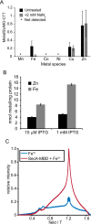
The SecA MBD copurifies with iron and binds to iron in vitro. A, cells producing SUMO-CTT were incubated in the absence (untreated) or presence (+N3) of 2 mm NaN3 for 10 min. SUMO-CTT was purified from the cell lysates using streptactin beads and washed extensively with buffer. The iron content of SUMO-CTT was determined using MS (ICP-MS) and normalized to the protein concentration in the eluted protein. Confidence intervals are 1 S.D. B, DRH839 cells (ΔsecA ptrc-secA-biotin) were grown in the presence of 10 μm or 1 mm IPTG to induce expression of SecA-biotin. Cells were rapidly lysed by cell disruption, incubated with streptavidin-Sepharose, and washed extensively with buffer. The zinc and iron content of the EDTA eluate was determined using ICP-OES and normalized to the amount of protein bound to the streptavidin beads. An estimation of the concentrations of SecA and streptavidin in the purified protein suggested that SecA copurified with stoichiometric amounts of iron. C, the echo-detected field swept EPR spectrum of oxidized FeSO4 was determined in the absence (blue trace) or presence (red trace) of SecA-MBD.
Figure 3.
Figure 3.
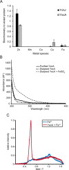
Spectroscopic analysis of metal binding by YecA and YchJ. A, His-SUMO-YecA and His-SUMO-YchJ were purified using nickel-affinity chromatography, and the amount of co-purifying Zn, Mn, Co, Cu, and Fe were determined by ICP-MS. The concentrations of the extracted metals were normalized to the amount of purified protein. Confidence intervals are 1 S.D. B, buffer-subtracted absorbance spectra of purified YecA (solid black), purified YecA that was dialyzed against EDTA (dash-dot), and EDTA-dialyzed YecA to which an equimolar concentration of FeSO4 was added (dashed). The absorbance spectra were normalized to the concentration of YecA. C, the echo-detected field swept EPR spectrum of FeCl3 was determined in the absence (blue trace) or presence (red trace) of YecA.
Figure 4.
Figure 4.
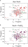
YecA binds to iron via its MBD. A, TROSY spectrum of metal-free 15N- and 13C-labeled YecA. The 1H, 15N, and 13C resonances of the polypeptide backbone for the C-terminal 20 amino acids (excluding prolines 206 and 208) were assigned using triple resonance experiments. Resonances of amino acids in the MBD are indicated with arrowheads. Amino acids producing two resonance peaks are labeled in green. B, overlay of the TROSY spectra of metal-free YecA (red) and YecA in the presence of equimolar concentrations of FeSO4 (blue). The absence of a blue peak suggests that the amino acid producing the resonance is in the proximity of the bound iron. Amino acids are represented as single letter abbreviations.
Figure 5.
Figure 5.
1H NMR analysis of metal binding by SecA-MBD and SecA-MBDS889A. A, 6.5–10 ppm region of the 1H NMR spectra of SecA-MBD in the absence (blue) or presence of equimolar concentrations of FeSO4 (red) or ZnSO4 (green). The appearance of resonances in the ∼8.5–10 ppm region in the presence of ZnSO4 indicates the formation of secondary structure. The strong quenching of the resonances in the presence of FeSO4 is due to proximity to the bound iron ion. B, 0.2–1.0 ppm region of the 1H NMR spectra of SecA-MBD in the absence (blue) or presence of equimolar concentrations of FeSO4 (red) or ZnSO4 (green). Binding of SecA-MBD to zinc results in a shift of the resonances corresponding to the methyl 1Hs of valine, and binding of SecA-MBD to iron results in a significant broadening and flattening of these resonances. C and D, 0.2–1.0 ppm region of the 1H NMR spectra of SecA-MBD (C) or SecA-MBDS889A (D) in the absence of metal (blue), in the presence of equimolar FeSO4 (red), and after the addition of competing concentrations of ZnSO4 to the iron-bound peptide after >10 min of equilibration at room temperature (purple).
Figure 6.
Figure 6.
Structural models of octahedral metal coordination. Ribbon diagrams of example structural models of the MBD coordinating a hexavalent metal using the δ-nitrogen (A) and ϵ-nitrogen (B) of His-897 generated using molecular dynamic simulations. Example structures from each 1 ns of the final 10 ns of each 100-ns simulation can be found in the supporting Data. The side chains of the metal-coordinating amino acids are depicted as sticks (yellow, sulfur; blue, nitrogen; red, oxygen). Images were rendered using UCSF Chimera version 1.12 (72).

Similar articles

Cited by

References

    1. Beckwith J. 2013) The Sec-dependent pathway. Res. Microbiol. 164, 497–504 10.1016/j.resmic.2013.03.007 - DOI - PMC - PubMed
    1. Tsirigotaki A., De Geyter J., Šoštaric N., Economou A., and Karamanou S.. 2017) Protein export through the bacterial Sec pathway. Nat. Rev. Microbiol. 15, 21–36 10.1038/nrmicro.2016.161 - DOI - PubMed
    1. Park E., and Rapoport T. A.. 2012) Mechanisms of Sec61/SecY-mediated protein translocation across membranes. Annu. Rev. Biophys. 41, 21–40 10.1146/annurev-biophys-050511-102312 - DOI - PubMed
    1. Cranford Smith T., and Huber D.. 2018) The way is the goal: how SecA transports proteins across the cytoplasmic membrane in bacteria. FEMS Microbiol. Lett. 365 10.1093/femsle/fny093 - DOI - PMC - PubMed
    1. Collinson I., Corey R. A., and Allen W. J.. 2015) Channel crossing: how are proteins shipped across the bacterial plasma membrane? Philos. Trans. R. Soc. Lond. B Biol. Sci. 370, pii.20150025 - PMC - PubMed

Publication types

LinkOut - more resources