. 2020 May 22;295(21):7224-7234.
doi: 10.1074/jbc.RA120.012738. Epub 2020 Apr 2.

Metal ion coordination delays amyloid-β peptide self-assembly by forming an aggregation-inert complex

Affiliations

Metal ion coordination delays amyloid-β peptide self-assembly by forming an aggregation-inert complex

Cecilia Wallin et al. J Biol Chem. .

Abstract

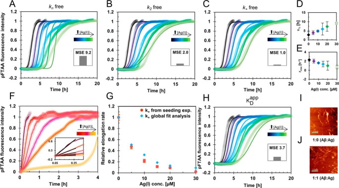
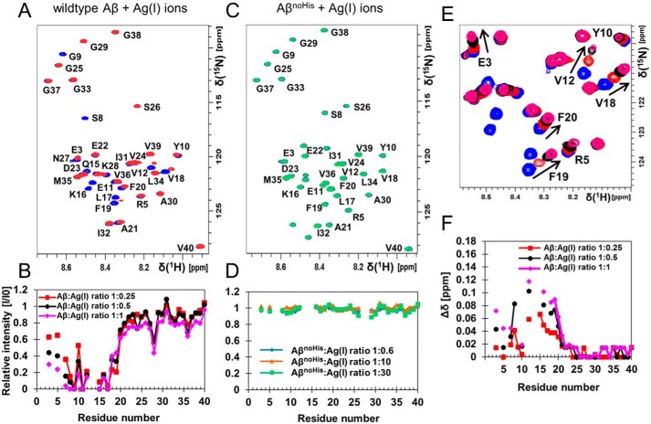
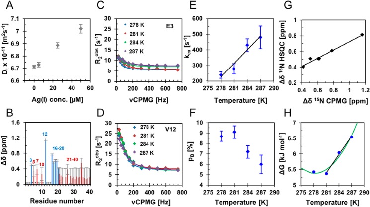
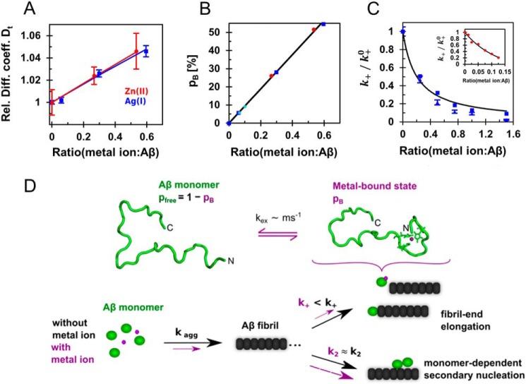
A detailed understanding of the molecular pathways for amyloid-β (Aβ) peptide aggregation from monomers into amyloid fibrils, a hallmark of Alzheimer's disease, is crucial for the development of diagnostic and therapeutic strategies. We investigate the molecular details of peptide fibrillization in vitro by perturbing this process through addition of differently charged metal ions. Here, we used a monovalent probe, the silver ion, that, similarly to divalent metal ions, binds to monomeric Aβ peptide and efficiently modulates Aβ fibrillization. On the basis of our findings, combined with our previous results on divalent zinc ions, we propose a model that links the microscopic metal-ion binding to Aβ monomers to its macroscopic impact on the peptide self-assembly observed in bulk experiments. We found that substoichiometric concentrations of the investigated metal ions bind specifically to the N-terminal region of Aβ, forming a dynamic, partially compact complex. The metal-ion bound state appears to be incapable of aggregation, effectively reducing the available monomeric Aβ pool for incorporation into fibrils. This is especially reflected in a decreased fibril-end elongation rate. However, because the bound state is significantly less stable than the amyloid state, Aβ peptides are only transiently redirected from fibril formation, and eventually almost all Aβ monomers are integrated into fibrils. Taken together, these findings unravel the mechanistic consequences of delaying Aβ aggregation via weak metal-ion binding, quantitatively linking the contributions of specific interactions of metal ions with monomeric Aβ to their effects on bulk aggregation.

Keywords: Alzheimer disease; amyloid; amyloid-beta (AB); metal; metal ion-protein interaction; monovalent ion; neurodegeneration; nuclear magnetic resonance (NMR); protein aggregation; silver; zinc.


Conflict of interest statement

The authors declare that they have no conflicts of interest with the contents of this article

Figures

Figure 1.
Figure 1.
Aβ fibrillization retardation in the presence of Ag(I) ions. A–C, global fit analysis of aggregation traces of 20 μm monomeric Aβ40 incubated in the presence of 0–30 μm Ag(I) in 10 mm MOPS buffer, pH 7.2, at +37 °C under quiescent conditions. The global fits were constrained such that only one nucleation rate (kn, k2, or k+) is the single free fit parameter, revealing the best fit for k+ (normalized mean squared error (MSE), 1.0), followed by k2 (normalized MSE, 2.0) and kn (normalized MSE, 9.2). D and E, parameters from sigmoidal curve fitting of the kinetic traces in A–C. The error bars represent standard deviation values from individual fits to four replicates (Fig. S1). F, elongation rates obtained from the initial slopes of highly seeded aggregation kinetics experiments, where 1 μm seeds were added to 20 μm monomeric Aβ40 with 0–30 μm Ag(I). The weighted average values calculated from four replicates are shown in A–C and F. G, relative k+ values obtained from the global fit analysis in C compared with values from seeding experiments in F, showing the same Ag(I) dependence of k+. H, global fit analysis applying a model, where the apparent free Aβ monomer concentration is determined by the dissociation constant KDapp, revealing KDapp = 14.5 ± 0.2 μm (normalized MSE, 3.7). I and J, images from solid-state AFM of samples taken after fibrillization kinetic experiments showing similar Aβ40 fibril structures in the absence (I) and presence (J) of Ag(I) ions at 1:1 ratio. The scale bar represents 1 μm.
Figure 2.
Figure 2.
Ag(I) ions bind specifically to the N-terminal part of monomeric Aβ peptide. A, 1H-15N HSQC spectra of 80 μm monomeric 13C-15N-labeled Aβ40 peptides alone (blue) and with 20 μm Ag(I) ions, 4:1 Aβ40:Ag(I) (red) in 20 mm sodium phosphate buffer, pH 7.4, recorded at 278 K. B, relative intensities determined from the Ag(I) ion titration. C and D, 1H-15N HSQC spectra of 15N-labeled Aβ40 H6A,H13A,H14A mutant peptides (AβnoHis) (blue) and in the presence of 1:30 Aβ40:Ag(I) ions (green), showing no changes in cross-peak intensities (D) and chemical shifts (C) in the presence of Ag(I) ions. E, magnification of the chemical shift changes observed in A. F, combined chemical shift changes from data in A. Hence, signal attenuation and chemical shift changes upon addition of Ag(I) are most prominent for the N-terminal residues.
Figure 3.
Figure 3.
Chemical exchange between free Aβ and an Aβ–Ag(I) ion complex. A, translational diffusion coefficients of 80 μm monomeric Aβ40 in the absence and presence of 5–50 μm Ag(I) ions at 281 K. The average and standard deviation values were determined from five repeated measurements. B–F, relaxation dispersion was measured for 80 μm 15N-labeled Aβ40 with two different Ag(I) concentrations (4 and 6 μm). B, chemical shift differences from an individual data set with 6 μm Ag(I). Blue bars show the residues exhibiting significant chemical exchange (p < 0.01), which were included in the full analysis, whereas red bars correspond to residues with no significant chemical exchange. Residues marked with an open circle were not observed because of low signal intensity, and residues marked with an asterisk exhibited too fast exchange with the solvent or spectral overlap. C and D, relaxation dispersion profiles from two selected N-terminal residues at four different temperatures with 6 μm Ag(I). E and F, the temperature dependence of the global fit parameters, the chemical exchange rate, kex, and bound population, pB. G, chemical shift changes from 1H-15N HSQC experiments plotted against the chemical shift differences from relaxation dispersion, displayed in B, revealing good correlation. H, Gibbs free energy for the four different temperatures shown for 6 μm Ag(I). At higher temperatures (≥281 K) the data points could be fitted linearly, whereas the whole data set exhibits a nonlinear dependence (green), described by an equation including a heat capacity difference (supporting text).
Figure 4.
Figure 4.
Model for the role of Aβ:metal ion complexes in Aβ fibrillization using substoichiometric concentrations of Ag(I) and Zn(II) ions. A, global fit of diffusion data where data from Ag(I) (blue) were fitted together with previously reported data for Zn(II) (14) (red) using a two-state model, revealing an increased relative diffusion coefficient (Rel. Diff. coeff.) of the bound/folded state by DB/Dfree = 1.087 ± 0.002. B, the metal-bound populations, pB, are determined by the apparent dissociation constants for the respective metal ions and exhibit a linear dependence on the metal ion:Aβ ratio, here shown for the population corresponding to the diffusion data in A. The bound populations from relaxation dispersion experiments of Ag(I) (turquoise) confirm this relation. C, the relative elongation rates, k+, of Zn(II) (red) and Ag(I), from seeding (blue triangles) and global fit analysis (blue squares), are plotted against the metal ion:Aβ ratio, and fitted with Equation 5, revealing an apparent dissociation constant of KDapp = 4.1 ± 0.4 μm for Ag(I) ions and KDapp = 1.2 ± 0.2 μm for Zn(II) ions (inset). These values are similar as determined by other methods for Ag(I) (Table S2) and determined previously for Zn(II) (28). These findings indicate that Ag(I) and Zn(II) interact with monomeric Aβ in a remarkably similar manner. D, monomeric Aβ binds transition metal ions at the N terminus, forming a compact, histidine-coordinated fold. This metal–peptide complex is not stable and exchanges with the free peptide on the millisecond time scale. Aβ fibrils are formed through secondary nucleation mechanisms, in addition to fibril-end elongation, where the latter is predominantly attenuated by the presence of metal ions. Hence, these binding processes reduce the apparent available pool of free monomeric Aβ, resulting in a retardation of the overall fibrillization.

References

    1. Dudev T., and Lim C.. 2008) Metal binding affinity and selectivity in metalloproteins: insights from computational studies. Annu. Rev. Biophys. 37, 97–116 10.1146/annurev.biophys.37.032807.125811 - DOI - PubMed
    1. Glenner G. G., and Wong C. W.. 1984) Alzheimer's disease: initial report of the purification and characterization of a novel cerebrovascular amyloid protein. Biochem. Biophys. Res. Commun. 120, 885–890 10.1016/S0006-291X(84)80190-4 - DOI - PubMed
    1. Haass C., and Selkoe D. J.. 2007) Soluble protein oligomers in neurodegeneration: lessons from the Alzheimer's amyloid β-peptide. Nat. Rev. Mol. Cell Biol. 8, 101–112 10.1038/nrm2101 - DOI - PubMed
    1. Meisl G., Michaels T. C. T., Arosio P., Vendruscolo M., Dobson C. M., and Knowles T. P. J.. 2019) Dynamics and control of peptide self-assembly and aggregation. Adv. Exp. Med. Biol. 1174, 1–33 10.1007/978-981-13-9791-2_1 - DOI - PubMed
    1. Cohen S. I., Vendruscolo M., Dobson C. M., and Knowles T. P.. 2012) From macroscopic measurements to microscopic mechanisms of protein aggregation. J. Mol. Biol. 421, 160–171 10.1016/j.jmb.2012.02.031 - DOI - PubMed

Publication types

LinkOut - more resources