. 2020 Apr 3;368(6486):85-89.
doi: 10.1126/science.aaw9872.

Cysteine depletion induces pancreatic tumor ferroptosis in mice

Affiliations

Cysteine depletion induces pancreatic tumor ferroptosis in mice

Michael A Badgley et al. Science. .

Abstract

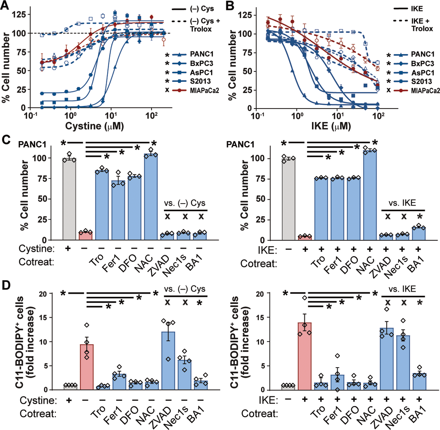
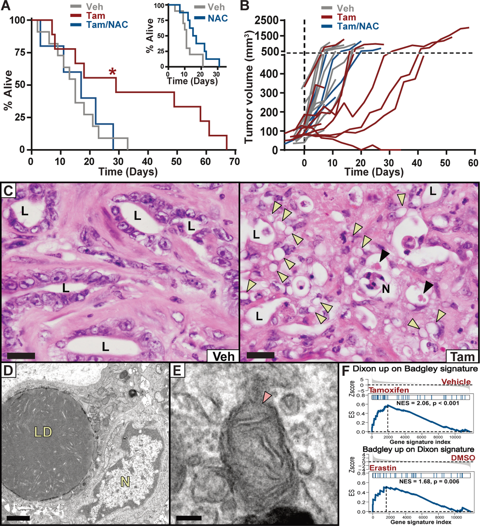
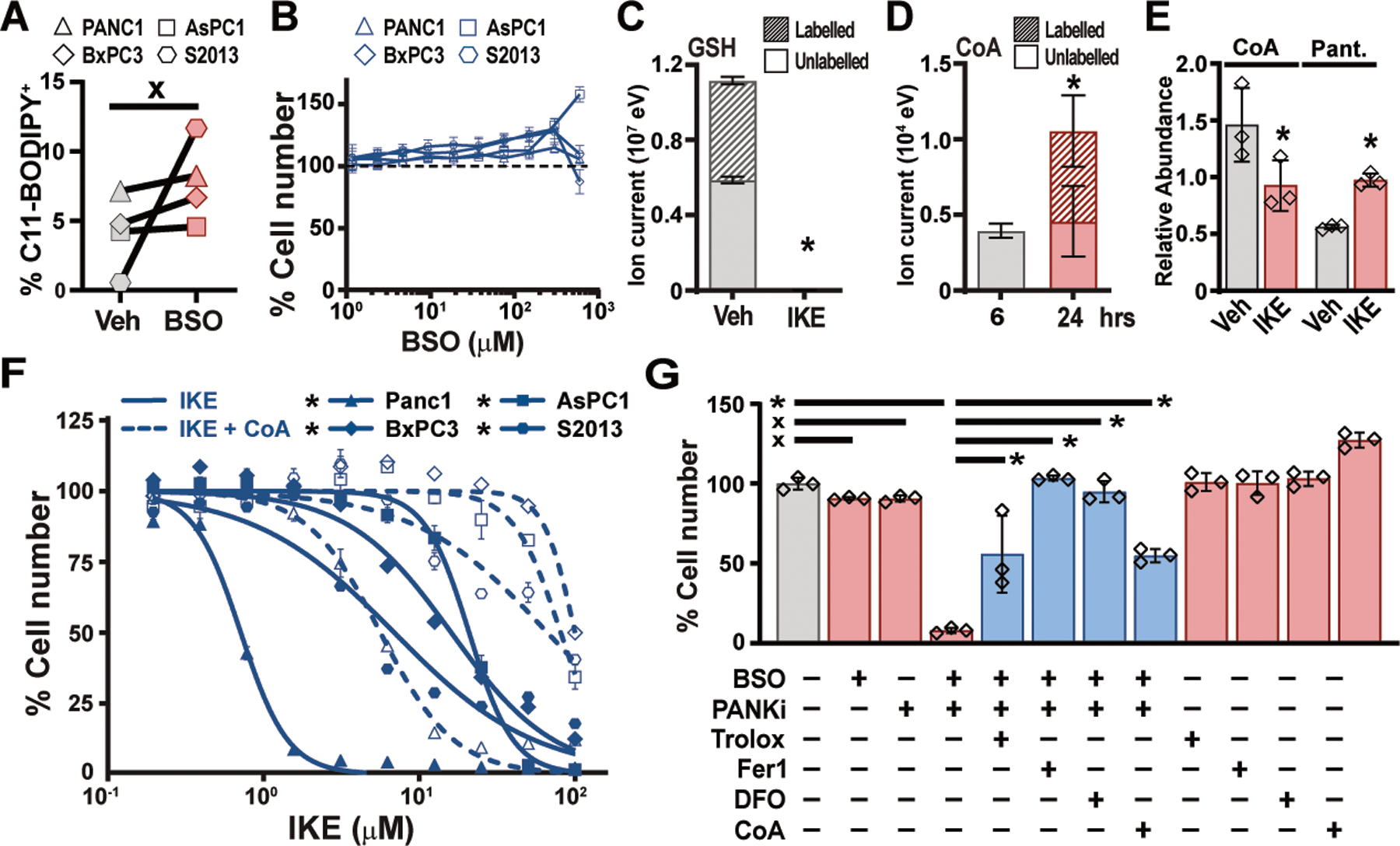
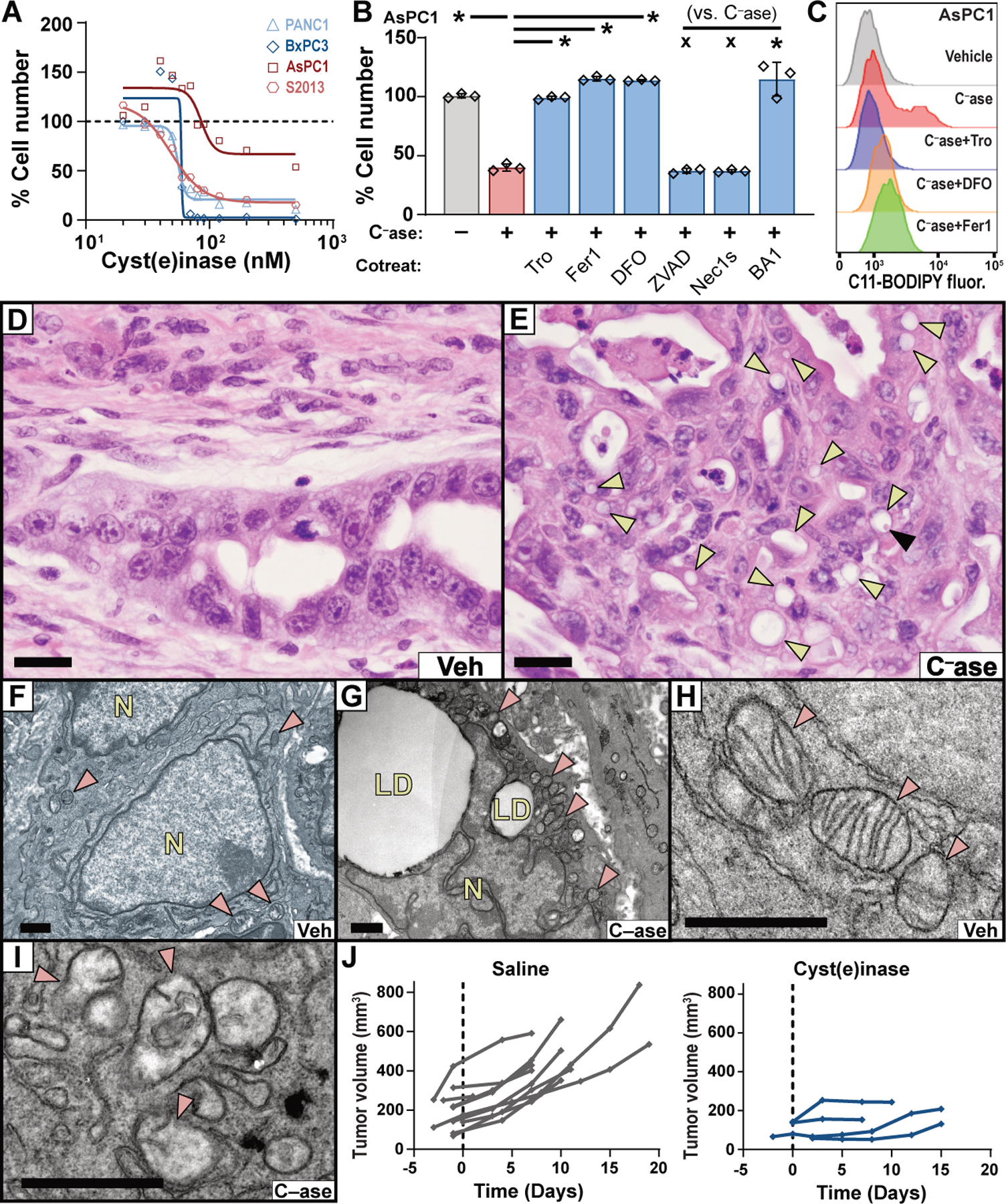
Ferroptosis is a form of cell death that results from the catastrophic accumulation of lipid reactive oxygen species (ROS). Oncogenic signaling elevates lipid ROS production in many tumor types and is counteracted by metabolites that are derived from the amino acid cysteine. In this work, we show that the import of oxidized cysteine (cystine) via system xC - is a critical dependency of pancreatic ductal adenocarcinoma (PDAC), which is a leading cause of cancer mortality. PDAC cells used cysteine to synthesize glutathione and coenzyme A, which, together, down-regulated ferroptosis. Studying genetically engineered mice, we found that the deletion of a system xC - subunit, Slc7a11, induced tumor-selective ferroptosis and inhibited PDAC growth. This was replicated through the administration of cyst(e)inase, a drug that depletes cysteine and cystine, demonstrating a translatable means to induce ferroptosis in PDAC.


Conflict of interest statement

Competing interests: B.R.S. holds equity in and serves as a consultant to Inzen Therapeutics and is an inventor on patents and applications related to ferroptosis, including the following U.S. patents and corresponding applications and patents in other countries, all submitted by Columbia University: 10,259,775, 10,233,171, 9,580,398, 20190292135, 20190135782, 20170233370, 20160297748, 20150079035, and 20080299076, 9,938,245, 20160332974, 9,695,133, 20150175558, 8,546,421, 20100081654, 8,535,897, 20110008803, 8,518,959, 20090214465, 8,124,365, 7,358,262, 20080220454, 7,615,554, 20190315681, and 20070161644. G.G. and E.S. have an equity interest in Aeglea Biotherapeutics, a company that has licensed the commercial development of Cyst(e)inase, and are inventors on U.S. patents and corresponding applications and patents in other countries, all submitted by the University of Texas, related to L-cyst(e)inase: 10,363,311, 20180327734. The other authors declare no competing financial interests.

Figures

Fig. 1.
Fig. 1.. Pancreatic cancer cells require exogenous cystine to avert ferroptosis.
(A and B) Viability of human PDAC lines after 24 hours culture in varying concentrations of cystine (A) or IKE (B), alone or in combination with 100 μM Trolox. Student’s t-test comparing maximal cytotoxicity ± Trolox. (C) Viability of PANC-1 cells cultured for 24 hours in cystine-free media or treated with 10 μM IKE, alone or in combination with 100 μM Trolox (Tro), 500 nM ferrostatin-1 (Fer1), 100 μM deferoxamine (DFO), 1mM N-acetyl cysteine (NAC), 50 μM ZVAD-FMK, 1 nM Bafilomycin A1 (BA1) or 10 μM Necrostatin-1s (Nec1s). Tukey test. (D) Flow cytometry of C11-BODIPY fluorescence in PANC-1, AsPC-1, BxPC-3, and S-2013 cells after 6–8 hours of treatment with conditions from panel C. Tukey test. All data are means ±SEM of three independent experiments. * p < 0.05, x = no significant difference.
Fig. 2.
Fig. 2.. Deletion of Slc7a11 in KPC mice induces tumor ferroptosis and extends survival.
(A) Survival of KPFSR mice treated with vehicle (n = 11, median 15 days), tamoxifen (n = 9, median 29 days), or tamoxifen/NAC (n = 5, median 17 days). * p < 0.0295, log-rank. INSET: Survival of KPC mice treated with NAC alone (n = 8, median = 16 days) versus historical saline-treated controls (n = 10, median = 11 days). (B) Growth curves for each KPFSR tumor. (C) Hematoxylin and eosin (H&E) stained sections of tumor tissue from KPFSR mice treated with vehicle or tamoxifen. L = lumen of malignant epithelium, N = necrosis, yellow arrowheads = lipid droplets, black arrowheads = megamitochondria. Scale = 20 μm. (D and E) TEM images from tamoxifen-treated KPFSR tumors. LD = lipid droplets, N = nucleus, arrow indicates damaged mitochondrion. (D) scale = 1μm; (E) scale = 100 nm. (F) Gene set enrichment analysis. Top panel depicts enrichment of a published ferroptosis expression signature (Dixon) among genes differentially expressed in tamoxifen-treated KPFSR epithelia (Badgley) (p < 0.001). Bottom panel depicts the reciprocal comparison (p < 0.006).
Fig. 3.
Fig. 3.. Combination GSH and CoA inhibition induces ferroptosis in human PDAC cells.
(A) Flow cytometry for C11-BODIPY fluorescence in four human PDAC lines treated for 6 hours with 150 μM BSO. Paired t-test. (B) Viability of human PDAC cells treated for 24 hours with indicated concentrations of BSO. (C) Liquid chromatography time-of-flight mass spectrometry (LC-TOF-MS) analysis of GSH in PANC-1 cells labelled for 6 hours with 13C-cystine combined with vehicle or 5μM IKE. Student’s t-test. (D) LC-TOF-MS analysis of CoA in PANC-1 cells labelled for 6 hours with 13C-cystine, after 6 or 24 hours. Student’s t-test. (E) Liquid chromatography triple quadrupole tandem mass spectrometry (LC-QqQ-MS) measurements of CoA and pantothenate (Pant.) levels in Panc-1 cells treated with vehicle and IKE for six hours. Student’s t-test. (F) Viability of human PDAC cell lines treated with IKE, alone or in combination 200 μM CoA. Student’s t-test comparing maximal cytotoxicity ± CoA. (G) PANC-1 cells treated for 24 hours with combinations of 300 μM BSO and 5 μM PANKi along with Trolox, Fer-1, DFO, or CoA as described in Fig. 1C. Tukey test. In all panels, * p < 0.05, x = not significantly different. In panels A, B, E, and F, data are means ±SEM from three independent experiments. In panels C and D, data are means ± SD from three biological replicates. In (E), * p < 0.05 comparing maximal cytotoxicity in CoA treated vs. untreated conditions for each line, Student’s t-test. In (F), * p < 0.05, one-way ANOVA with posthoc Tukey test.
Fig. 4.
Fig. 4.. Cyst(e)inase treatment induces tumor-selective ferroptosis in KPC mice.
(A) Viability of human PDAC lines cultured with varying concentrations of cyst(e)inase for 48 hours (AsPC1) or 72 hours (PANC-1, BxPC3, S2–013). (B) Viability of AsPC1 cells treated with 90 nM cyst(e)inase (Case) for 72 hours, alone or in combination with indicated agents, under conditions described in Fig. 1C. (C) C11-BODIPY fluorescence was measured by flow cytometry in AsPC-1 cells after 24 hours of treatment with 90 nM cyst(e)inase, alone or in combination with indicated agents, under conditions described in Fig. 1C. (D and E) H&E stained sections of pancreatic tumors from KPC mice treated with vehicle or cyst(e)inase. Yellow arrowheads indicate lipid droplets. Black arrowhead indicates megamitochondrion. Bars = 20 μm. (F–I) TEM of pancreatic tumors from the KPC model treated with vehicle (F and H) or cyst(e)inase (G and I). Red arrowheads indicate mitochondria; LD = lipid droplets; N = nucleus. Bars = 1 μm. (J) Tumor growth curves from KPC mice treated with saline (historical controls) or 100 mg/kg cyst(e)inase, q.2.d., i.p.. For panels A–B, data depict mean of 3 biological replicates. * p < 0.05, x = not significant.

Similar articles

Cited by

References

    1. DeNicola GM et al., Oncogene-induced Nrf2 transcription promotes ROS detoxification and tumorigenesis. Nature 475, 106–109 (2011). - PMC - PubMed
    1. Sato H et al., Redox imbalance in cystine/glutamate transporter-deficient mice. J. Biol. Chem 280, 37423–37429 (2005). - PubMed
    1. Yang WS et al., Regulation of ferroptotic cancer cell death by GPX4. Cell 156, 317–331 (2014). - PMC - PubMed
    1. Dixon SJ et al., Ferroptosis: an iron-dependent form of nonapoptotic cell death. Cell 149, 1060–1072 (2012). - PMC - PubMed
    1. Gao M et al., Ferroptosis is an autophagic cell death process. Cell Res. 26, 1021–1032 (2016). - PMC - PubMed

Publication types

MeSH terms