. 2020 Apr 2;11(1):1642.
doi: 10.1038/s41467-020-15483-7.

ASK1 inhibits browning of white adipose tissue in obesity

Affiliations

ASK1 inhibits browning of white adipose tissue in obesity

Fabrizio C Lucchini et al. Nat Commun. .

Abstract

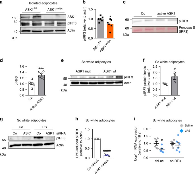
Increasing energy expenditure via induction of adipose tissue browning has become an appealing strategy to treat obesity and associated metabolic complications. Herein, we identify adipocyte-expressed apoptosis signal-regulating kinase 1 (ASK1) as regulator of adipose tissue browning. High fat diet-fed adipocyte-specific ASK1 knockout mice reveal increased UCP1 protein levels in inguinal adipose tissue concomitant with elevated energy expenditure, reduced obesity and ameliorated glucose tolerance compared to control littermates. In addition, ASK1-depletion blunts LPS-mediated downregulation of isoproterenol-induced UCP1 in subcutaneous fat both in vitro and in vivo. Conversely, adipocyte-specific ASK1 overexpression in chow-fed mice attenuates cold-induced UCP1 protein levels in inguinal fat. Mechanistically, ASK1 phosphorylates interferon regulatory factor 3 (IRF3) resulting in reduced Ucp1 expression. Taken together, our studies unravel a role of ASK1 in mediating the inhibitory effect of caloric surplus or LPS-treatment on adipose tissue browning. Adipocyte ASK1 might be a pharmacological target to combat obesity and associated morbidities.


Conflict of interest statement

The authors declare no competing interests.

Figures

Fig. 1
Fig. 1. LPS treatment blunts UCP1 expression in subcutaneous adipocytes ASK1-dependently.
a, b Western blot and quantified protein levels of UCP1 in total lysates of inguinal white adipose tissue harvested from cold-exposed C57BL/6J mice chronically treated with or without LPS. n = 3 mice per group. **p = 0.004. c Ucp1 mRNA expression in inguinal fat explants harvested from C57BL/6J mice were treated with or without 100 ng/ml LPS and subsequently with 10 µM isoproterenol for 8 h. n = 3 biological replicates. *p = 0.036. d Ucp1 mRNA expression in subcutaneous adipocytes pre-treated with or without 100 ng/ml LPS for 24 h followed by stimulation with 0.1 µM isoproterenol for 6 h. n = 7 (LPS) or n = 8 (control) biological replicates. **p = 0.007. e, f Western blot and quantified protein levels of ASK1 in total lysates of inguinal white adipose tissue harvested from C57BL/6J mice fed a chow or HFD for 20 weeks. n = 8 (chow) and n = 7 (HFD) mice per group. *p = 0.037. g Ask1 mRNA expression in subcutaneous adipocytes treated with or without 100 ng/ml LPS treatment for 24 h. n = 7 biological replicates. *p = 0.046. h Ucp1 mRNA expression in subcutaneous adipocytes transfected with control shRNA lentivirus (shLuc) or shRNA lentivirus targeting ASK1 (shASK1) pre-treated with 100 ng/ml LPS for 24 h followed by stimulation with 0.1 µM isoproterenol for 6 h. n = 11 (shASK1 control and shLuc LPS) or n = 12 (shASK1 LPS and shLuc control) biological replicates. *p = 0.027. Values are expressed as mean ± SEM. Statistical tests used: two-sided t-tests for (bd, fh). Source data are provided as a Source Data file.
Fig. 2
Fig. 2. Reduced body weight and improved glucose tolerance in HFD-fed ASK1Δadipo mice.
a Protein levels of ASK1 in lysate of isolated epididymal adipocytes harvested from ASK1F/F and ASK1Δadipo mice (n = 3 mice per group). b, c Body weight and body weight gain during 12 weeks of chow (ASK1F/F, n = 20 mice; ASK1∆adipo, n = 13 mice) or HFD feeding (ASK1F/F, n = 52 mice; ASK1∆adipo, n = 58 mice). **p = 0.02, ***p < 0.001. d, e Intraperitoneal glucose-tolerance test (chow-fed: ASK1F/F, n = 10 mice; ASK1∆adipo, n = 8 mice; HFD-fed: ASK1F/F, n = 13 mice; ASK1∆adipo, n = 14 mice) at 18 weeks of age. **p = 0.03, ***p < 0.001. f Glucose infusion rate (GIR) during hyperinsulinemic-euglycemic clamps in HFD-fed ASK1F/F (n = 6) and ASK1∆adipo (n = 8) mice. *p = 0.011. g Glucose uptake into respective tissues during hyperinsulinemic-euglycemic clamps in HFD-fed ASK1F/F (n = 5 (ing WAT, epiWAT and skeletal muscle) or n = 6 (BAT)) and ASK1∆adipo (n = 4 (BAT), n = 6 (ingWAT) or n = 7 (epiWAT and skeletal muscle)) mice. *p = 0.034, #p = 0.062. h Endogenous glucose production (EGP) during hyperinsulinemic-euglycemic clamps in HFD-fed ASK1F/F (n = 6) and ASK1∆adipo (n = 8) mice. *p = 0.040. i Liver triglyceride levels determined in HFD-fed ASK1F/F (n = 6) and ASK1∆adipo (n = 6) mice. *p = 0.012. j Mesenteric adipose tissue mRNA expression of respective targets determined in HFD-fed ASK1F/F (n = 5 (Il-10, F4/80) or n = 8 (TNFα, Il-6, Mcp1, Il-1β) and ASK1∆adipo (n = 7 (Il-10, F4/80) or n = 9 (TNFα, Il-6, Mcp1, Il-1β)) mice. Values are expressed as mean ±  SEM. Statistical tests used: two-sided t-tests for (f, g) (without adjustments for multiple comparisons), h, i ANOVA for (be). AUC: area under the curve. Source data are provided as a Source Data file.
Fig. 3
Fig. 3. Increased heat production in HFD-fed ASK1Δadipo mice.
a Light (day) and dark (night) oxygen consumption in HFD-fed ASK1F/F (n = 8) and ASK1Δadipo (n = 11) mice during a 24-h period at RT. **p = 0.020 (light), **p = 0.010 (dark). b Light (day) and dark (night) oxygen consumption in HFD-fed ASK1F/F (n = 7) and ASK1Δadipo (n = 6) mice during a 24-h period after 20 days housing at thermoneutrality. #p = 0.073, *p = 0.042. c Light (day) and dark (night) heat production in HFD-fed ASK1F/F (n = 8) and ASK1Δadipo (n = 11) mice during a 24-h period at RT. **p = 0.009. d Rectal temperature measured in HFD-fed ASK1F/F (n = 7) and ASK1Δadipo (n = 7) mice. *p = 0.015. Values are expressed as mean ± SEM. Statistical tests used: two-sided t-tests for (ad). Source data are provided as a Source Data file.
Fig. 4
Fig. 4. Increased adipose tissue browning in HFD-fed ASK1Δadipo mice.
a, b Fat pad mass as well as total body fat mass (chow-fed: ASK1F/F, n = 13; ASK1∆adipo, n = 10; HFD-fed: ASK1F/F, n = 10; ASK1∆adipo, n = 16) in mice at 18 weeks of age. **p = 0.005; ***p < 0.001. c Representative image of inguinal adipose tissue of a HFD-fed ASK1F/F and ASK1∆adipo mouse (scale bar: 10 mm). d Representative immunohistochemistry for UCP1 in inguinal adipose tissue of a HFD-fed ASK1F/F (n = 3 mice) and ASK1∆adipo mouse (n = 3 mice) (scale bar: 50 µm). e mRNA expression of respective genes in inguinal adipose tissue of HFD-fed ASK1F/F (n = 8) and ASK1Δadipo (n = 9) mice. *p = 0.027 (Ucp1), *p = 0.028 (Cidea). f, g Representative western blot and quantification of UCP1 and PGC1α protein levels in inguinal adipose tissue harvested from HFD-fed ASK1F/F (n = 8) and ASK1Δadipo (n = 10) mice. *p = 0.031 (Ucp1), *p = 0.015 (Cidea). h, i Representative western blot and quantified protein levels of UCP1 in total lysates of inguinal white adipose tissue harvested from cold-exposed mice chronically treated with or without LPS (saline ASK1F/F, n = 6 mice; saline ASK1∆adipo, n = 5 mice; LPS ASK1F/F, n = 7 mice; LPS ASK1∆adipo, n = 6 mice). **p = 0.003. Values are expressed as mean ±  SEM. Statistical tests used: two-sided t-tests for, e (Cidea), i two-sided Mann–Whitney for e (UCP1), g ANOVA for (a and b). Source data are provided as a Source Data file.
Fig. 5
Fig. 5. Adipocyte-specific ASK1 overexpression decreases adipose tissue browning.
a Protein levels of ASK1 in lysate of isolated adipocytes harvested from CAG-ASK1F/F and CAG-ASK1+adipo mice (n = 2 mice per group). b mRNA expression of respective genes in inguinal adipose tissue of cold-exposed CAG-ASK1F/F (n = 5) and CAG-ASK1+adipo (n = 6) mice. #p = 0.094, *p = 0.042, *p = 0.017. c, d Representative western blot and quantification of UCP1 protein levels in inguinal adipose tissue harvested from cold-exposed CAG-ASK1F/F (n = 7) and CAG-ASK1+adipo (n = 8) mice. *p = 0.037. Values are expressed as mean ± SEM. Statistical tests used: two-sided t-tests for (b) (Ucp1 and Pgc1α), d two-sided Mann–Whitney for (b) (Cidea). Source data are provided as a Source Data file.
Fig. 6
Fig. 6. ASK1 phosphorylates IRF3.
a, b Representative western blot and quantification of pIRF3 protein levels in isolated adipocytes of HFD-fed ASK1F/F and ASK1Δadipo mice. n = 6 mice per group. *p = 0.048. c, d Representative western blot and quantification of pIRF3 upon incubation of recombinant IRF3 with or without active ASK1. n = 6 biological replicates. ***p = 0.0006. e, f Western blot and quantification of pIRF3 protein levels in subcutaneous adipocytes transfected with lentivirus expressing wild-type (pLV-CMV-ASK1; ASK1 wt) or kinase-negative (pLV-CMV-ASK1-K716R; ASK1 mut) ASK1. n = 3 biological replicates. #p = 0.070. g, h Representative western blot and quantification of pIRF3 protein levels in subcutaneous adipocytes transfected with control siRNA (Co siRNA) or siRNA targeting ASK1 (ASK1 siRNA) treated with or without 100 ng/ml LPS for 6 h. n = 4 biological replicates. ****p < 0.0001. i Ucp1 mRNA expression in subcutaneous adipocytes transfected with control shRNA lentivirus (shLuc) or shRNA lentivirus targeting IRF3 (shIRF3) pre-treated with 100 ng/ml LPS for 24 h followed by stimulation with 0.1 µM isoproterenol for 6 h. n = 6 biological replicates. *p = 0.019. Values are expressed as mean ± SEM. Statistical tests used: two-sided t-tests for (b, d, f, i), two-sided one sample t-test for (h). Source data are provided as a Source Data file.

References

    1. Betz MJ, Enerback S. Human brown adipose tissue: what we have learned so far. Diabetes. 2015;64:2352–2360. doi: 10.2337/db15-0146. - DOI - PubMed
    1. van Marken Lichtenbelt WD, et al. Cold-activated brown adipose tissue in healthy men. N. Engl. J. Med. 2009;360:1500–1508. doi: 10.1056/NEJMoa0808718. - DOI - PubMed
    1. Virtanen KA, et al. Functional brown adipose tissue in healthy adults. N. Engl. J. Med. 2009;360:1518–1525. doi: 10.1056/NEJMoa0808949. - DOI - PubMed
    1. Hayakawa R, Hayakawa T, Takeda K, Ichijo H. Therapeutic targets in the ASK1-dependent stress signaling pathways. Proc. Jpn Acad. Ser. B Phys. Biol. Sci. 2012;88:434–453. doi: 10.2183/pjab.88.434. - DOI - PMC - PubMed
    1. Krauss S, Zhang CY, Lowell BB. The mitochondrial uncoupling-protein homologues. Nat. Rev. Mol. Cell Biol. 2005;6:248–261. doi: 10.1038/nrm1592. - DOI - PubMed

Publication types