. 2020 May 28;29(8):1340-1352.
doi: 10.1093/hmg/ddaa061.

Nemo-like kinase reduces mutant huntingtin levels and mitigates Huntington's disease

Affiliations

Nemo-like kinase reduces mutant huntingtin levels and mitigates Huntington's disease

Mali Jiang et al. Hum Mol Genet. .

Abstract

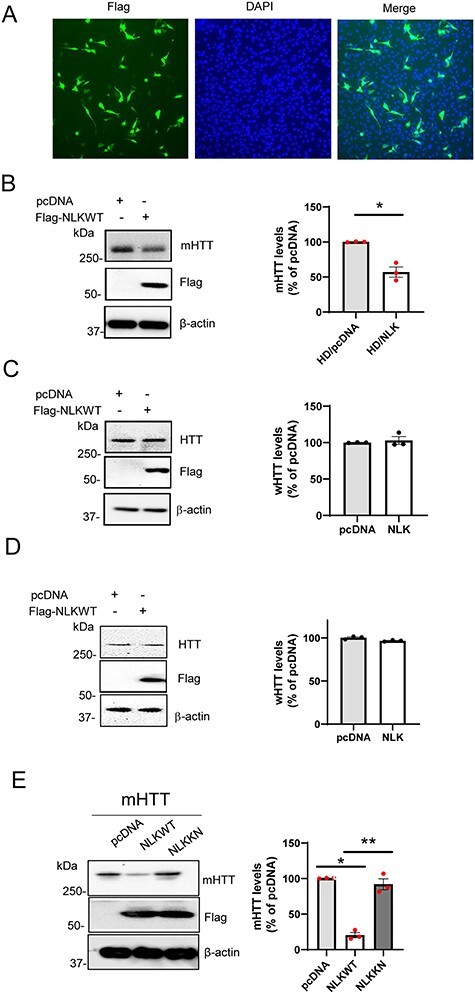
Nemo-like kinase (NLK), an evolutionarily conserved serine/threonine kinase, is highly expressed in the brain, but its function in the adult brain remains not well understood. In this study, we identify NLK as an interactor of huntingtin protein (HTT). We report that NLK levels are significantly decreased in HD human brain and HD models. Importantly, overexpression of NLK in the striatum attenuates brain atrophy, preserves striatal DARPP32 levels and reduces mutant HTT (mHTT) aggregation in HD mice. In contrast, genetic reduction of NLK exacerbates brain atrophy and loss of DARPP32 in HD mice. Moreover, we demonstrate that NLK lowers mHTT levels in a kinase activity-dependent manner, while having no significant effect on normal HTT protein levels in mouse striatal cells, human cells and HD mouse models. The NLK-mediated lowering of mHTT is associated with enhanced phosphorylation of mHTT. Phosphorylation defective mutation of serine at amino acid 120 (S120) abolishes the mHTT-lowering effect of NLK, suggesting that S120 phosphorylation is an important step in the NLK-mediated lowering of mHTT. A further mechanistic study suggests that NLK promotes mHTT ubiquitination and degradation via the proteasome pathway. Taken together, our results indicate a protective role of NLK in HD and reveal a new molecular target to reduce mHTT levels.


Figures

Figure 1
Figure 1
NLK interacts with HTT. (A) HEK293-FT cells were co-transfected with Flag-NLK and FL-HTT with 23Q (wHTT) or 123Q (mHTT), or pcDNA as a control. Cells were collected for immunoprecipitation (IP). Western blotting analysis was performed with MW1 antibody to mHTT, MAB2166 antibody to HTT (both wHTT and mHTT) or anti-Flag antibody (to transgene NLK). (B) HEK293-FT cells were co-transfected with Flag-NLK and Myc-N171-HTT with 18Q or 82Q. Cells were collected for the IP experiments. Western blotting was performed with anti c-Myc (to N-HTT) or Flag (to NLK) antibodies. Arrow indicated the specific band for Flag-NLK. (C) NLK and HTT are detected in both nuclear and cytoplasmic compartments. HEK293-FT cells were co-transfected with Flag-tagged NLK and full-length HTT with either 23Q or 82Q. Cell extracts from indicated groups were subject to subcellular fractionation. Nuclear fraction and cytoplasm compartment were subjected to Western blotting. Transfected HTT and NLK were expressed in both compartments. (D) Mouse cerebral cortex samples from 6-month-old homozygous zQ175 HD or WT mice were subjected to co-IP with anti-NLK antibody and then blotting with HTT antibodies (MW1 for mHTT, MAB 2166 for HTT). Alternatively, samples were subjected to co-IP with indicated HTT antibodies and then western blotting was performed with anti-NLK antibody.
Figure 2
Figure 2
NLK protein levels are decreased in HD. (A) NLK levels in the human postmortem control (Con) and HD motor cortex A4. Western blotting was performed with NLK and β-actin antibodies. Intensity of blots was quantified by NIH Image J software and normalized by β-actin loading controls. n = 5. (B) NLK levels in the striatum of N171-82Q HD and WT mice at 6 months age. n = 3. (C) NLK levels in the striatum of HdhQ 250Q HD and WT mice at 12 months age. n = 3. (D) NLK levels in the striatal cells expressing wild type (WT, STHdhQ7/Q7) or mHTT (HD, STHdhQ111/Q111). The values are expressed as Mean ± SE, *P < 0.05 between HD and control group by unpaired Student’s t-test.
Figure 3
Figure 3
Overexpression of NLK attenuates brain pathology in HD mice. N171-82Q mice were injected with AAV2-NLK or AAV2-GFP control (CON) to the striatum at the age of 10 weeks. (A) Expression of AAV-mediated NLK (GFP-tagged) in the striatum was evidenced at 3 weeks after AAV injection. Scale bar is 50 μm. Western blots of striatal samples from indicated groups suggest that AAV-NLK was expressed. Note that the lower bands (blue arrow) represent the endogenous NLK (about 55KDa), and the upper bands are NLK transgene (GFP-tagged, orange arrow). (B) In vivo structural MRI was conducted at 16 weeks of age. Values are Mean ± SE, n = 4–5. *P < 0.05 compared with the value of WT/CON, **P < 0.05 compared with the value of HD by one-way ANOVA with Holm–Sidak post hoc test. (C) Representative western blot of DARPP32 and quantification data. Values are Mean ± SE, n = 3. *P < 0.05 compared with the value of WT/CON, **P < 0.05 compared with the value of HD by one-way ANOVA with Holm–Sidak post hoc test. (D) Representative images from HD mouse striatum immunostained with EM48 antibody. Scale bar is 50 μm. Number of cells with mHTT aggregates were quantified per microscope field. Mean ± SE. n = 3. *P < 0.05 versus HD/CON group by unpaired Student’s t-test.
Figure 4
Figure 4
Reduction of NLK exacerbates brain pathology in HD mice. HdhQ250 mice were crossbred with NLKKO mice. (A) Reduction of NLK increases mHtt levels. HdhQ250 mice were crossbreeding with the heterozygous NLKKO mice. Western Blotting was performed with MW1 (mHtt), NLK and β-actin antibodies in the cortex of 2-month-old HD and HD/NLKKO mice. n = 3. The values are expressed as mean ± SE. *p < 0.05 compared with the values of the NLK+/+ group by unpaired Student’s t-test. (B) Reduction of NLK does not decrease wHtt levels. Western Blotting was performed with 2166 antibody in the cortex of 2-month-old WT and NLKKO mice. (C) Representative MRI images from indicated groups at age of 12 months (left panel). Brain volumes were determined by structural MRI (right panel). n = 5. (D) DARPP32 levels were evaluated in the striatum of 12-month-old mice. n = 3–6. Densitometry analysis was performed by NIH Image J. *P < 0.05 compared with the values of the WT group, **P < 0.05 compared with the values of the HD group by one-way ANOVA with Dunn’s or Holm–Sidak post hoc test.
Figure 5
Figure 5
NLK selectively decreases endogenous mHTT protein levels in a kinase-dependent manner. (A) Mouse striatal precursor neurons expressing endogenous levels of mHTT (111Q) were transfected with Flag-NLK. Cells were immunostained with anti-Flag (NLK) antibody, DAPI stains nucleus. (B) mHTT expressing mouse striatal cells were transfected with pcDNA or NLK. Cells were collected for western blot. n = 3. The values are expressed as Mean ± SE. *P < 0.05 versus HD/pcDNA group by unpaired Student’s t-test. (C) Mouse striatal cells expressing wHTT (Q7) were transfected with pcDNA, or Flag-NLK. Cells were collected and western blotting was performed with MAB 2166 (HTT), Flag (NLK) and actin antibodies. n = 3. (D) Human HEK 293 cells were transfected with pcDNA or Flag-NLKWT. Cells were collected for western blot. The values are expressed as Mean ± SE, n = 3. (E) Human HEK 293 cells were co-transfected with mutant HTT (mHTT) with pcDNA or Flag-NLKWT or kinase null mutant NLK (Flag-NLK KN). Cells were collected for western blot. The values are expressed as Mean ± SE, n = 3. *P < 0.05 compared with the values of the pcDNA group, **P < 0.05 compared with the values of the NLKWT group by one-way ANOVA with Holm–Sidak post hoc test.
Figure 6
Figure 6
The effects of NLK on mHTT levels are mediated by HTT phosphorylation at S120. (A) NLK phosphorylates mHTT at S/T-P sites. HEK293-FT cells were co-transfected with pcDNA or Flag-NLK and full-length mHTT (123Q). Cells were collected for co-IP with mHTT antibody. Representative western blot with MPM2 (anti-phospho-S/T-P), MW1 (mHTT) and 2166 (total HTT) antibodies. The values are expressed as Mean ± SE. n = 3. (B) HEK293-FT cells were co-transfected with HTT (N586-20Q or 82Q), and Flag-NLK WT, or the kinase inactive mutant NLK-KN or pcDNA control. The values are expressed as Mean ± SE, *P < 0.05 compared with the values of the pcDNA group, **P < 0.05 compared with the values of the NLK-WT group by one-way ANOVA with Holm–Sidak post hoc test. (C) NLK had no effect on mHTT protein levels when serine 120 was mutated to alanine (S120A, phospho-resistant). HEK293-FT cells were co-transfected with pcDNA or Flag-NLK and N586-S120A mHTT, n = 3. (D) HEK293 FT cells were co-transfected with pcDNA or Flag-NLK and full-length mHTT with S1201A or S1181A mutation, and western blotting was conducted. n = 3. *P < 0.05 compared with the values of the pcDNA group by unpaired Student’s t-test.
Figure 7
Figure 7
NLK decreases mHTT level via ubiquitin–proteasome pathway. (A) HEK293-FT cells were co-transfected with mHTT (N586-82Q), pcDNA, or NLK-WT for 24 h. Cells were treated with DMSO, or 50 nM epoxomicin. mHTT levels were determined at 24 h after treatment. The values are Mean ± SE. n = 3. *P < 0.05 compared with the values of the pcDNA group, **P < 0.05 compared with the values of the NLK WT group by one-way ANOVA with Holm–Sidak post hoc test. (B) HEK293-FT cells were co-transfected with mHTT and pcDNA or Flag-NLK. Cells were collected at 24 h after transfection for co-IP with mHTT antibody followed by western blot with anti-ubiquitin antibody. The values are Mean ± SE, n = 3. *P < 0.05 compared with the values of the pcDNA group by unpaired Student’s t-test. (C) HEK293-FT cells were co-transfected with mHTT and pcDNA or Flag-NLK. Cells were collected at 24 h after transfection for co-IP with mHTT antibody followed by western blot with anti-ubiquitin antibody. The values are Mean ± SE, n = 3. *P < 0.05 compared with the values of the pcDNA group by unpaired Student’s t-test.

References

    1. The Huntington's Disease Collaborative Research Group (1993) A novel gene containing a trinucleotide repeat that is expanded and unstable on Huntington's disease chromosomes. Cell, 72, 971–983. - PubMed
    1. Miller, J., Arrasate, M., Brooks, E., Libeu, C.P., Legleiter, J., Hatters, D., Curtis, J., Cheung, K., Krishnan, P., Mitra, S.et al. (2011) Identifying polyglutamine protein species in situ that best predict neurodegeneration. Nat. Chem. Biol., 7, 925–934. - PMC - PubMed
    1. Lu, B. and Palacino, J. (2013) A novel human embryonic stem cell-derived Huntington's disease neuronal model exhibits mutant huntingtin (mHTT) aggregates and soluble mHTT-dependent neurodegeneration. FASEB J., 27, 1820–1829. - PubMed
    1. Genetic Modifiers of Huntington's Disease Consortium. Electronic address, g.h.m.h.e. and Genetic Modifiers of Huntington's Disease, C (2019) CAG repeat not Polyglutamine length determines timing of Huntington's disease onset. Cell, 178, 887–900 e814. - PMC - PubMed
    1. Wanker, E.E., Ast, A., Schindler, F., Trepte, P. and Schnoegl, S. (2019) The pathobiology of perturbed mutant huntingtin protein-protein interactions in Huntington's disease. J. Neurochem., 151, 507–519. - PubMed

Publication types

MeSH terms

Substances