Review
. 2020 Aug;237(2):209-224.
doi: 10.1111/joa.13196. Epub 2020 Apr 3.

Geometric description for the anatomy of the mitral valve: A review

Affiliations
Review

Geometric description for the anatomy of the mitral valve: A review

Diana Oliveira et al. J Anat. 2020 Aug.

Abstract

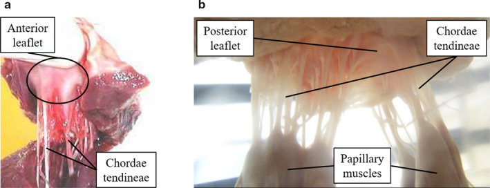
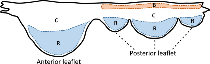
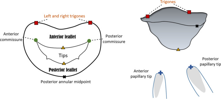
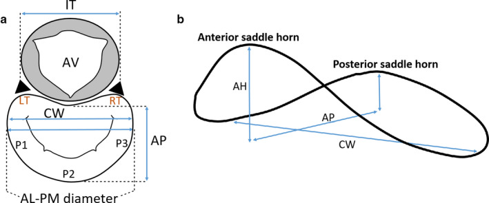
The mitral valve is a complex anatomical structure whose physiological functioning relies on the biomechanical properties and structural integrity of its components. Their compromise can lead to mitral valve dysfunction, associated with morbidity and mortality. Therefore, a review on the morphometry of the mitral valve is crucial, more specifically on the importance of valve dimensions and shape for its function. This review initially provides a brief background on the anatomy and physiology of the mitral valve, followed by an analysis of the morphological information available. A characterisation of mathematical descriptions of several parts of the valve is performed and the impact of different dimensions and shape changes in disease is then outlined. Finally, a section regarding future directions and recommendations for the use of morphometric information in clinical analysis of the mitral valve is presented.

Keywords: biomechanics; computational anatomy; mitral valve; mitral valve disease; morphology analysis.


Conflict of interest statement

None.

Figures

FIGURE 1
FIGURE 1
Porcine mitral valve apparatus: (a) displays the anterior leaflet and chordae tendineae and (b) focuses on the subvalvular apparatus, where chordae arise from both PMs and insert into the rough region of the posterior leaflet
FIGURE 2
FIGURE 2
Human MV leaflet tissue areas. C, clear zone; R, rough zone; B, basal zone
FIGURE 3
FIGURE 3
Atrial and lateral views of a mitral valve (MV) schematic model based upon nine landmarks. The annulus, anterior and posterior leaflets and papillary muscle tips can be observed
FIGURE 4
FIGURE 4
Atrial (a) and lateral (b) views of important mitral annular dimensions. AV, aortic valve; LT, left trigone; RT, right trigone; P1, P2, P3, posterior leaflet scallops; AL–PM diameter, anterolateral‐posteromedial diameter; AP, antero‐posterior distance; CW, commissural width; IT, inter‐trigonal distance
FIGURE 5
FIGURE 5
Circle representing the annulus, according to Okamoto et al. (2007), with relevant dimensions highlighted. LT, left trigone; RT, right trigone; AL–PM diameter, anterolateral‐posteromedial diameter; CW, commissural width; IT distance, inter‐trigonal distance; θTg, trigonal angle; θCom, commissural angle
FIGURE 6
FIGURE 6
Hyperboloids representing the mitral valve (MV) saddle shape and defined by Equation 11 (Park et al., 2019) are displayed, created from literature values for CW (33.3 mm), AP diameter (28 mm), AL–PM diameter (31 mm) and AHCWR (0.237). Different commissural heights (b) and posterior annular depth (c) in comparison with a pure hyperboloid (a) can be observed, shown by changes in transformation parameters
FIGURE 7
FIGURE 7
Coaptation triangle. The tenting height divides the base of the triangle (AP diameter) into two segments—the anterior and the posterior cords. α A, anterior leaflet angle; α P, posterior leaflet angle
FIGURE 8
FIGURE 8
Parameters of human MV leaflet geometry, with an extended (a) and closed (b) leaflet representation. A‐B, anterolateral commissure; B‐C, anterior leaflet; C‐D, posteromedial commissure; D‐A′, posterior leaflet; D‐E, posteromedial commissural scallop (P1); E‐F, middle scallop (P2); F‐A′, anterolateral commissural scallop (P3)
FIGURE 9
FIGURE 9
Measurements describing PM morphology. ALPM, anterolateral PM; PMPM, posteromedial PM
FIGURE 10
FIGURE 10
Distances between PM tips and o’clock points of mitral annulus, in axial (left) and isometric (right) views (adapted from Sakai et al., 1999; Yamaura et al., 2008). ALPM, anterolateral PM; PMPM, posteromedial PM; 0 o’clock, midpoint of the anterior annular circumference; 2 o’clock, right trigone; 10 o’clock, left trigone; 4 o’clock, separation of posterior and middle scallops of posterior leaflet; 8 o’clock, separation of anterior and middle scallops of posterior leaflet
FIGURE 11
FIGURE 11
Axial view of C‐shape representation for the PMs, as present in the literature and suggested by expert surgeons, as well as a simplified depiction of chordae tendineae insertion patterns. Blue, green and orange chordae are those inserting into the anterior leaflet, posterior leaflet and paracommissural zones, respectively
FIGURE 12
FIGURE 12
Example of Reeb graph employed to characterise chordae tendineae topology in Khalighi et al. (2017), illustrating the connectivity between branches. Green, blue and orange vertices correspond to chordal origin (at the PM level), bifurcation points and insertion points into the leaflets, respectively
FIGURE 13
FIGURE 13
Hyperboloids representing: (a) a mitral annular normal saddle configuration; (b) a configuration associated with type I dysfunction, defined as in Park et al. (2019). The first configuration was created with the dimensions shown on Figure 6 and the diseased configuration on literature values for CW (42.2 mm), AP diameter (38.8 mm), AL–PM diameter (39 mm) and AHCWR (.132)

References

    1. Al‐Atabi, M. , Espino, D.M. , Hukins, D.W. and Buchan, K.G. (2012) Biomechanical assessment of surgical repair of the mitral valve. Proceedings of the Institution of Mechanical Engineers. Part H. Journal of Engineering in Medicine, 226, 275–287. - PubMed
    1. Baillargeon, B. , Rebelo, N. , Fox, D.D. , Taylor, R.L. and Kuhl, E. (2014) The Living Heart Project: a robust and integrative simulator for human heart function. European Journal of Mechanics – A/Solids, 48, 38–47. - PMC - PubMed
    1. Bloodworth, C.H.T. , Pierce, E.L. , Easley, T.F. , Drach, A. , Khalighi, A.H. , Toma, M. , et al. (2017) Ex vivo methods for informing computational models of the mitral valve. Annals of Biomedical Engineering, 45, 496–507. - PMC - PubMed
    1. Bouma, W. and Gorman, R.C. (2019) Commentary: Three‐dimensional P3 tethering angle at the heart of future surgical decision making in ischemic mitral regurgitation. Journal of Thoracic and Cardiovascular Surgery, 157, 1806–1807. - PubMed
    1. Calleja, A. , Poulin, F. , Woo, A. , Meineri, M. , Jedrzkiewicz, S. , Vannan, M.A. , et al. (2015) Quantitative modeling of the mitral valve by three‐dimensional transesophageal echocardiography in patients undergoing mitral valve repair: correlation with intraoperative surgical technique. Journal of the American Society of Echocardiography, 28, 1083–1092. - PubMed

Publication types

LinkOut - more resources