Review
. 2020 May;295(1):54-67.
doi: 10.1111/imr.12848. Epub 2020 Apr 3.

Cell-intrinsic metabolic regulation of mononuclear phagocyte activation: Findings from the tip of the iceberg

Affiliations
Review

Cell-intrinsic metabolic regulation of mononuclear phagocyte activation: Findings from the tip of the iceberg

Nikki van Teijlingen Bakker et al. Immunol Rev. 2020 May.

Abstract

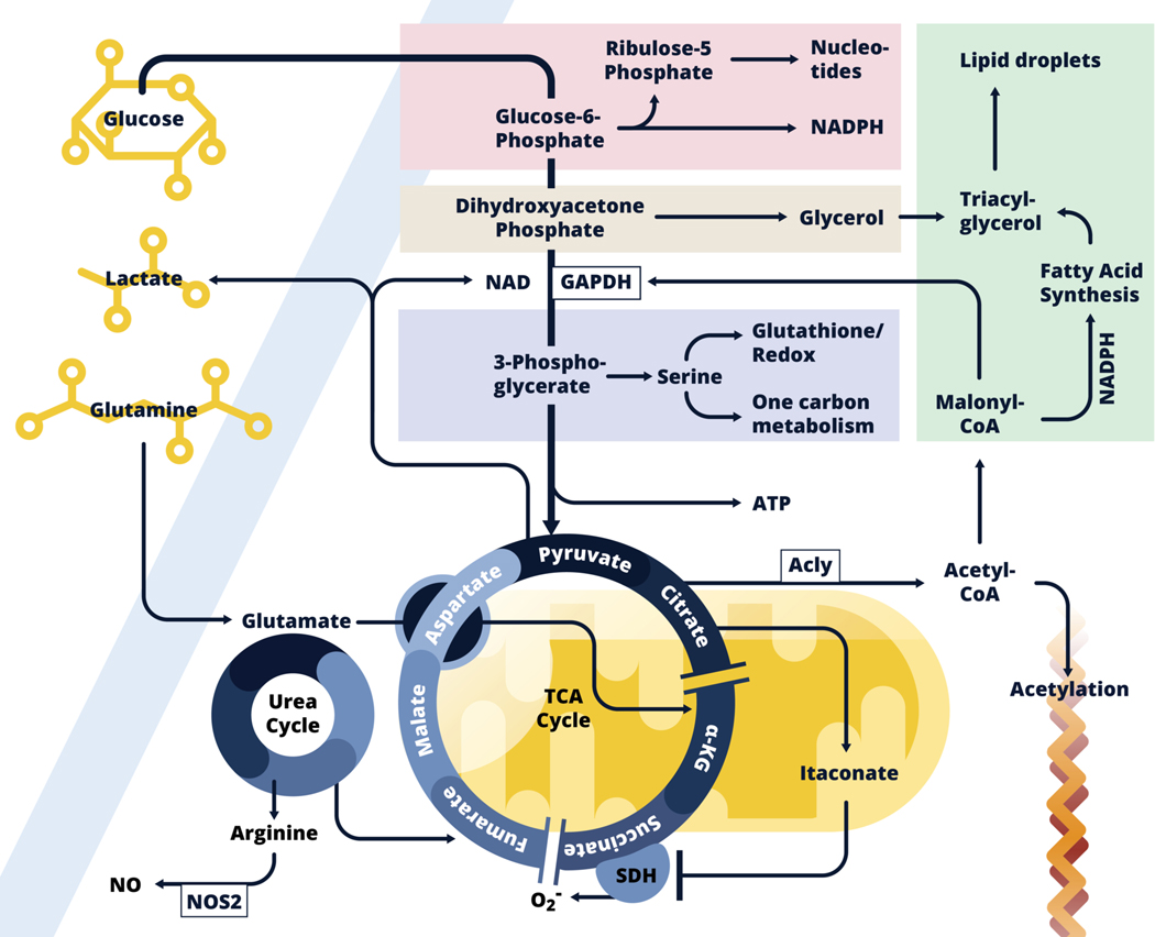
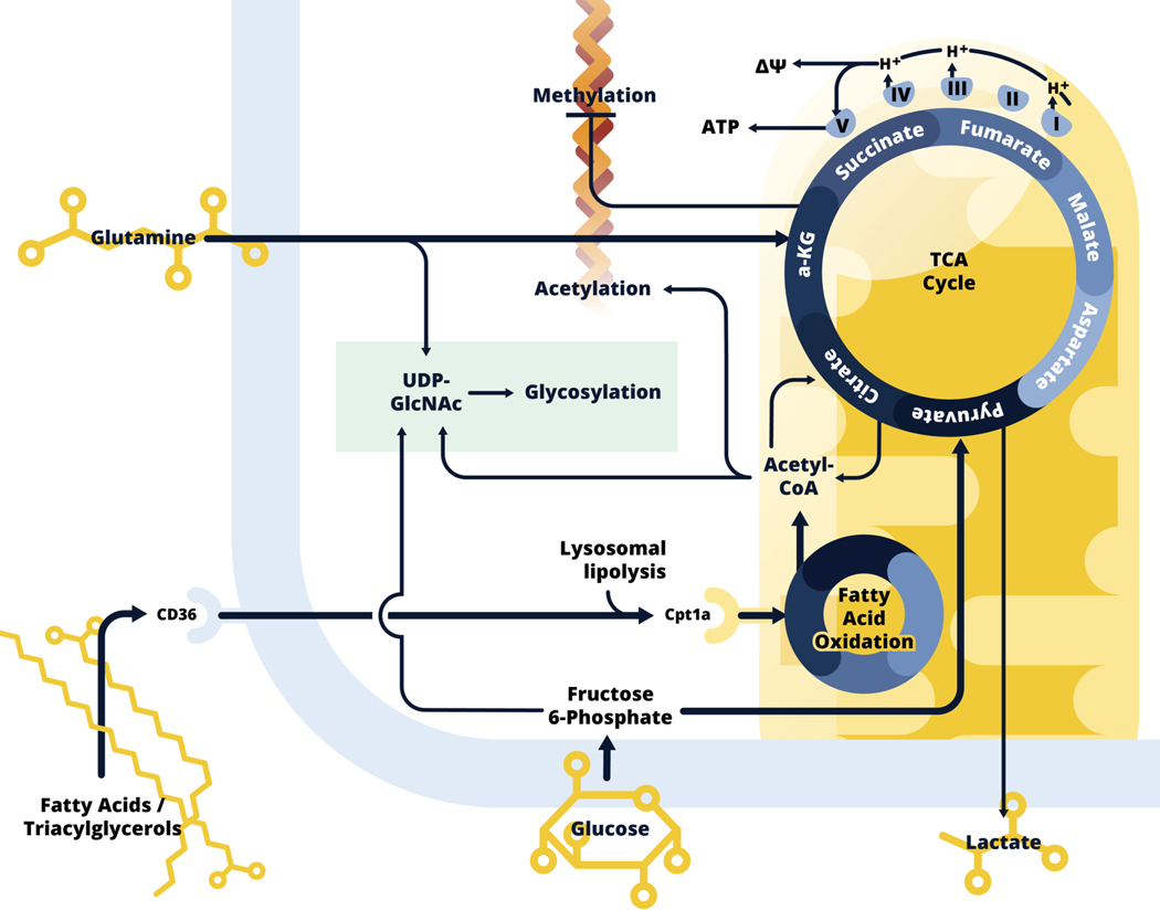
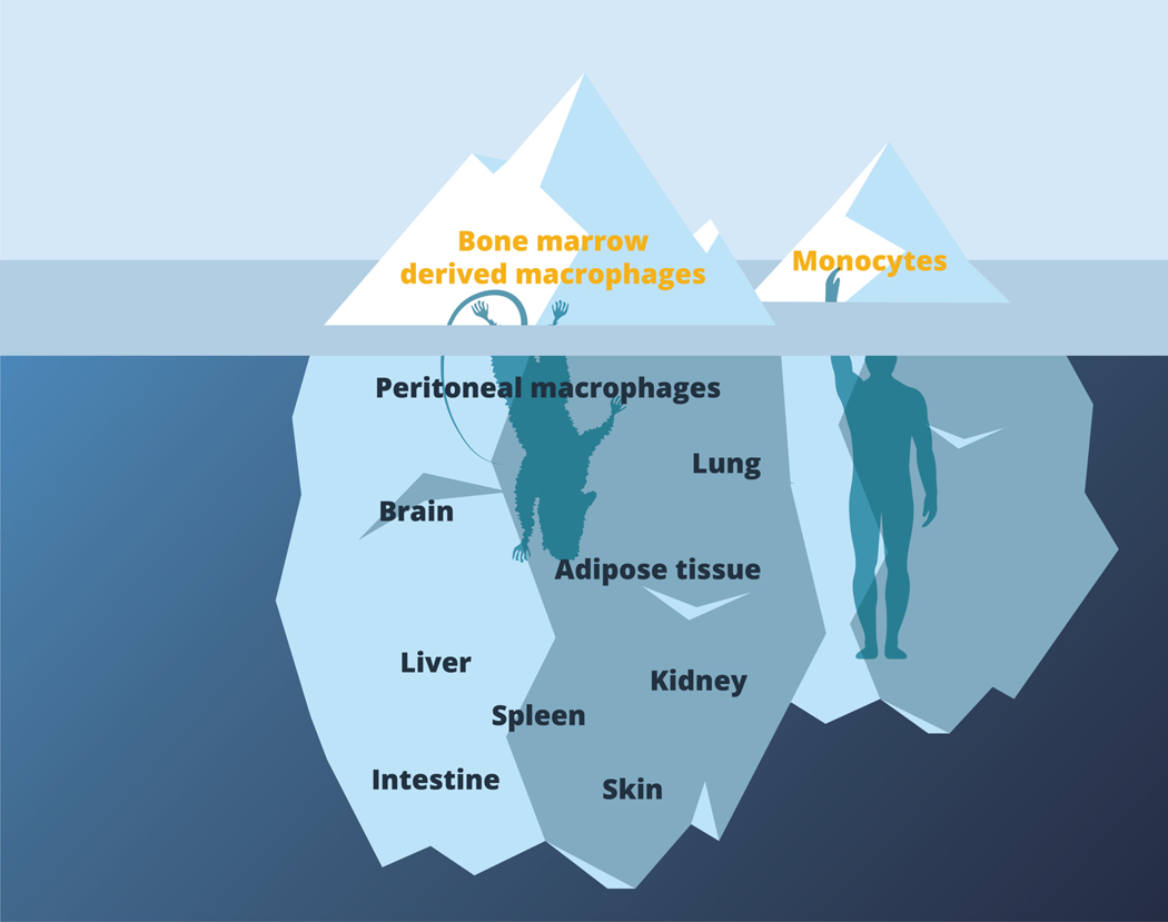
We have only recently started to appreciate the extent to which immune cell activation involves significant changes in cellular metabolism. We are now beginning to understand how commitment to specific metabolic pathways influences aspects of cellular biology that are the more usual focus of immunological studies, such as activation-induced changes in gene transcription, post-transcriptional regulation of transcription, post-translational modifications of proteins, cytokine secretion, etc. Here, we focus on metabolic reprogramming in mononuclear phagocytes downstream of stimulation with inflammatory signals (such as LPS and IFNγ) vs alternative activation signals (IL-4), with an emphasis on work on dendritic cells and macrophages from our laboratory, and related studies from others. We cover aspects of glycolysis and its branching pathways (glycogen synthesis, pentose phosphate, serine synthesis, hexose synthesis, and glycerol 3 phosphate shuttle), the tricarboxylic acid pathway, fatty acid synthesis and oxidation, and mitochondrial biology. Although our understanding of the metabolism of mononuclear phagocytes has progressed significantly over the last 10 years, major challenges remain, including understanding the effects of tissue residence on metabolic programming related to cellular activation, and the translatability of findings from mouse to human biology.

Keywords: IL-4; LPS; TCA cycle; alternative activation; cytokines; dendritic cells; glycolysis; immunometabolism; inflammation; macrophages; metabolism; mitochondria.


Conflict of interest statement

Conflict of interest

EJP is a founder of Rheos Medicines.

Figures

Figure 1:
Figure 1:
Inflammatory dendritic cell and macrophage metabolism. Glycolysis allows glucose carbon to be taken up and used not only to fuel the TCA cycle, but also to feed into the pentose phosphate pathway (Pink), the glycerol phosphate shuttle (Beige), and the serine synthesis pathway (Lavender), all of which are implicated in inflammatory activation. Core functions associated with these pathways are noted. Downstream in the glycolysis pathway, pyruvate is converted into lactate, which allows ATP generation in the absence of respiration, or enters into mitochondria (Yellow) where it is converted into acetyl-CoA which enters the TCA cycle. In inflammatory macrophages, expression of Idh, the enzyme that converts citrate to α-KG is suppressed, and citrate levels build and fuel both itaconate production within mitochondria, and following export into the cytoplasm, the production of acetyl-CoA which is used for fatty acid synthesis (FAS, Green), and as a donor for the acetylation of proteins, including histones. Fatty acids (synthesized and acquired) are used with glycerol for triacylglycerol synthesis and lipid droplet formation. Malonyl-CoA, an intermediate in the FAS pathway posttranslationally modifies GAPDH, the central enzyme in the glycolysis pathway, to minimize its RNA-binding properties thereby promoting the release and translation of Tnf mRNA. Succinate dehydrogenase (SDH), which catalyzes the conversion of succinate to fumarate, is inhibited by itaconate. Succinate accumulates and promotes inflammatory activation through reverse electron transport at Complex I to Complex II, with the accompanying generation of ROS. This leads to HIF1-α activation which promotes glycolysis. Itaconate also promotes activation of Nrf2, which allows parallel induction of anti-inflammatory effects (not shown). Induced Nos2 expression leads to the production of NO which results in the cessation of respiration due to the inhibition of components of the electron transport chain. Under these conditions, production of ATP by glycolysis is required for cell survival. Aspartate generated from glutamate, fuels the urea cycle in which nitrogens are donated to arginine, the substrate for NO production by NOS2. The urea cycle feeds the TCA cycle by donating carbons in the form of fumarate, which can generate malate, aspartate and eventually citrate. Glutamine also back-fills the TCA cycle at α-KG downstream.
Figure 2.
Figure 2.
Alternatively activated macrophage metabolism. Alternatively activated macrophages use fatty acids, glutamine and glucose to fuel the TCA cycle, and respiration is generally increased in these cells. Exogenous fatty acids and triacylglycerols are taken up by CD36, and lysosomal lipolysis is used to generate fatty acids for fatty acid oxidation (FAO). Activated fatty acids enter the mitochondria, mediated by Cpt1a, to engage in fatty acid oxidation, which generates acetyl-CoA, to contribute to the TCA cycle. Glucose carbon is also critical for citrate synthesis. Unlike in inflammatory macrophages, Idh is expressed in alternatively activated macrophages, and in addition to being used for the production of acetyl-CoA, which is used for histone acetylation, citrate is converted to α-KG, which is also generated from glutamine by glutaminolysis. α-KG inhibits DNA methylases, and therefore the process of methylation, and in this way facilitates increased expression of genes and downstream processes associated with alternative activation. α-KG cycles through the TCA cycle by conversion into succinate, fumarate, malate, aspartate, and eventually back to citrate. These steps of the TCA cycle, as well as FAO, generate NADH and FADH2 (not shown) which can donate electrons to Complex I and II of the electron transport chain (ETC), while complex I, III and IV pump protons into the intermembrane space, to generate mitochondrial membrane potential (ΔΨ), which provides the motive force for ATP generation by complex V. The cells integrate fructose 6-phosphate (Glc), glutamine (N) and acetyl-CoA (Ac) for emphasized synthesis of UDP-GlcNAc, a sugar donor for glycosylation (green).
Figure 3.
Figure 3.
Bone marrow and monocyte-derived macrophages are the tip of mononuclear phagocyte iceberg. Most work to date has been performed using mouse bone marrow derived macrophages or human monocyte derived macrophages. These provide important, tractable primary cell populations that can be considered to be representative of macrophages that derive from monocytes in acute inflammatory settings. However, there is a broad range of tissue-resident macrophages that remain relatively uncharacterized in terms of their baseline and adaptive metabolic states. These are represented within the body of the iceberg beneath the surface. Future work will need to explore metabolism in these cells during health and disease, and to confirm the relatedness of findings from mice and humans.

Similar articles

Cited by

References

    1. Pearce EL, Pearce EJ. Metabolic pathways in immune cell activation and quiescence. Immunity. 2013;38(4):633–43. - PMC - PubMed
    1. Kapsenberg ML. Dendritic-cell control of pathogen-driven T-cell polarization. Nat Rev Immunol. 2003;3(12):984–93. - PubMed
    1. Joffre O, Nolte MA, Spörri R, Sousa CRE. Inflammatory signals in dendritic cell activation and the induction of adaptive immunity. Immunol Rev. 2009;227(1):234–47. - PubMed
    1. Wynn TA, Chawla A, Pollard JW. Macrophage biology in development, homeostasis and disease. Nature. 2013;496(7446):445–55. - PMC - PubMed
    1. Jones RG, Thompson CB. Revving the engine: signal transduction fuels T cell activation. Immunity. 2007. Aug;27(2):173–8. - PubMed

Publication types