. 2020 Apr;7(4):527-542.
doi: 10.1002/acn3.51023. Epub 2020 Apr 3.

Reduced neurosteroid potentiation of GABAA receptors in epilepsy and depolarized hippocampal neurons

Affiliations

Reduced neurosteroid potentiation of GABAA receptors in epilepsy and depolarized hippocampal neurons

Suchitra Joshi et al. Ann Clin Transl Neurol. 2020 Apr.

Abstract

Objective: Neurosteroids regulate neuronal excitability by potentiating γ-aminobutyric acid type-A receptors (GABARs). In animal models of temporal lobe epilepsy, the neurosteroid sensitivity of GABARs is diminished and GABAR subunit composition is altered. We tested whether similar changes occur in patients with epilepsy and if depolarization-induced increases in neuronal activity can replicate this effect.

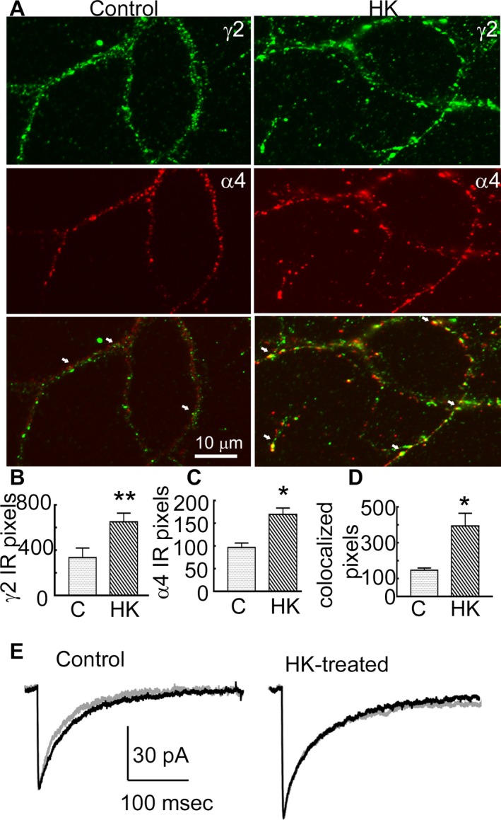
Methods: We determined GABAR α4 subunit expression in cortical tissue resected from pediatric epilepsy patients. Modulation of human GABARs by allopregnanolone and Ro15-4513 was measured in Xenopus oocytes using whole-cell patch clamp. To extend the findings obtained using tissue from epilepsy patients, we evaluated GABAR expression and modulation by allopregnanolone and Ro15-4513 in cultured rat hippocampal neurons exposed to high extracellular potassium (HK) to increase neuronal activity.

Results: Expression of α4 subunits was increased in pediatric cortical epilepsy specimens encompassing multiple pathologies. The potentiation of GABA-evoked currents by the neurosteroid allopregnanolone was decreased in Xenopus oocytes expressing GABARs isolated from epilepsy patients. Furthermore, receptors isolated from epilepsy but not control tissue were sensitive to potentiation by Ro15-4513, indicating higher expression of α4 βx γ2 subunit-containing receptors. Correspondingly, increasing the activity of cultured rat hippocampal neurons reduced allopregnanolone potentiation of miniature inhibitory postsynaptic currents (mIPSCs), increased modulation of tonic GABAR current by Ro15-4513, upregulated the surface expression of α4 and γ2 subunits, and increased the colocalization of α4 and γ2 subunit immunoreactivity.

Interpretation: These findings suggest that seizure activity-induced upregulation of α4 βx γ2 subunit-containing GABARs could affect the anticonvulsant actions of neurosteroids.


Conflict of interest statement

The authors do not have any conflict of interest.

Figures

Figure 1
Figure 1
Increased expression of α4 subunit‐containing GABARs in human epilepsy. (A) GABAR 4 subunit protein expression in control and epilepsy cortical tissue. A representative blot from three control, three epilepsy‐ganglioglioma, and three epilepsy‐FCD IIa patients is shown. 30 μg of protein was used for each well and β‐tubulin expression was used as a loading control. Anti‐α4 subunit (1:250 dilution, Novus Biologicals NB300‐194, Littleton, CO) and anti‐β‐tubulin (1:2000, Novus Biologicals NB600‐1514) antibodies were used. (B) The expression of α4 subunits in epilepsy tissue (336.1 ± 42, n = 19) relative to that in control tissue (102.5 ± 12.9, n = 10) ***P < 0.001, unpaired t‐test.
Figure 2
Figure 2
Diminished neurosteroid modulation of GABARs in human epilepsy. (A) Potentiation of GABA‐evoked currents by increasing concentrations of allopregnanolone recorded from oocytes incorporating control and epilepsy GABARs, n = 22–26 cells expressing receptors isolated from four control and epilepsy tissues each (*P < 0.05 vs. corresponding control, repeated measures two‐way ANOVA followed by Bonferroni post hoc analysis). At 1 μmol/L allopregnanolone, the potentiation was 730 ± 90% in control tissue and 510 ± 50% in epileptic tissue, and at 3 μmol/L allopregnanolone it was 970 ± 80% versus 660 ± 70%. (B) Representative traces of currents evoked by GABA (10 μmol/L) in the presence or absence of allopregnanolone (3 μmol/L) in an oocyte expressing control or epilepsy GABARs.
Figure 3
Figure 3
Enhanced modulation of GABAR currents by Ro15‐4513 in human epilepsy. (A) Differential effects of Ro15‐4513 on GABA (10 μmol/L)‐evoked currents in oocytes incorporating control and epilepsy GABARs; n = 26–31 cells expressing receptors isolated from four control and epilepsy tissues each. *P < 0.05 versus corresponding control, repeated measures two‐way ANOVA followed by Bonferroni post hoc analysis. (B) Representative traces of currents evoked by GABA (10 μmol/L) in the presence or absence of Ro15‐4513 (1 μmol/L) in an oocyte expressing control or epilepsy GABARs. Ro15‐4513 enhanced the current in oocytes incorporating epilepsy GABARs by 22 ± 5%, whereas it had no effect (−3 ± 5%) on control GABARs.
Figure 4
Figure 4
Reduced neurosteroid modulation of synaptic currents in HK‐treated neurons. (A) Representative voltage clamp recordings showing the effect of allopregnanolone (10 nmol/L) in cultured dissociated hippocampal neurons either untreated or treated with HK (10 mmol/L, 48 h). (B) Averaged current from representative control and HK‐treated neurons before (black) and after application of allopregnanolone (gray). Bath application of allopregnanolone (10 nmol/L) prolonged the decay from 35 ± 2 msec to 43 ± 3 msec (n = 13, P < 0.0005, paired t‐test) in control cultures. In contrast, the decay was only prolonged from 29 ± 2 msec to 34 ± 2 msec in the HK‐treated neurons (n = 12, P < 0.005, paired t‐test). All values are presented as mean ± SEM. (C) The allopregnanolone modulation of tonic current in HK‐treated and control neurons. The arrows represent the start of bath application of allopregnanolone (10 nmol/L) and picrotoxin (50 μmol/L). Total tonic current in HK‐treated and control cultures was 25 ± 4 pA, n = 5 and 28 ± 3 pA, n = 6 neurons, P > 0.05. Allopregnanolone enhanced the tonic current by 26 ± 5 pA in HK‐treated neurons (n = 14), similar to that in control neurons, 24 ± 3 pA, n = 12, P > 0.05, t‐test.
Figure 5
Figure 5
Increased surface expression of γ2 and α4 subunits in HK‐treated cultures. (A) Representative western blots showing the surface and total expression of the γ2, α4, and δ subunits in HK‐treated (HK) and control (C) organotypic hippocampal slice cultures. The expression of β‐actin is shown to demonstrate the purity of the surface samples. Mouse monoclonal anti‐γ2 subunit antibody 10F10‐C1‐B8 (2 μg/mL dilution), 49 rabbit polyclonal anti‐α4 subunit antibody (1:400 dilution, Millipore, Billerica, MA), anti‐ subunit antibody (1:1000, Millipore, Billerica, MA), and anti‐β‐actin (1:5000, clone AC‐40, Sigma‐Aldrich) were used. Lane M shows molecular weight marker. (B) Total (T) and surface (S) expression of γ2, α4, and δ subunits in HK‐treated slice cultures. Data represent average and standard error from n = 11 for γ2 subunits, n = 6 for δ subunits and n = 4 for α4 subunits, *P < 0.05. The subunit protein expression in HK‐treated cultures was normalized to that in control cultures which were run in parallel in every experiment. The total γ2 subunit expression in HK‐treated cultures was 179 ± 25% of that in control cultures (P < 0.05, Mann–Whitney test), the δ subunit expression was 91 ± 10% of that in controls (P > 0.05), and that of the α4 subunit was 139 ± 56% of that in controls (P > 0.05). The normalized surface expression (S) of γ2 subunits with HK treatment was 153 ± 11% of that in controls (P < 0.05, Mann–Whitney test, Fig. 4B), the α4 subunit surface expression in HK‐treated neurons was 158 ± 7%, P < 0.05, and the δ surface expression of the subunit in HK‐treated neurons was 121 ± 16%, P > 0.05.
Figure 6
Figure 6
HK treatment increased co‐localization between γ2 and α4 subunits. (A) Images demonstrating surface immunoreactivity of the γ2 (green) and α4 (red) subunits and their colocalization over dendrites of representative control and HK‐treated neurons. Arrows mark colocalization of the immunoreactive puncta. (B and C) Number of pixels corresponding to the γ2 and α4 subunit immunoreactivity in control and HK‐treated neurons, respectively, n = 16 control neurons from four batches of cultures and n = 26 HK‐treated neurons from 4 batches of cultures, *P < 0.05, **P < 0.005. The average number of γ2 puncta per neuron in controls was 338 ± 84 (n = 16 neurons from 4 replicate cultures), and in HK‐treated neurons it was 654 ± 76 (n = 26 from 4 replicate cultures, P < 0.005, t‐test). The number of α4 subunit puncta was also higher with 170 ± 14 in HK‐treated neurons compared to 97 ± 9 in control neurons (P < 0.05). (D) The number of pixels at which the γ2 and α4 subunit immunoreactivities colocalized, the number of replicates is identical as that in panel B and C, *P < 0.05. The average number of colocalized puncta was 146 ± 13 in control neurons and 395 ± 71 in HK‐treated neurons. (E) Averaged currents illustrating the effect of Ro15‐4513 (300 nmol/L, grey trace) on mIPSCs recorded from a representative control and HK‐treated neuron. The black trace illustrates current before the application of Ro15‐4513. In the control neurons decay shortened from 42 ± 5 msec to 37 ± 5 msec (n = 6 neurons/6 replicate cultures, P < 0.05, paired t‐test), whereas it remained stable in HK‐treated neurons (40 ± 4 msec and 42 ± 6 msec, n = 7 neurons/6 replicate cultures, P > 0.05, paired t‐test). The average change in decay was −5.6 ± 1.5 msec in control neurons and 1.7 ± 1.1 msec in HK‐treated neurons (P < 0.05).

Similar articles

Cited by

References

    1. Belelli D, Lambert JJ. Neurosteroids: endogenous regulators of the GABAA receptor. Nat Rev Neurosci 2005;6:565–575. - PubMed
    1. Joshi S, Kapur J. Neurosteroid regulation of seizures: role of GABAA receptor plasticity In: Talevi A., Rocha L., eds. Antiepileptic drug discovery: novel approaches. 1st ed. pp. 127–46. New York, NY: Springer, 2016.
    1. Kokate TG, Svensson BE, Rogawski MA. Anticonvulsant activity of neurosteroids: correlation with GABA‐evoked chloride current potentiation. J Pharmacol Exp Ther 1994;270:1223–1229. - PubMed
    1. Herzog AG, Frye CA. Seizure exacerbation associated with inhibition of progesterone metabolism. Ann Neurol 2003;53:390–391. - PubMed
    1. Reddy DS, Kim HY, Rogawski MA. Neurosteroid withdrawal model of perimenstrual catamenial epilepsy. Epilepsia 2001;42:328–336. - PubMed

Publication types

MeSH terms

LinkOut - more resources