. 2020 May 26;14(5):5298-5313.
doi: 10.1021/acsnano.9b04926. Epub 2020 Apr 13.

Surface Tethering of Inflammation-Modulatory Nanostimulators to Stem Cells for Ischemic Muscle Repair

Affiliations

Surface Tethering of Inflammation-Modulatory Nanostimulators to Stem Cells for Ischemic Muscle Repair

Jiayu Leong et al. ACS Nano. .

Abstract

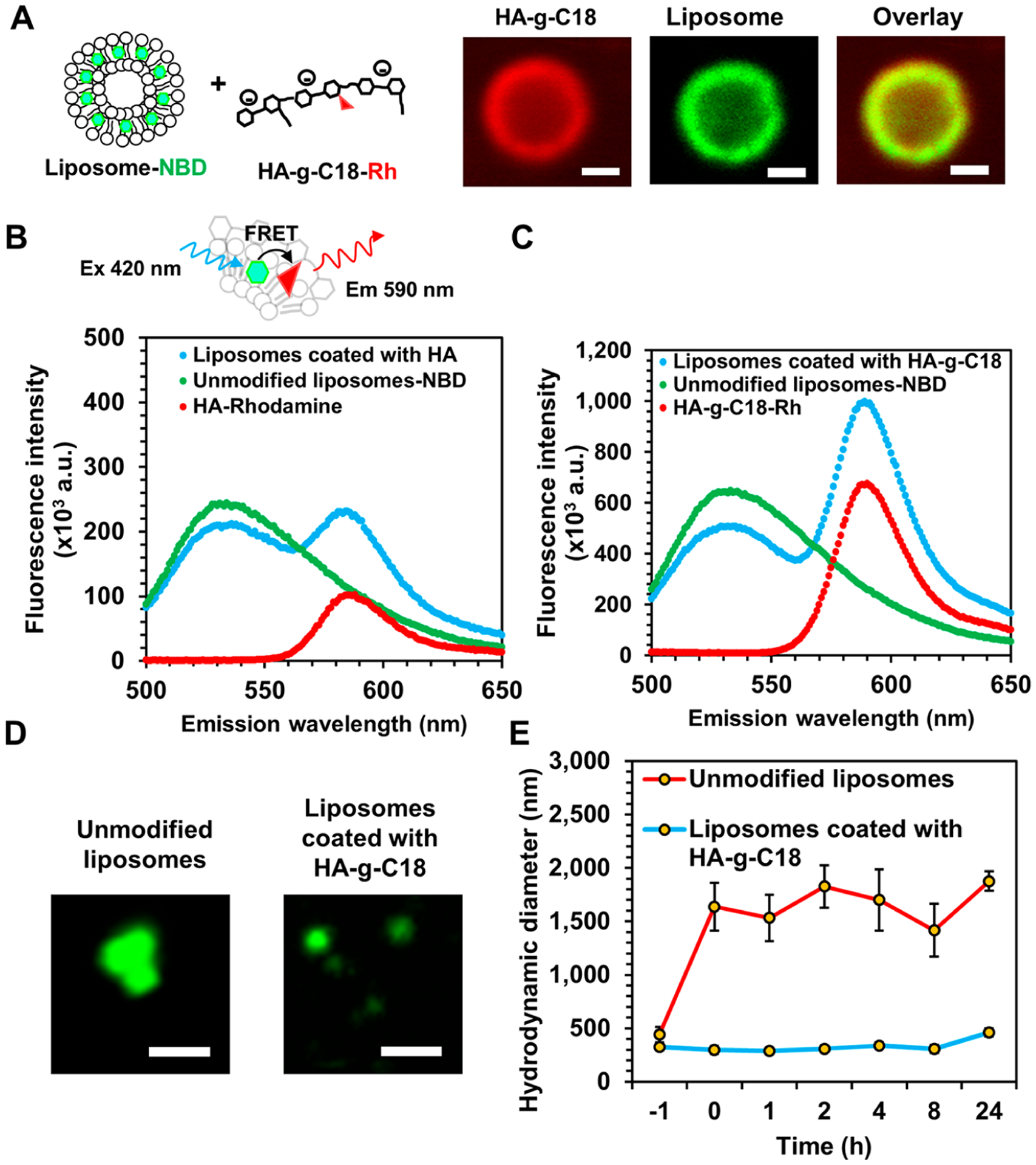
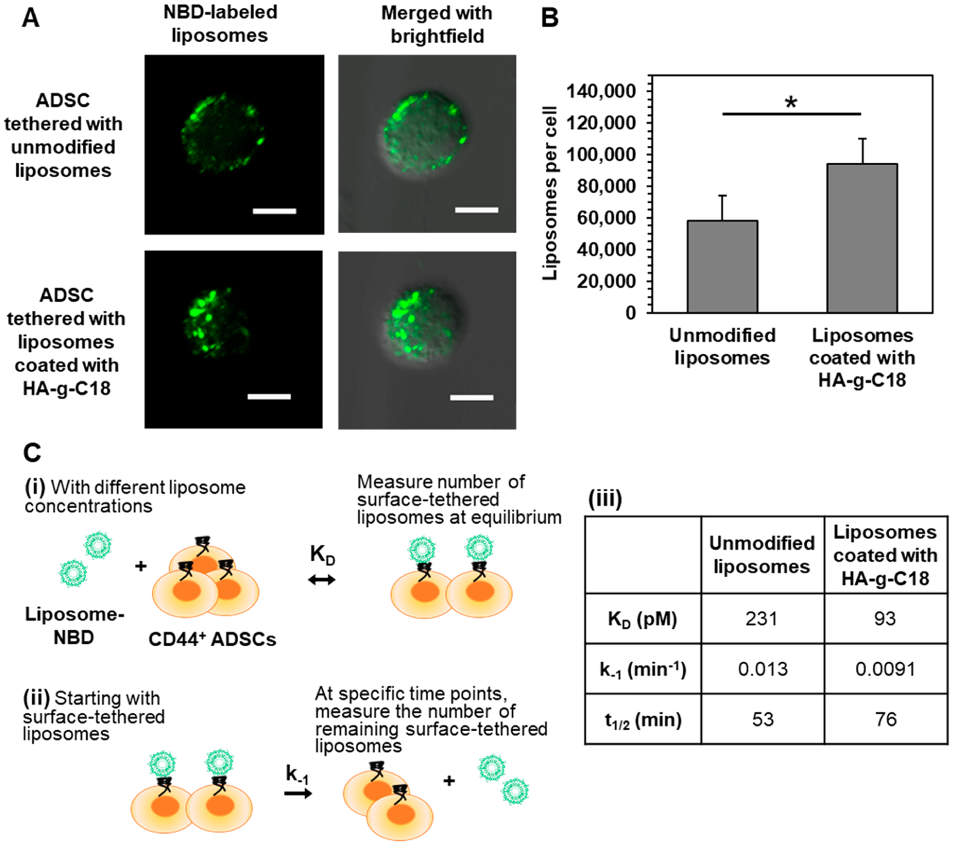
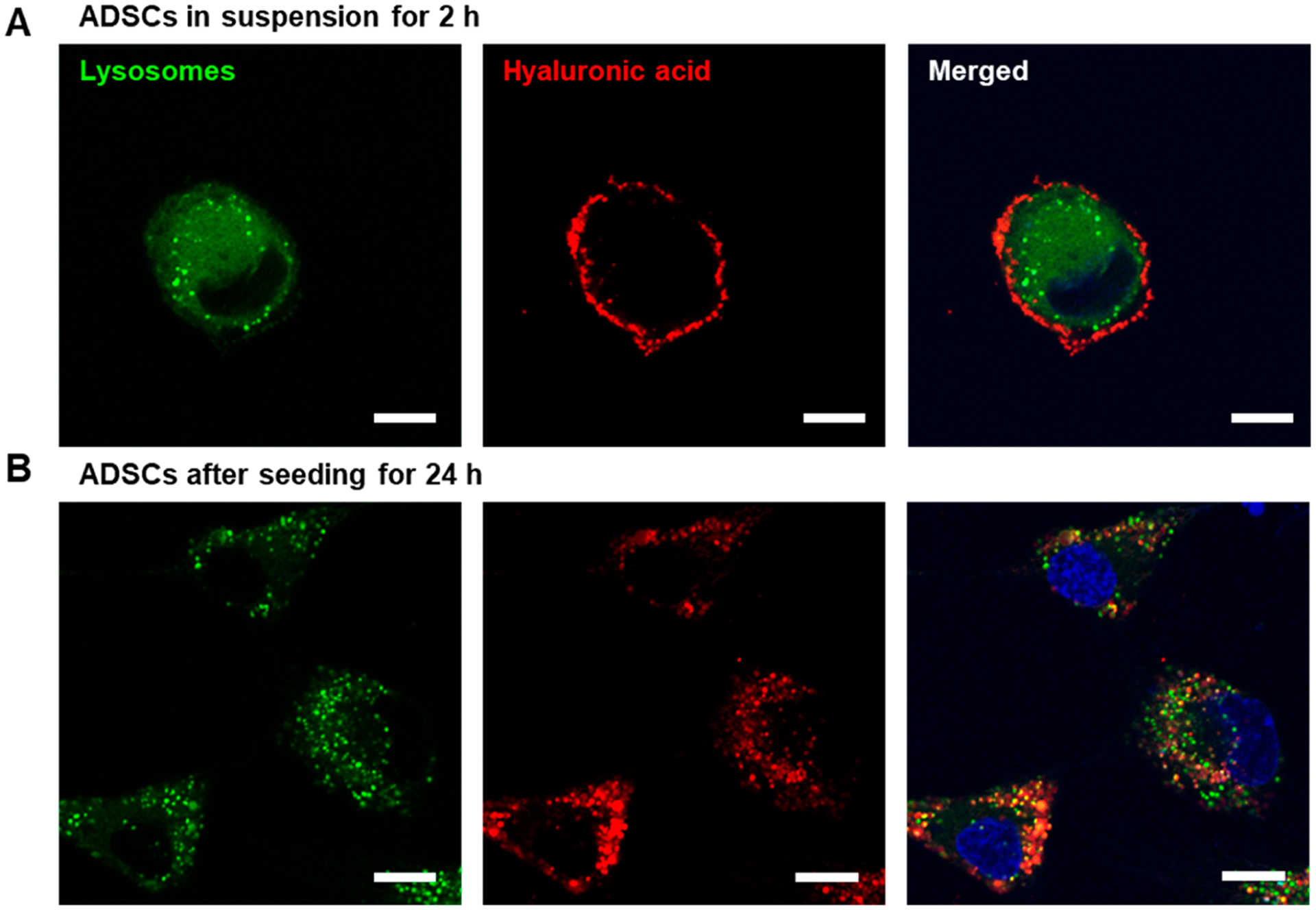
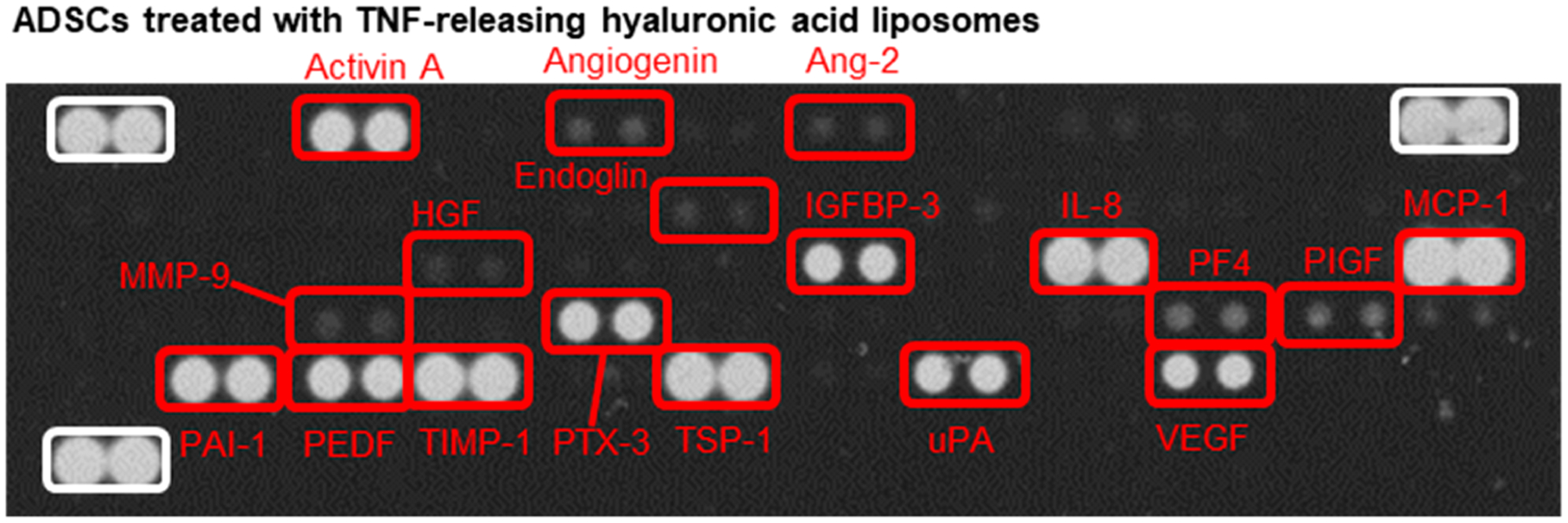
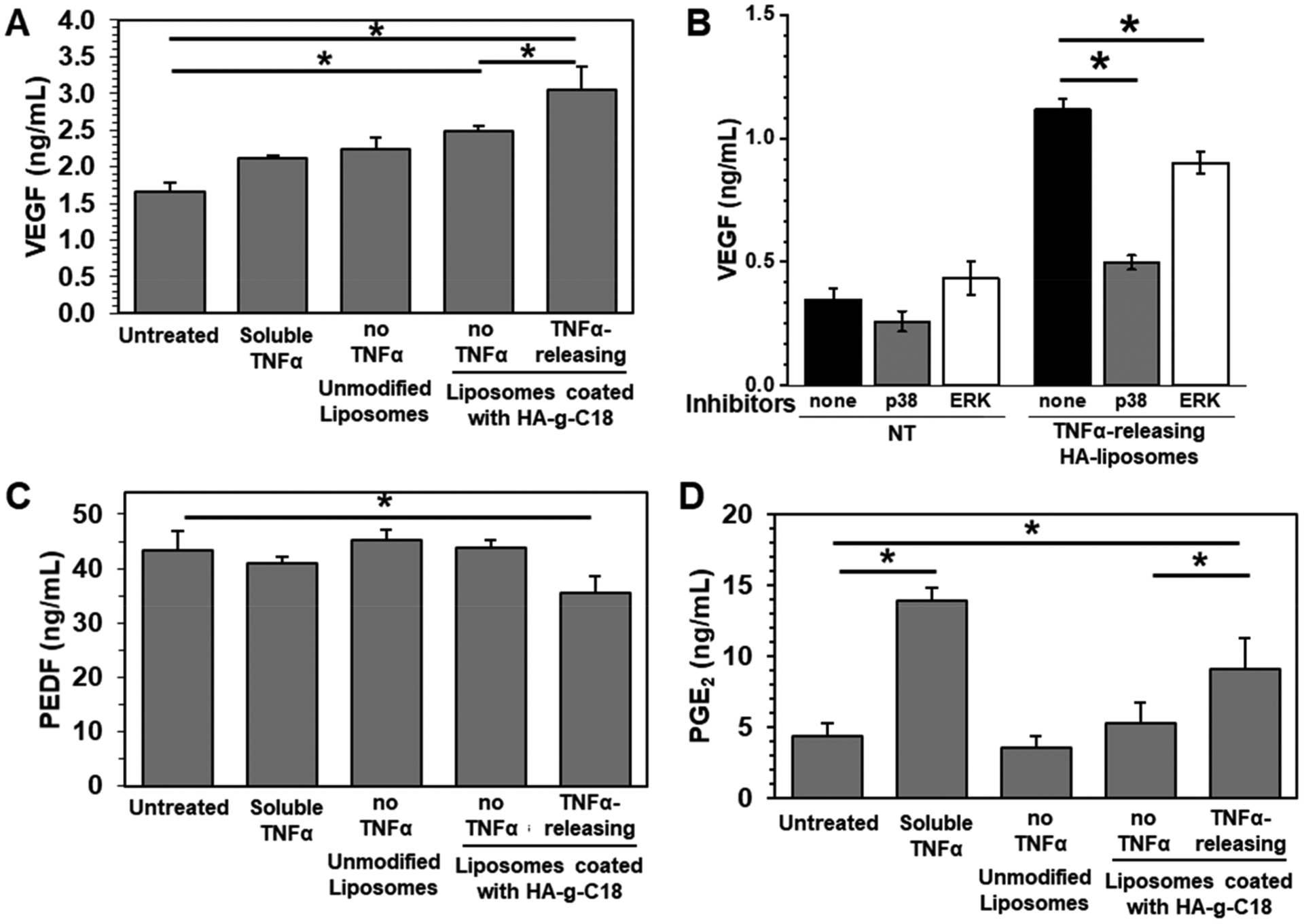
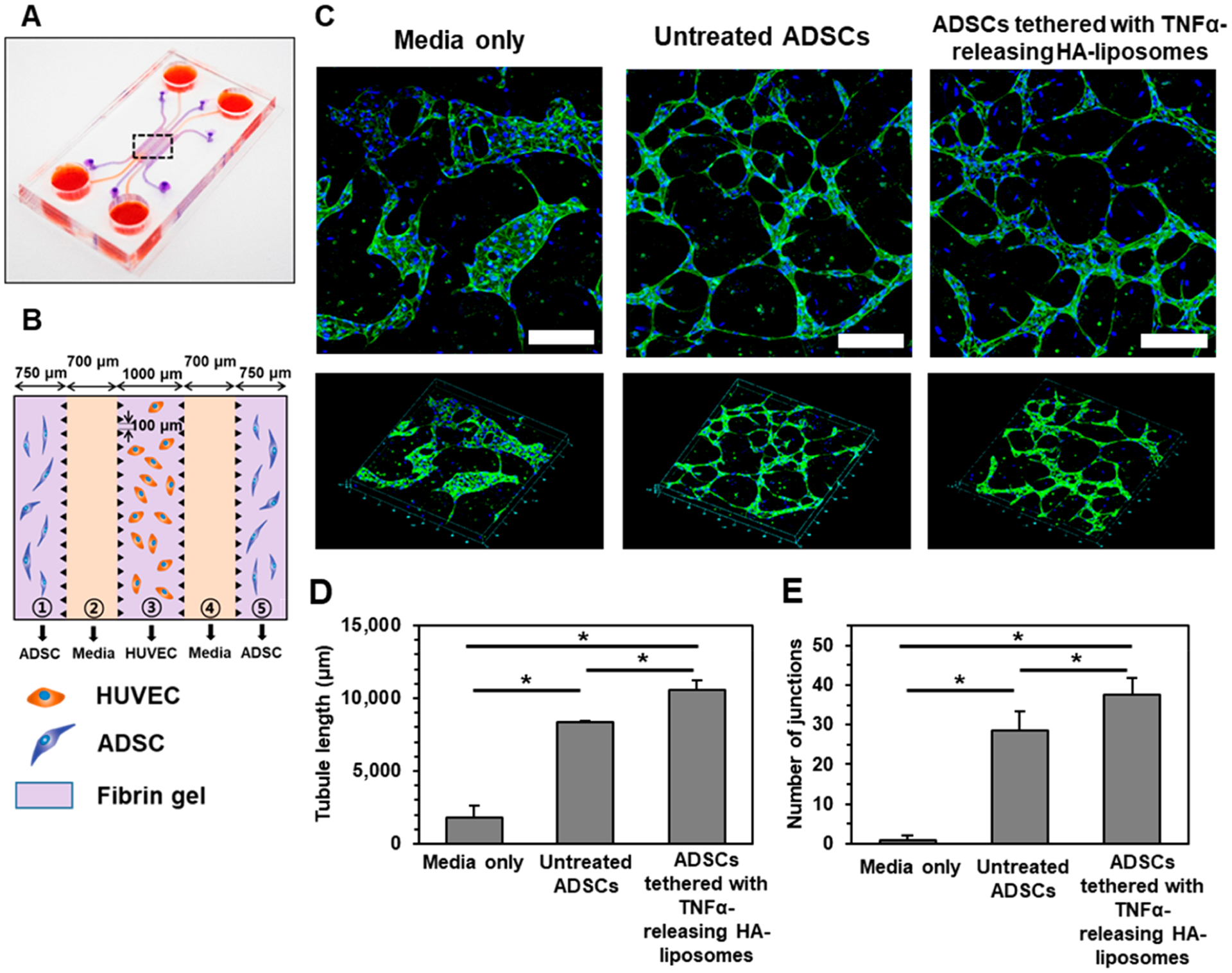
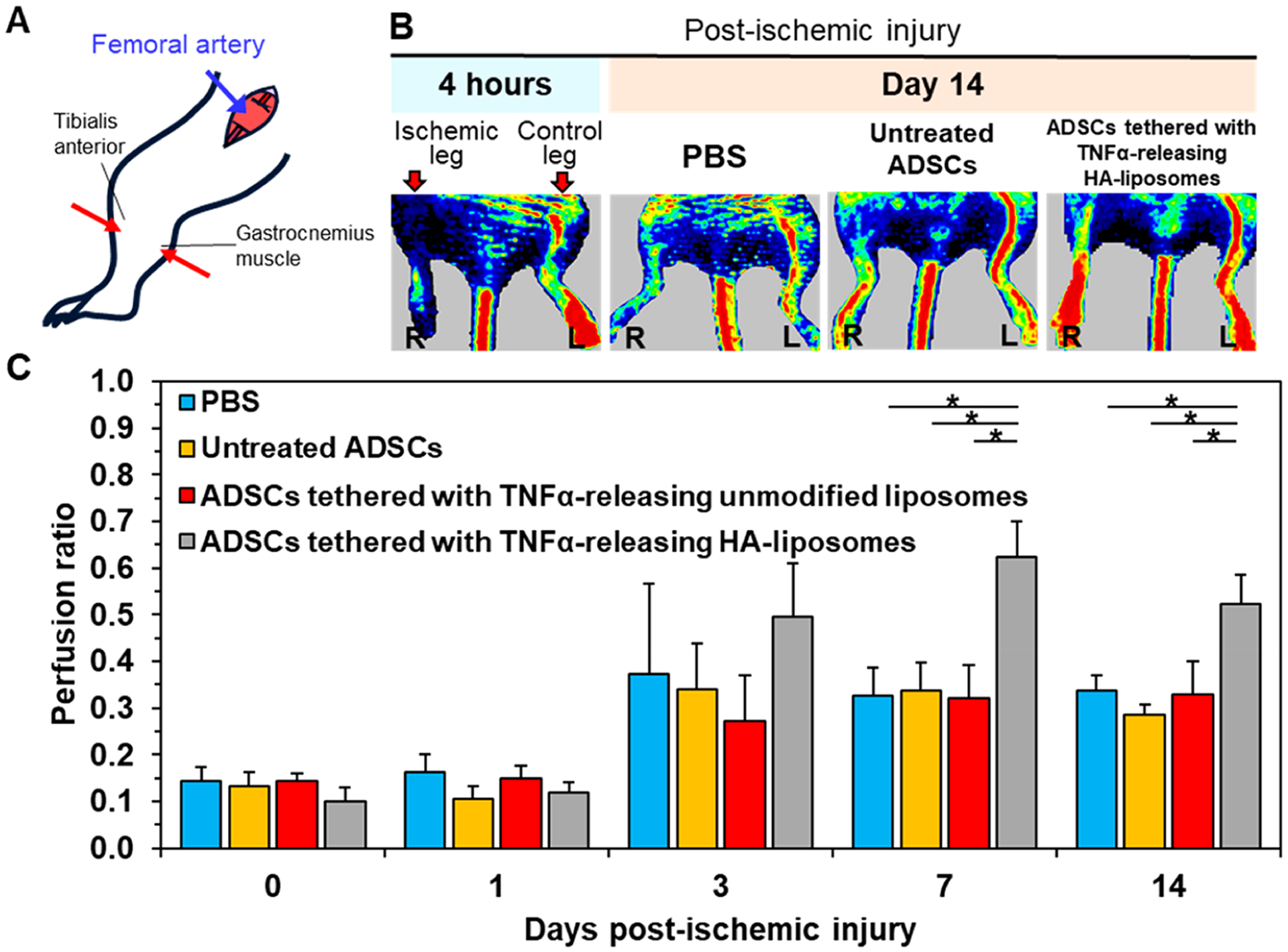
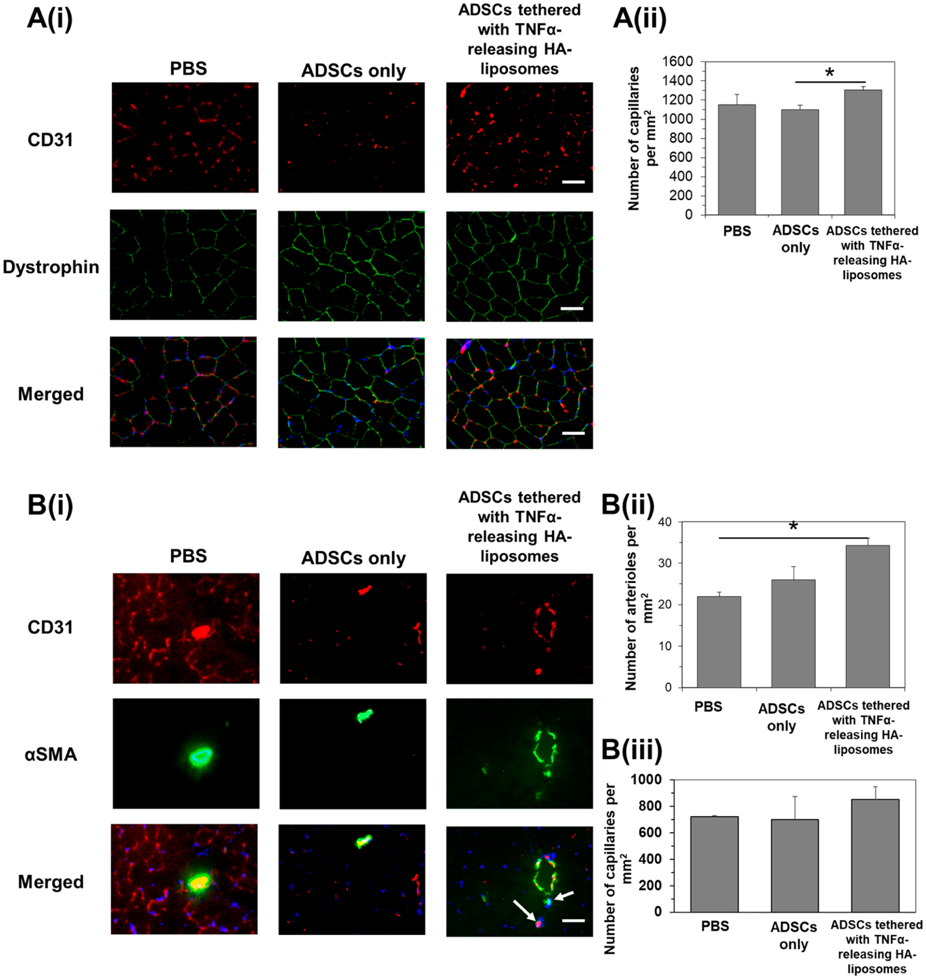
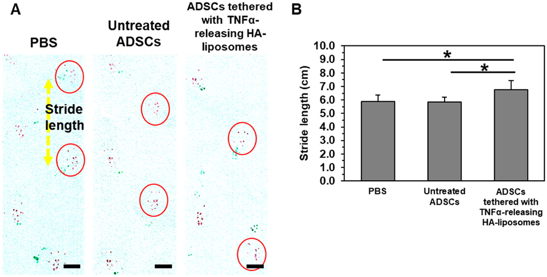
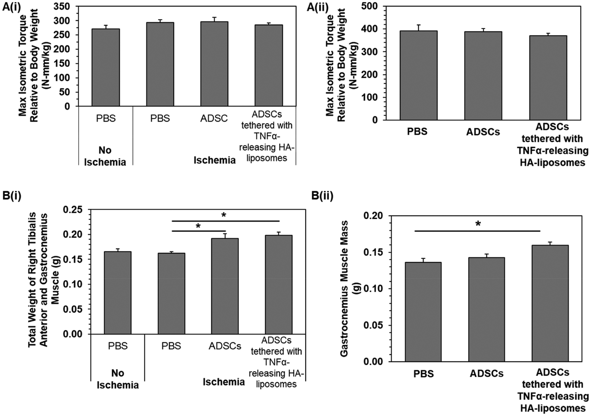
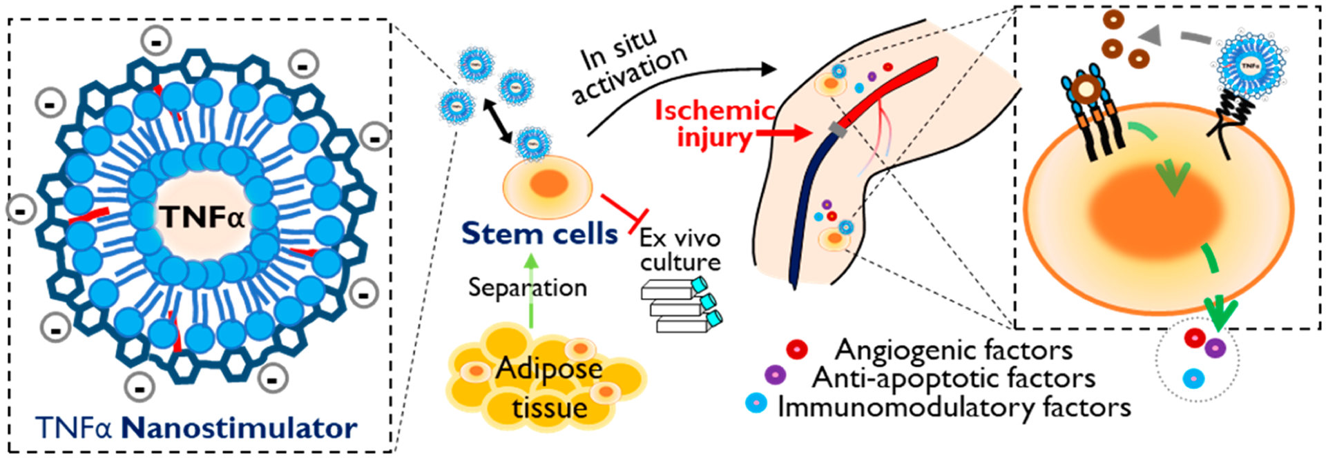
Stem cell transplantation has been a promising treatment for peripheral arterial diseases in the past decade. Stem cells act as living bioreactors of paracrine factors that orchestrate tissue regeneration. Prestimulated adipose-derived stem cells (ADSCs) have been proposed as potential candidates but have been met with challenges in activating their secretory activities for clinical use. Here, we propose that tethering the ADSC surface with nanoparticles releasing tumor necrosis factor α (TNFα), named nanostimulator, would stimulate cellular secretory activity in situ. We examined this hypothesis by complexing octadecylamine-grafted hyaluronic acid onto a liposomal carrier of TNFα. Hyaluronic acid increased the liposomal stability and association to CD44 on ADSC surface. ADSCs tethered with these TNFα carriers exhibited up-regulated secretion of proangiogenic vascular endothelial growth factor and immunomodulatory prosteoglandin E2 (PGE2) while decreasing secretion of antiangiogenic pigment epithelium-derived factors. Accordingly, ADSCs tethered with nanostimulators promoted vascularization in a 3D microvascular chip and enhanced recovery of perfusion, walking, and muscle mass in a murine ischemic hindlimb compared to untreated ADSCs. We propose that this surface tethering strategy for in situ stimulation of stem cells would replace the costly and cumbersome preconditioning process and expedite clinical use of stem cells for improved treatments of various injuries and diseases.

Keywords: adipose-derived stem cells; angiogenesis; hyaluronic acid; liposome; muscle; vascular endothelial growth factor.


Conflict of interest statement

The authors declare no competing financial interest.

Figures

Figure 1.
Figure 1.
Physical characterization of liposomes coated with hyaluronic acid-g-C18 (HA-g-C18). (A) Fluorescence images of liposomes coated with HA-g-C18 where HA-g-C18 were labeled with rhodamine B (Rh in red) and the lipid layer was labeled with nitrobenzoxadiazole (NBD) (in green). Lipids were covalently bound to NBD. The overlay panel shows the merged images. Images were taken after mixing HA-g-C18 and liposomes at a mass ratio of 2:1. Scale bars represent 1 μm. (B) Förster resonance energy transfer (FRET) assay to evaluate the association between HA-g-C18 and liposomes or (C) unmodified HA and liposomes. All three samples were excited at wavelength of 460 nm. (D) Fluorescence images of unmodified liposomes and liposomes coated with HA-g-C18 incubated in media supplemented with 10% serum. Scale bar represents 1 μm. (E) Hydrodynamic diameter of liposomes with (in blue) and without HA-g-C18 (in red) incubated in media supplemented with 10% serum at 37 °C. Data points represent the mean, and error bars represent standard deviations. N = 3.
Figure 2.
Figure 2.
Characterization of the association of liposomes with ADSCs. (A) Confocal microscope images of ADSCs after incubation with unmodified liposomes or liposomes coated with HA-g-C18. Liposomes were fluorescently labeled using the covalently bound NBD fluorophore (in green). Scale bar represents 10 μm. (B) Quantification of the number of liposomes on the surface of ADSCs. Data points represent the mean, and error bars represent standard deviations. N = 3, * represents the statistical significance in the number of liposomes between the unmodified liposomes and liposomes coated with HA-g-C18. *p < 0.05. (C) Schematic illustration of the kinetic analysis of the liposome tethering to ADSCs. (i) At a given liposome concentration, the number of liposomes tethered to cells was measured to quantify the dissociation equilibrium constant, KD. (ii) The dissociation rate constant, k−1, the half-time of liposome dissociation, t1/2, and the number of remaining surface-tethered liposomes were measured by counting the liposomes detached from cells at different time points. (iii) Tabulation of KD, k−1, and t1/2 of unmodified liposomes and liposomes coated with HA-g-C18.
Figure 3.
Figure 3.
Confocal microscope analysis to determine cellular internalization of liposomes coated with HA-g-C18. Lysosomes were labeled with LysoTracker Green, while liposomes were labeled with rhodamine B-conjugated HA-g-C18 (red). (A) ADSCs were incubated in suspension for 2 h. (B) ADSCs were incubated on type I collagen hydrogels for 24 h. The third column shows the merged images of the first two columns. Scale bars represent 10 μm.
Figure 4.
Figure 4.
Analysis of the secretory activity of ADSCs using the angiogenesis antibody array. Images of angiogenesis antibody array membrane incubated with media from the ADSC culture after 24 h. ADSCs tethered with TNFα-releasing HA-liposomes secrete growth factors and cytokines involved in proangiogenesis, antiangiogenesis, fibrosis, antiapoptosis, and tissue remodeling.
Figure 5.
Figure 5.
Quantification of proangiogenic VEGF, antiangiogenic PEDF, and immunomodulatory PGE2 levels secreted by ADSCs using ELISA. (A) VEGF secretion level. (B) Amount of VEGF in the media cultured with untreated ADSCs and ADSCs tethered with TNFα-releasing HA-liposomes after 24 h of incubation in the absence or presence of p38 MAPK or ERK inhibitors. (C) PEDF secretion level and (D) PGE2 secretion level. Data points represent the mean and error bars represent standard deviations. N = 3, * represents the statistical significance between the conditions indicated. *p < 0.05.
Figure 6.
Figure 6.
In vitro angiogenesis assay with a 3D microvascular device. (A) Schematic illustration of the device fabricated with polydimethylsiloxane using soft lithography. (B) The central portion features five channels. ADSCs in fibrin gels were seeded in the outer channels ① and ⑤; cell culture medium was filled in channels ② and ④; and HUVECs in the fibrin gel were seeded in the center channel ③. (C) Confocal laser scanning microscope images of immunostained HUVECs with CD31 (in green) in channel ③ after 5 days of incubation with the cell culture media only, with untreated ADSCs only, or ADSCs tethered with TNFα-releasing HA-liposomes. Cell nuclei were stained with Hoechst dye (in blue). Scale bar represents 200 μm. The lower panels display an overview of the selected region. (D) Quantification of the tubule length and (E) the number of interconnected junctions. Data points represent the mean, and error bars represent standard deviations. N = 3, * represents the statistical significant between the conditions indicated. *p < 0.05.
Figure 7.
Figure 7.
Laser Doppler perfusion imaging of mice induced with ischemic hindlimb injury. (A) Schematic diagram of the surgery to induce limb ischemia followed by cell injection into the limb. (B) LDPI images of mice after the ischemic hindlimb surgery. Ischemia was introduced by ligating the femoral artery in the right leg. The color scale represents the relative intensity of perfusion with red representing the highest intensity. The ischemic leg imaged after 24 h and after it was treated for 14 days with 1 million ADSCs, 1 million untreated ADSCs, and 1 million ADSCs tethered with TNFα-releasing HA-liposomes. (C) Quantification of the mean perfusion ratio defined as the perfusion in the ischemic limb divided by the perfusion in the nonischemic limb (N = 7 mice, *p < 0.05).
Figure 8.
Figure 8.
Immunohistological analysis of muscle tissues 14 days after ischemic injury and injection of ADSCs only or ADSCs tethered with TNFα-releasing HA liposomes. (A(i)) The tibialis anterior muscle was stained with antibodies against CD31 (in red) and dystrophin (in green). Merged images on the bottom row show cell nuclei (in blue), CD31 (in red), and dystrophin (in green). Scale bar represents 50 μm. (A(ii)) Quantification of the capillary density in the tibialis anterior. * represents significant difference between the two groups, *p < 0.05. (B(i)) The gastrocnemius muscle was stained with antibodies against CD31 (in red) and alpha smooth muscle actin (αSMA, in green). Merged images on the bottom row show cell nuclei (in blue), CD31 (in red), and αSMA (in green). White arrows point to smaller arterioles. Scale bar represents 50 μm. Quantification of the (ii) total arteriole and (iii) capillary density in the gastrocnemius muscle. * represents significant difference between the two groups, *p < 0.05.
Figure 9.
Figure 9.
Gait analysis of recovered mice on day 26. (A) Footpads of mice, forepaws and hindpaws, were dipped in green and red inkpads, respectively. Images were treated for higher image contrast of the red ink. Scale bar represents 2 cm. (B) Quantification of the average stride length (N = 7, 8 mice, *p < 0.05).
Figure 10.
Figure 10.
Muscle force measurements of recovered mice at the end points, (i) day 14 and (ii) day 28, respectively. (A) Maximum isometric torque relative to body weight was determined by measuring the muscle force and dividing it by the mouse body weight. (B) Weight of the right muscles, tibialis anterior and gastrocnemius muscle. Bars represent the average value, and error bars indicate standard error. * represents significant difference between the two groups, *p < 0.05 (N = 7, 8).
Scheme 1.
Scheme 1.
In situ stimulation of adipose-derived stem cells (ADSCs) for cellular therapy in ischemic tissue recovery. Nanostimulator consists of a TNFα-releasing liposome coated with hyaluronic acid-graft-octadecylamine (HA-g-C18). HA drives the liposomes tethered to the stem cell surface via specific binding with CD44. The nanostimulator on the stem cell surface up-regulates cellular secretion levels of angiogenic and immunomodulatory factors for revascularization and the prevention of muscle damage. Also, the nanostimulator removes ex vivo culture for pre-conditioning of stem cells, thus allowing for isolation and injection in an operating room.

Similar articles

Cited by

References

    1. Liew A; O’Brien T Therapeutic Potential for Mesenchymal Stem Cell Transplantation in Critical Limb Ischemia. Stem Cell Res. Ther 2012, 3, 28. - PMC - PubMed
    1. Tsuji W; Rubin JP; Marra KG Adipose-Derived Stem Cells: Implications in Tissue Regeneration. World J. Stem Cells 2014, 6, 312–321. - PMC - PubMed
    1. Wietecha MS; Król MJ; Michalczyk ER; Chen L; Gettins PG; DiPietro LA Pigment Epithelium-Derived Factor (PEDF) as a Multifunctional Regulator of Wound Healing. Am. J. Physiol.: Heart Circ. Physiol 2015, 309, H812–26. - PMC - PubMed
    1. Ito WD; Arras M; Winkler B; Scholz D; Schaper J; Schaper W Monocyte Chemotactic Protein-1 Increases Collateral and Peripheral Conductance after Femoral Artery Occlusion. Circ. Res 1997, 80, 829–837. - PubMed
    1. Spaggiari GM; Abdelrazik H; Becchetti F; Moretta L MSCs Inhibit Monocyte-Derived DC Maturation and Function by Selectively Interfering with the Generation of Immature DCs: Central Role of MSC-Derived Prostaglandin E2. Blood 2009, 113, 6576–6583. - PubMed

Publication types

Substances