

Case Reports
doi: 10.1161/CIRCULATIONAHA.120.047164.
Epub 2020 Apr 3.
The Variety of Cardiovascular Presentations of COVID-19
Affiliations
- PMID: 32243205
- PMCID: PMC7314498
- DOI: 10.1161/CIRCULATIONAHA.120.047164
Item in Clipboard
Case Reports
The Variety of Cardiovascular Presentations of COVID-19
Circulation.
.
No abstract available
Keywords: COVID-19; SARS virus; coronavirus; myocarditis; severe acute respiratory syndrome coronavirus 2; transplant recipients.
Figures
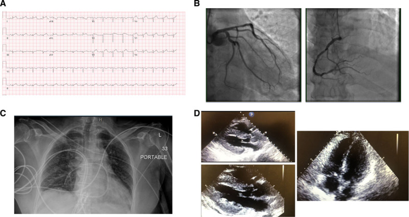
Chest pain and ST elevation. Initial ECG showed sinus tachycardia, low-voltage QRS complexes in the limb leads, and diffuse ST elevation in leads I, II, aVL, and leads V2–V6 (A). Coronary angiogram demonstrated mild disease in the left anterior descending artery and left circumflex artery and 40% stenosis in the mid-right coronary artery (B). Chest radiography demonstrated clear lungs (C). Transthoracic echocardiogram with severe increased left ventricular wall thickness and left ventricular ejection fraction approximately 30% with trace circumferential pericardial effusion (D).
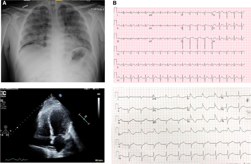
Cardiogenic shock rescued by veno-arterial-venous extracorporeal membrane oxygenation. Chest radiograph showed diffuse ill-defined airspace opacities bilaterally (A). Initial ECG (top) demonstrated sinus tachycardia with incomplete right bundle-branch block. Repeat ECG (bottom) demonstrated accelerated idioventricular rhythm (B). Transthoracic echocardiogram demonstrated left ventricular end-diastolic diameter of 4.5 cm, left ventricular ejection fraction 20% to 25%, with akinesis of mid-left ventricular segments (C).
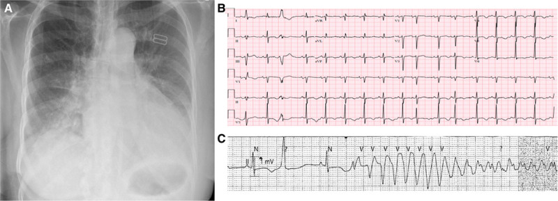
Decompensated heart failure. Chest radiography shows pulmonary vascular congestion, patchy airspace opacities at bases, and bilateral pleural effusions (A). ECG shows sinus rhythm with premature atrial and ventricular complexes, lateral T-wave inversions, and a prolonged QT interval (B). Telemetry strip shows prolonged QT interval and torsades de pointes after R-on-T phenomenon (C).
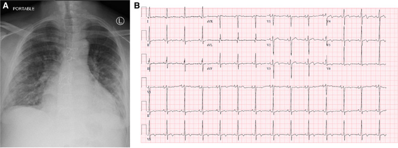
Heart transplant recipient. Chest radiography with multifocal bilateral patchy airspace opacities (A). ECG with normal sinus rhythm with new nonspecific ST changes and T wave inversions in inferior and lateral leads (B).
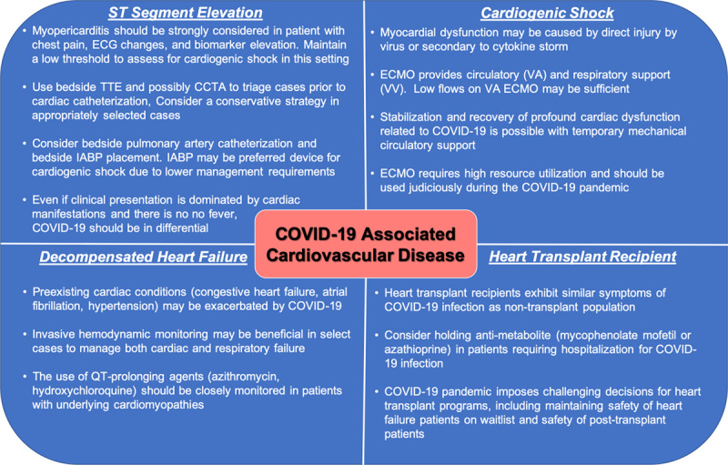
Important messages from each cardiovascular presentation of COVID-19. CCTA indicates cardiac computed tomographic angiography; COVID-19, corona virus disease 2019; ECMO, extracorporeal membrane oxygenation; IABP, intraaortic balloon pump; TTE, transthoracic echocardiogram; VA, veno-arterial; and VV, veno-venous.
References
-
- Welt FGP, Shah PB, Aronow HD, Bortnick AE, Henry TD, Sherwood MW, Young MN, Davidson LJ, Kadavath S, Mahmud E, et al. Catheterization laboratory considerations during the coronavirus (COVID-19) pandemic: from ACC’s Interventional Council and SCAI. Journal of the American College of Cardiology. 2020;75:2372–75. doi: 10.1016/j.jacc.2020.03.021. - PMC - PubMed
-
- Clerkin KJ, Fried JA, Raikhelkar J, Sayer G, Griffin JM, Masoumi A, Jain SS, Burkhoff D, Kumaraiah D, Rabbani L, et al. Coronavirus disease 2019 (COVID-19) and cardiovascular disease [published online March 21, 2020]. Circulation. doi: 10.1161/circulationaha.120.046941. - PubMed
-
- MacLaren G, Fisher D, Brodie D. Preparing for the most critically ill patients with COVID-19: the potential role of extracorporeal membrane oxygenation. JAMA. 2020;323:1245–46. doi:10.1001/jama.2020.2342. - PubMed
Publication types
MeSH terms
Substances
Grants and funding
LinkOut - more resources
Full Text Sources
Medical
Miscellaneous
