

Chest CT Features of COVID-19 in Rome, Italy
- PMID: 32243238
- PMCID: PMC7194020
- DOI: 10.1148/radiol.2020201237
Chest CT Features of COVID-19 in Rome, Italy
Abstract
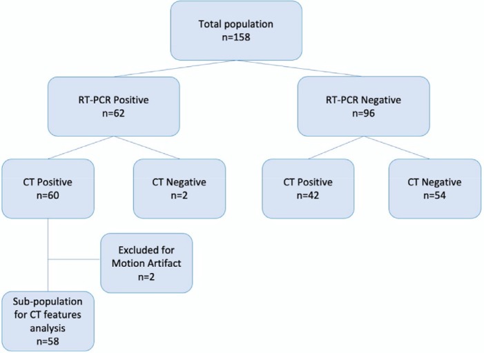
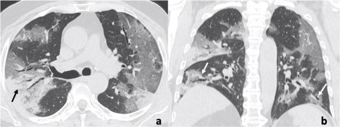
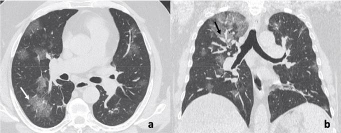
Background The standard for diagnosis of severe acute respiratory syndrome coronavirus 2 is a reverse transcription polymerase chain reaction (RT-PCR) test, but chest CT may play a complimentary role in the early detection of Coronavirus Disease 2019 (COVID-19) pneumonia. Purpose To investigate CT features of patients with COVID-19 in Rome, Italy, and to compare the accuracy of CT with that of RT-PCR. Materials and Methods In this prospective study from March 4, 2020, until March 19, 2020, consecutive patients suspected of having COVID-19 infection and respiratory symptoms were enrolled. Exclusion criteria were contrast material-enhanced chest CT performed for vascular indications, patients who refused chest CT or hospitalization, and severe CT motion artifact. All patients underwent RT-PCR and chest CT. Diagnostic performance of CT was calculated using RT-PCR as the reference standard. Chest CT features were calculated in a subgroup of patients with positive RT-PCR and CT findings. CT features of hospitalized patients and patients in home isolation were compared using the Pearson χ2 test. Results The study population included 158 consecutive participants (83 male, 75 female; mean age, 57 years ± 17 [standard deviation]). Of the 158 participants, fever was observed in 97 (61%), cough was observed in 88 (56%), dyspnea was observed in 52 (33%), lymphocytopenia was observed in 95 (60%), increased C-reactive protein level was observed in 139 (88%), and elevated lactate dehydrogenase level was observed in 128 (81%). Sensitivity, specificity, and accuracy of CT were 97% (95% confidence interval [CI]: 88%, 99%) (60 of 62), 56% (95% CI: 45%, 66%) (54 of 96), and 72% (95% CI: 64%, 78%) (114 of 158), respectively. In the subgroup of 58 participants with positive RT-PCR and CT findings, ground-glass opacities were present in all 58 (100%), both multilobe and posterior involvement were present in 54 (93%), bilateral pneumonia was present in 53 (91%), and subsegmental vessel enlargement (>3 mm) was present in 52 (89%). Conclusion The typical pattern of COVID-19 pneumonia in Rome, Italy, was peripheral ground-glass opacities with multilobe and posterior involvement, bilateral distribution, and subsegmental vessel enlargement (>3 mm). Chest CT had high sensitivity (97%) but lower specificity (56%). © RSNA, 2020.
Figures
Comment in
-
The tug-of-war between coagulopathy and anticoagulant agents in patients with COVID-19.Eur Heart J Cardiovasc Pharmacother. 2020 Jul 1;6(4):262-264. doi: 10.1093/ehjcvp/pvaa048. Eur Heart J Cardiovasc Pharmacother. 2020. PMID: 32383737 Free PMC article. No abstract available.
References
-
- Novel Coronavirus (2019-nCoV) situation reports. 2020.
-
- Interim Guidance : Healthcare Professionals 2019-nCoV | CDC. 2020.
-
- Yang Y, Yang M, Shen C, et al. Evaluating the accuracy of different respiratory specimens in the laboratory diagnosis and monitoring the viral shedding of 2019-nCoV infections. 2020. doi: 10.1101/2020.02.11.20021493
MeSH terms
LinkOut - more resources
Full Text Sources
Research Materials
