

Cell-Block cytology in diagnosis of primary central nervous system lymphoma: A case report
- PMID: 32243381
- PMCID: PMC7440305
- DOI: 10.1097/MD.0000000000019598
Cell-Block cytology in diagnosis of primary central nervous system lymphoma: A case report
Abstract
Introduction: Primary Central Nervous System Lymphoma (PCNSL) remains a diagnostic challenge due to the variable clinical manifestations. Liquid biopsies, particularly those involving cell-free DNA (cfDNA) from plasma, are rapidly emerging as important and minimally invasive adjuncts to traditional biopsies. However, conventional pathology may be still essential to obtain a diagnosis.
Patient concerns: A 56-year-old woman presented with a progressive headache, dizziness, blurred vision, and lower limbs weakness with dysesthesia. Atypical clinical and radiological presentations, previous empirical treatment in another hospital, together with the patient's refusal to stereotactic brain biopsy made it challenging to diagnose. Her status deteriorated continuously during hospitalization.
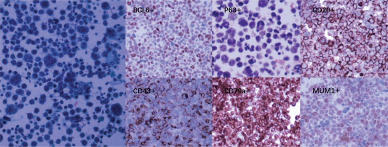
Diagnosis: Lumber punctual was performed, and CSF cytological analysis revealed malignancy cells with a high nuclear-cytoplasmic ratio. However, these cells were too loose to perform immunohistochemical stains. Genetic aberrations detections with CSF and peripheral blood sample were also inconclusive. We made a "cell-block" using the sedimentary cells collected from CSF collected through multiple aspirations via an Omaya reservoir. We further performed cytopathological and immunohistochemical analysis using this "cell-block," which finally confirmed the diagnosis of diffuse large-B cell PCNSL.
Interventions: Intracranial chemotherapy began afterwards (MTX 15 mg and dexamethasone 5 mg, twice per weeks).
Outcomes: Unfortunately, this patient was dead 2 weeks later due to severe myelosuppression and secondary septic shock.
Conclusion: We provided "cell-block" method, which collects cell components from large amount of CSF for cytology and immunohistochemical analysis. "Cell-block" cytology can be an alternative diagnostic method in diagnosis of PCNSL.
Conflict of interest statement
The authors have no conflicts of interest to disclose.
Figures
Similar articles
-
Diagnosis of central nervous system lymphoma via cerebrospinal fluid cytology: a case report.BMC Neurol. 2019 May 7;19(1):90. doi: 10.1186/s12883-019-1317-3. BMC Neurol. 2019. PMID: 31064334 Free PMC article.
-
Primary diffuse large B-cell lymphoma of the central nervous system identified with CSF biomarkers.BMC Neurol. 2024 Jul 22;24(1):250. doi: 10.1186/s12883-024-03761-6. BMC Neurol. 2024. PMID: 39039441 Free PMC article.
-
Overcoming missed diagnoses of primary central nervous system Lymphoma-The key role of cerebrospinal fluid cytology: a case report.Diagn Pathol. 2025 Mar 15;20(1):29. doi: 10.1186/s13000-025-01626-1. Diagn Pathol. 2025. PMID: 40089740 Free PMC article.
-
Molecular analysis in liquid biopsies for diagnostics of primary central nervous system lymphoma: Review of literature and future opportunities.Crit Rev Oncol Hematol. 2018 Jul;127:56-65. doi: 10.1016/j.critrevonc.2018.05.010. Epub 2018 May 17. Crit Rev Oncol Hematol. 2018. PMID: 29891112 Review.
-
Current strategies in the diagnosis of diffuse large B-cell lymphoma of the central nervous system.Br J Haematol. 2012 Feb;156(4):421-32. doi: 10.1111/j.1365-2141.2011.08928.x. Epub 2011 Nov 11. Br J Haematol. 2012. PMID: 22077417 Review.
Cited by
-
Diffuse Large B-cell Lymphoma with a Multilobated Nuclear Morphology Harboring t(14;18)(q32;q21) and t(3;22)(q27;q11).Intern Med. 2025 Jul 15;64(14):2234-2239. doi: 10.2169/internalmedicine.4719-24. Epub 2024 Dec 26. Intern Med. 2025. PMID: 39721682 Free PMC article.
References
-
- Royer-Perron L, Hoang-Xuan K, Alentorn A. Primary central nervous system lymphoma: time for diagnostic biomarkers and biotherapies? Curr Opin Neurol 2017;30:669–76. - PubMed
-
- Sinicrope K, Batchelor T. Primary Central Nervous System Lymphoma. Neurol Clin 2018;36:517–32. - PubMed
-
- Bataille B, Delwail V, Menet E, et al. Primary intracerebral malignant lymphoma: report of 248 cases. J Neurosurg 2000;92:261–6. - PubMed
Publication types
MeSH terms
LinkOut - more resources
Full Text Sources
