. 2020 Apr 3;15(4):e0230966.
doi: 10.1371/journal.pone.0230966. eCollection 2020.

Co-culture of induced pluripotent stem cells with cardiomyocytes is sufficient to promote their differentiation into cardiomyocytes

Affiliations

Co-culture of induced pluripotent stem cells with cardiomyocytes is sufficient to promote their differentiation into cardiomyocytes

Axel J Chu et al. PLoS One. .

Abstract

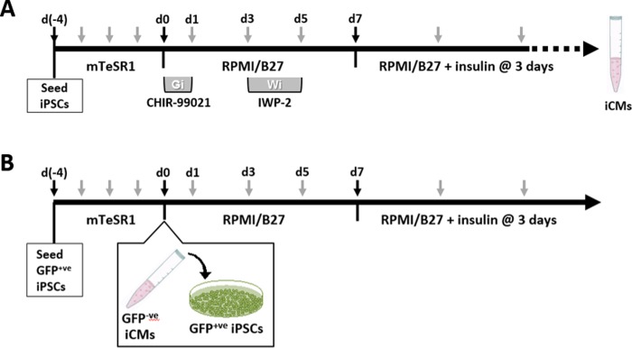
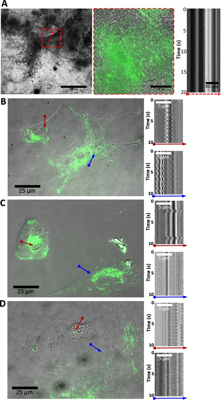
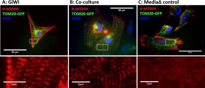
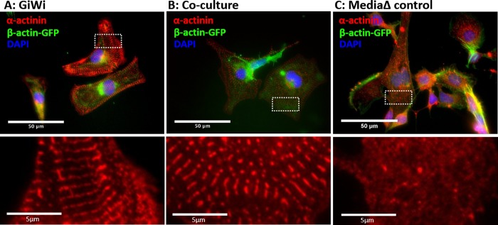
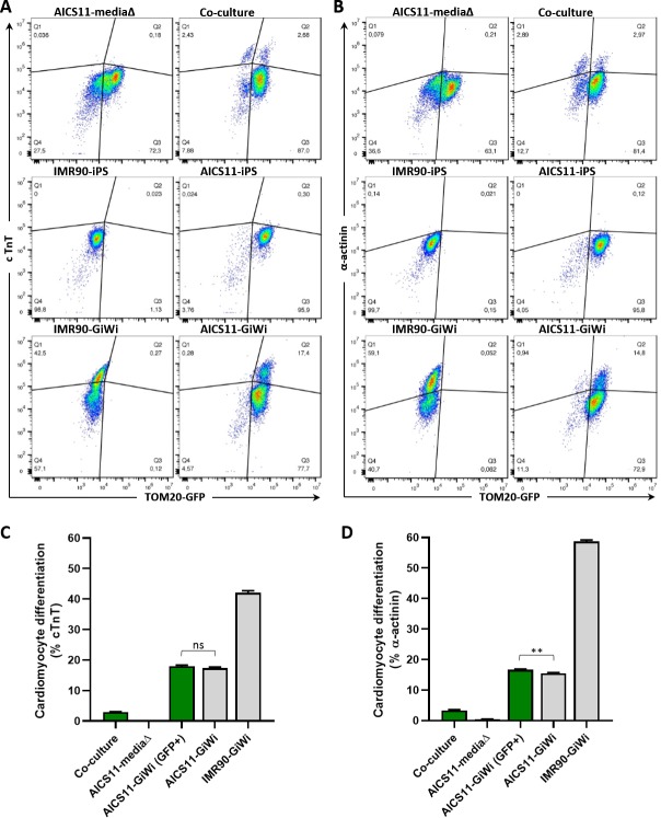
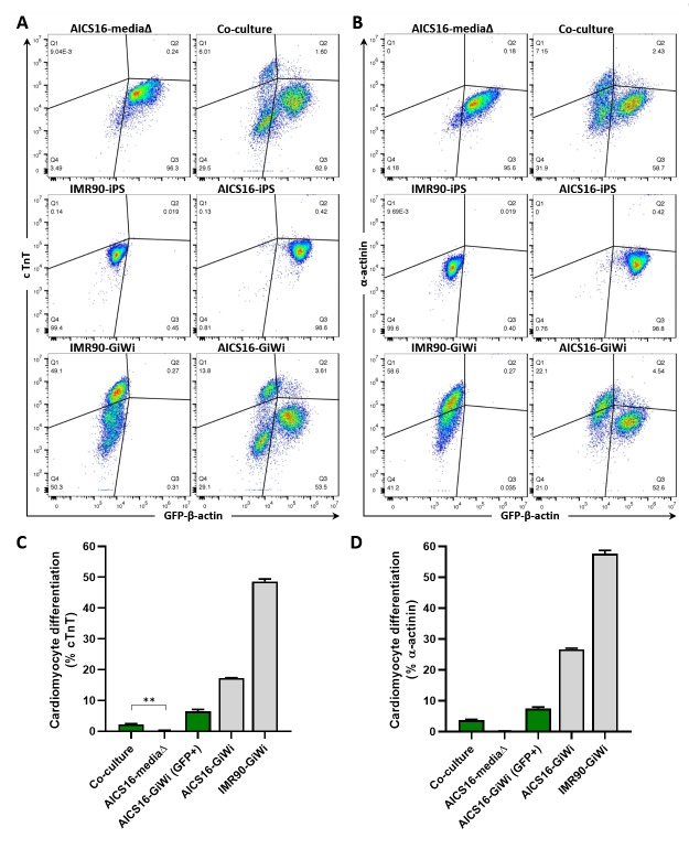
Various types of stem cells and non-stem cells have been shown to differentiate or transdifferentiate into cardiomyocytes by way of co-culture with appropriate inducer cells. However, there is a limited demonstration of a co-culture induction system utilizing stem cell-derived cardiomyocytes as a stimulatory source for cardiac reprogramming (of stem cells or otherwise). In this study, we utilized an inductive co-culture method to show that previously differentiated induced pluripotent stem (iPS) cell-derived cardiomyocytes (iCMs), when co-cultivated with iPS cells, constituted a sufficient stimulatory system to induce cardiac differentiation. To enable tracking of both cell populations, we utilized GFP-labeled iPS cells and non-labeled iCMs pre-differentiated using inhibitors of GSK and Wnt signaling. Successful differentiation was assessed by the exhibition of spontaneous self-contractions, structural organization of α-actinin labeled sarcomeres, and expression of cardiac specific markers cTnT and α-actinin. We found that iCM-iPS cell-cell contact was essential for inductive differentiation, and this required overlaying already adherent iPS cells with iCMs. Importantly, this process was achieved without the exogenous addition of pathway inhibitors and morphogens, suggesting that 'older' iCMs serve as an adequate stimulatory source capable of recapitulating the necessary culture environment for cardiac differentiation.


Conflict of interest statement

The authors have declared that no competing interests exist.

Figures

Fig 1
Fig 1. Schematic of GiWi and co-culture method used to initiate cardiac differentiation of iPS cells.
(A) For differentiation by GiWi, iPS cells were seeded onto Matrigel-coated plates and cultured to ~90% confluence in mTeSR1. The basal media was switched to RPMI/B27 at days 0–7, with addition of CHIR-99021 at days 0–1, and IWP-2 at days 3–5. Thereafter, the basal media was switched RPMI/B27/insulin. (B) For co-culture differentiation, GFP+ve iPS cells (AICS11 or AICS16) were similarly cultured to ~90% confluence in mTeSR1. At day 0, iCMs derived from non-labeled GiWi-differentiated iPS cells were dissociated and added to GFP+ve iPS cells. Cells are maintained in RPMI/B27 at days 0–7 (without GiWi), and subsequently in RPMI/B27/insulin. Black arrows indicate key steps, while gray arrows indicate the frequency of culture media replacement.
Fig 2
Fig 2. AICS11 and AICS16 iPS cells co-cultured IMR90 iCMs exhibit spontaneous self-contractions.
(A) DIC image of pre-culture on day 23 (scale bar: 500μm), red box shows magnified overlay of DIC and GFP images (scale bar: 100μm). Kymograph of contracting cluster indicated by red arrow (scale bar: 50μm). (B-D) Co-cultures were dissociated and reseeded at low cell density for imaging and kymograph analysis of isolated cells. Kymogram traces are for the indicated colored arrows. In (D), a non-contractile GFP+ve cell (blue arrow) is shown in proximity to a GFP-ve cell exhibiting self-contractions (red arrow). Scale bars: 25μm in overlays; 5μm in kymographs. Cells are AICS16 for A; AICS11 for B,C,D.
Fig 3
Fig 3. Staining of AICS11 (TOM20-GFP) cells for sarcomeric α-actinin.
AICS11 cells were differentiated using (A) GiWi protocol or (B) inductive co-culture with iCMs, and (C) basal media change alone (absent differentiation factors). Bottom panels show a magnified image of α-actinin staining for the area bounded by white rectangles. Scale bars are 50um for top panels and 5um for bottom panels.
Fig 4
Fig 4. Staining of AICS16 (β-actin-GFP) cells for sarcomeric α-actinin.
AICS16 cells were differentiated using (A) GiWi protocol or (B) inductive co-culture with iCMs, and (C) basal media change alone (absent differentiation factors). Bottom panels show a magnified image of α-actinin staining for the area bounded by white rectangles. Scale bars are 50um for top panels and 5um for bottom panels.
Fig 5
Fig 5. Cardiomyocyte differentiation efficiency of AICS11 co-cultured with iCMs evaluated by flow cytometry.
AICS11 were either co-cultured with IMR90-iCMs, or subjected to media change only as a control. Additional controls include non-differentiated iPS or GiWi-differentiated AICS11 or IMR90 cells. Total cells were harvested and immuno-stained for the cardiomyocyte markers (A) cTnT and (B) a-actinin, and analyzed by flow cytometry in conjunction with TOM20-GFP to distinguish AICS11 cells. As shown are representative flow plots for an experiment performed in triplicates. Calculation of cardiomyocyte differentiation efficiency for (C) cTnT and (D) a-actinin. Green bars indicate cells gated for GFP only, where % = Q2/(Q2+Q3)*100, while grey bars indicate total cells, where % = (Q1+Q2)/(Q1+Q2+Q3+Q4)*100. P-value for all pairwise comparison is ≤0.0001, except otherwise indicated, where ns is non-significant, and ** is <0.002.
Fig 6
Fig 6. Cardiomyocyte differentiation efficiency of AICS16 co-cultured with iCMs evaluated by flow cytometry.
Description for this experiment is identical to that for Fig 5, but with AICS16 cells (expressing GFP-b-actin) in place of AICS11. P-value for all pairwise comparison is ≤0.0001, except otherwise indicated, where ** is <0.003.

Similar articles

Cited by

References

    1. Lian X, Zhang J, Azarin SM, Zhu K, Hazeltine LB, Bao X, et al. Directed cardiomyocyte differentiation from human pluripotent stem cells by modulating Wnt/beta-catenin signaling under fully defined conditions. Nat Protoc. 2013;8(1):162–75. 10.1038/nprot.2012.150 - DOI - PMC - PubMed
    1. Machiraju P, Greenway SC. Current methods for the maturation of induced pluripotent stem cell-derived cardiomyocytes. World J Stem Cells. 2019;11(1):33–43. 10.4252/wjsc.v11.i1.33 - DOI - PMC - PubMed
    1. Mummery CL. Perspectives on the Use of Human Induced Pluripotent Stem Cell-Derived Cardiomyocytes in Biomedical Research. Stem Cell Reports. 2018;11(6):1306–11. 10.1016/j.stemcr.2018.11.011 - DOI - PMC - PubMed
    1. Mummery CL, Zhang J, Ng ES, Elliott DA, Elefanty AG, Kamp TJ. Differentiation of human embryonic stem cells and induced pluripotent stem cells to cardiomyocytes: a methods overview. Circ Res. 2012;111(3):344–58. 10.1161/CIRCRESAHA.110.227512 - DOI - PMC - PubMed
    1. Dunn KK, Palecek SP. Engineering Scalable Manufacturing of High-Quality Stem Cell-Derived Cardiomyocytes for Cardiac Tissue Repair. Front Med (Lausanne). 2018;5:110. - PMC - PubMed

Publication types

MeSH terms