. 2020 Apr 1;143(4):1158-1176.
doi: 10.1093/brain/awaa067.

Distinct patterns of structural damage underlie working memory and reasoning deficits after traumatic brain injury

Affiliations

Distinct patterns of structural damage underlie working memory and reasoning deficits after traumatic brain injury

Amy E Jolly et al. Brain. .

Abstract

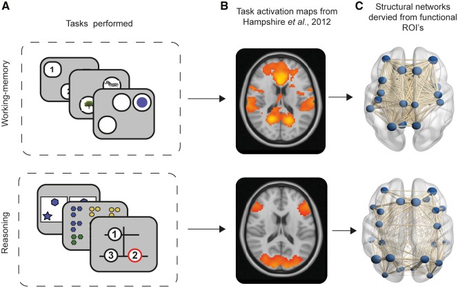
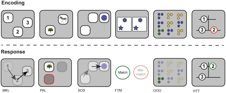
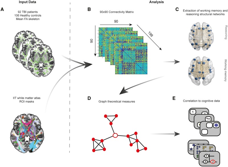
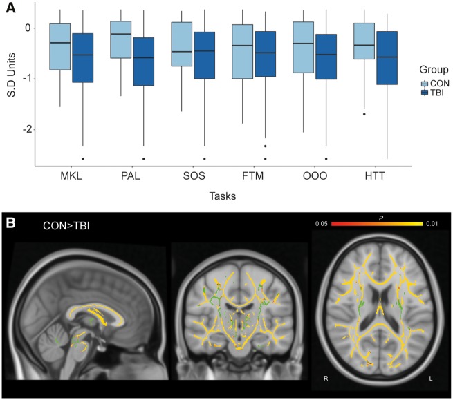
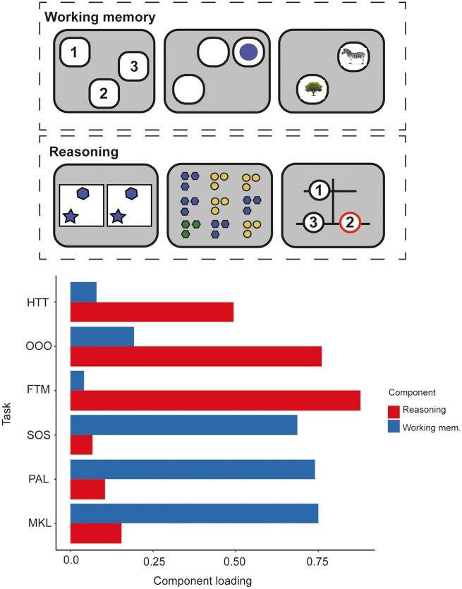
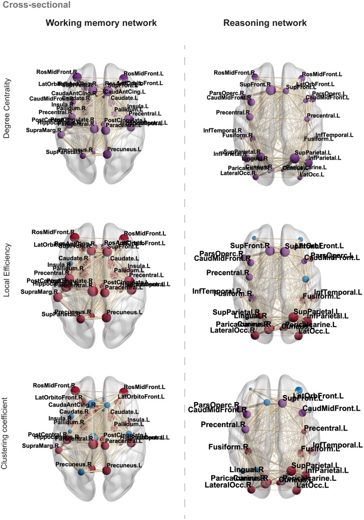
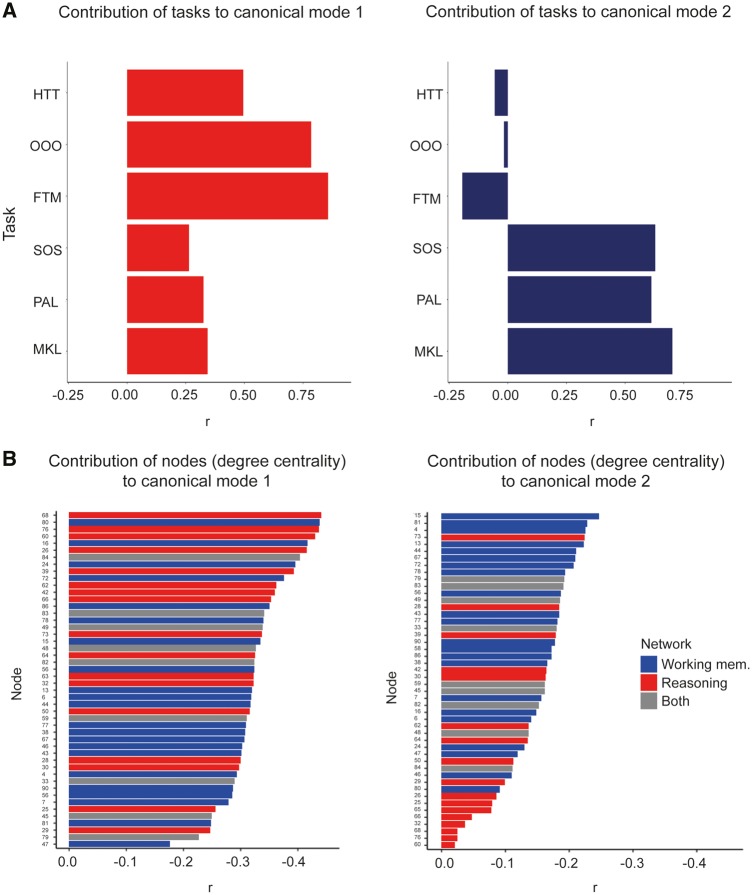
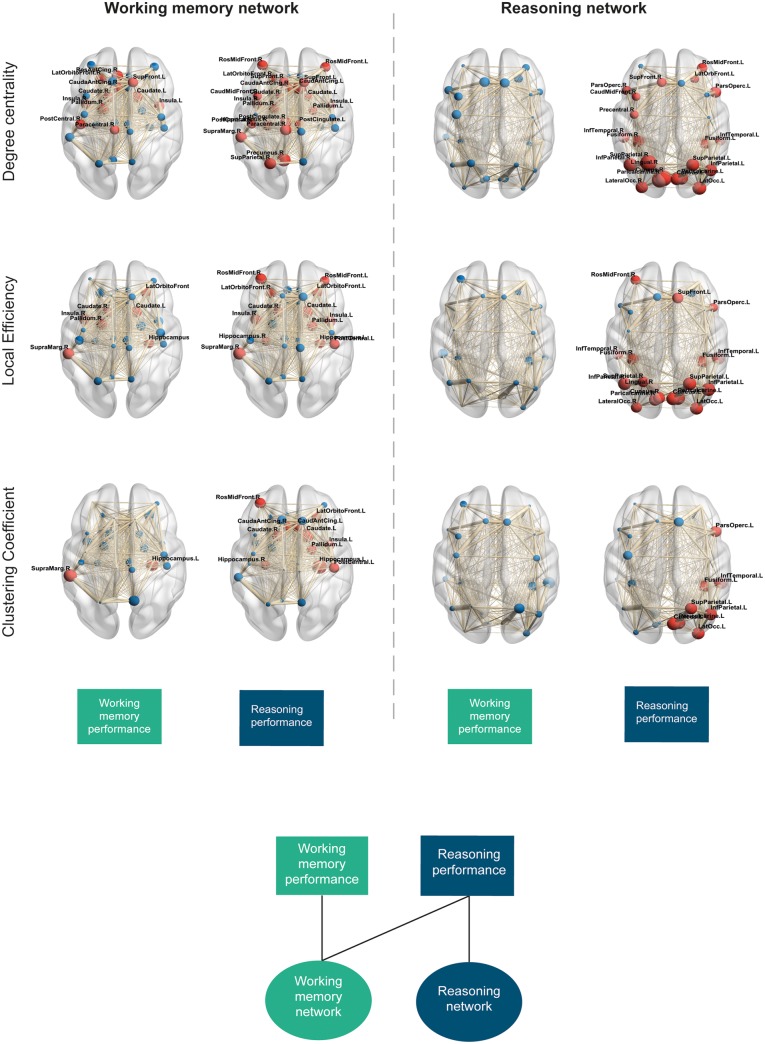
It is well established that chronic cognitive problems after traumatic brain injury relate to diffuse axonal injury and the consequent widespread disruption of brain connectivity. However, the pattern of diffuse axonal injury varies between patients and they have a correspondingly heterogeneous profile of cognitive deficits. This heterogeneity is poorly understood, presenting a non-trivial challenge for prognostication and treatment. Prominent amongst cognitive problems are deficits in working memory and reasoning. Previous functional MRI in controls has associated these aspects of cognition with distinct, but partially overlapping, networks of brain regions. Based on this, a logical prediction is that differences in the integrity of the white matter tracts that connect these networks should predict variability in the type and severity of cognitive deficits after traumatic brain injury. We use diffusion-weighted imaging, cognitive testing and network analyses to test this prediction. We define functionally distinct subnetworks of the structural connectome by intersecting previously published functional MRI maps of the brain regions that are activated during our working memory and reasoning tasks, with a library of the white matter tracts that connect them. We examine how graph theoretic measures within these subnetworks relate to the performance of the same tasks in a cohort of 92 moderate-severe traumatic brain injury patients. Finally, we use machine learning to determine whether cognitive performance in patients can be predicted using graph theoretic measures from each subnetwork. Principal component analysis of behavioural scores confirm that reasoning and working memory form distinct components of cognitive ability, both of which are vulnerable to traumatic brain injury. Critically, impairments in these abilities after traumatic brain injury correlate in a dissociable manner with the information-processing architecture of the subnetworks that they are associated with. This dissociation is confirmed when examining degree centrality measures of the subnetworks using a canonical correlation analysis. Notably, the dissociation is prevalent across a number of node-centric measures and is asymmetrical: disruption to the working memory subnetwork relates to both working memory and reasoning performance whereas disruption to the reasoning subnetwork relates to reasoning performance selectively. Machine learning analysis further supports this finding by demonstrating that network measures predict cognitive performance in patients in the same asymmetrical manner. These results accord with hierarchical models of working memory, where reasoning is dependent on the ability to first hold task-relevant information in working memory. We propose that this finer grained information may be useful for future applications that attempt to predict long-term outcomes or develop tailored therapies.

Keywords: graph theory; reasoning; structural connectome; traumatic brain injury; working memory.


Figures

Figure 1
Figure 1
Illustration of extraction of structural connectomes from task-activation maps produced by Hampshire et al. ( 2012 ). (A) Cognitive tasks used, and cognitive component structure created from PCA revealing a working memory versus reasoning component structure. (B) Working memory and reasoning activation maps derived from task-functional MRI relative to rest in healthy controls performing the cognitive tasks. (C) Construction of structural connectomes in current study using activation maps from Hampshire et al. (2012) intersected with Desikan-Killiany grey matter atlas to define nodes (regions of grey matter) and subsequent edges between nodes as white matter connectivity.
Figure 2
Figure 2
Schematic of the six computerized cognitive tasks used to assess TBI patients and controls. From left to right: working memory tasks MKL, PAL, SOS; and reasoning tasks FTM, OOO, and HTT. Encoding demonstrates the initial presentation of stimuli for each task and response demonstrates the method in which to complete the task correctly.
Fig 3
Fig 3
Overview of graph theory analysis methods. (A) Skeletonized fractional anisotropy (FA) maps for the 92 TBI patients and 106 healthy controls were intersected with region-to-region, thresholded and binarized track density images (TDIs) created using probabilistic tractography for the IIT white matter atlas. Mean fractional anisotropy for each region of interest (ROI) was therefore extracted for all patients and controls. (B) Mean fractional anisotropy for each region of interest derived from the 90 region-to-region connections were then used to create a 90 × 90 connectivity matrix for all 198 subjects and weighted by number of streamlines to account for weak connections. A thresholding approach using the mean and standard deviation of healthy controls for each connection was then applied to all controls and patients to remove weak connections. (C) Using task-functional MRI activation maps from Hampshire et al. (2012), working memory and reasoning structural networks were derived from the 90 × 90 connectivity matrices by selecting nodes that overlapped with activation maps and their respective connections. (D) Graph theoretical measures were subsequently derived from these two structural networks including global/local efficiency, degree centrality and clustering coefficient. (E) Graph theory metrics for each network were then correlated to performance on cognitive tasks associated with working memory and reasoning in patients.
Figure 4
Figure 4
Cross-sectional analysis of TBI patients and controls for cognitive performance and whole-brain fractional anisotropy. (A) Performance across the six cognitive tasks in TBI patients and healthy controls. Y-axis is in SD units. (B) Voxelwise analysis using TBSS, TFCE corrected with age as a covariate. Yellow indicates voxels with significantly higher fractional anisotropy in controls than patients (P <0.05). Green indicates the group averaged mean fractional anisotropy skeleton mask.
Figure 5
Figure 5
Principal component analysis of patient cognitive data. Component loadings for each task demonstrate higher loadings onto component 1 for reasoning-based tasks such as the HTT, OOO and FTM. Greater loadings to component two were observed in working memory-based tasks including MKL, PAL and SOS.
Figure 6
Figure 6
Cross-sectional analysis of local/nodal properties in TBI patients and controls. Blue nodes indicate non-significance, purple nodes indicate significantly lower values in patients compared to controls and red nodes indicate significantly greater values in patients compared to controls, FDR corrected. Node size = difference between patient and control local/nodal properties for each metric examined. Edge width = weighted by fractional anisotropy and no. streamlines. Significant nodes are labelled for reference.
Figure 7
Figure 7
Back projection of canonical variates to task and node data from canonical correlation analysis. Canonical correlation analysis produced two significant modes demonstrating two distinct relationships between cognitive variables and degree centrality of the 90 nodes defined in the whole brain connectome. (A) Back projection of cognitive canonical variates to cognitive tasks demonstrated greater contribution of reasoning tasks (HTT, OOO and FTM) to canonical mode 1 and greater contribution of working memory tasks (MKL, PAL, SOS) to canonical mode 2. (B) Back projection of node canonical variates to individual nodes and their measures of degree centrality revealed that dissociable patterns of nodes were associated with each canonical mode. Specifically, reasoning nodes (red bars) contributed the most to canonical mode 1 and working memory nodes (blue bars) contributed the most to canonical mode 2. Grey bars denote where nodes are present within both structural networks.
Figure 8
Figure 8
Local graph theory network measures and correlation to cognitive performance. Node size represents correlation coefficient (r, positive or negative). Red nodes demonstrate a significant correlation to cognitive components (P <0.05, FDR corrected), blue nodes demonstrate non-significant correlations. For measures of degree centrality, red nodes denote significant positive correlations, FDR corrected. For measures of local efficiency and clustering coefficient, red nodes denote significant negative correlations, FDR corrected. Edge thickness demonstrates weight of a given connection (fractional anisotropy and number of streamlines). Boxes at the bottom demonstrate which cognitive component is correlated to each of the networks and properties demonstrated. Bottom panel illustrates the dissociable relationship between cognitive components and working memory and reasoning structural network properties.

Similar articles

Cited by

References

    1. Achard S, Salvador R, Whitcher B, Suckling J, Bullmore E.. A resilient, low-frequency, small-world human brain functional network with highly connected association cortical hubs. J Neurosci 2006; 26: 63–72. - PMC - PubMed
    1. Bassett DS, Bullmore E.. Small-world brain networks. Neuroscientist 2006; 12: 512–23. - PubMed
    1. Beckmann CF, DeLuca M, Devlin JT, Smith SM.. Investigations into resting-state connectivity using independent component analysis. Phil Trans R Soc B 2005; 360: 1001–13. - PMC - PubMed
    1. Bonnelle V. Salience network integrity predicts default mode network function after traumatic brain injury. Proc Natl Acad Sci USA 2012; 109: 4690–5. - PMC - PubMed
    1. Braun U, Muldoon SF, Bassett DS.. On human brain networks in health and disease In: eLS. Chichester: John Wiley & Sons, Ltd; 2015.

Publication types