. 2020 Jun;251(2):200-212.
doi: 10.1002/path.5447. Epub 2020 May 21.

Ocular cytomegalovirus latency exacerbates the development of choroidal neovascularization

Affiliations

Ocular cytomegalovirus latency exacerbates the development of choroidal neovascularization

Jinxian Xu et al. J Pathol. 2020 Jun.

Abstract

Age-related macular degeneration (AMD) is a complex, multifactorial, progressive disease which represents a leading cause of irreversible visual impairment and blindness in older individuals. Human cytomegalovirus (HCMV), which infects 50-80% of humans, is usually acquired during early life and persists in a latent state for the life of the individual. In view of its previously described pro-angiogenic properties, we hypothesized that cytomegalovirus might be a novel risk factor for progression to an advanced form, neovascular AMD, which is characterized by choroidal neovascularization (CNV). The purpose of this study was to investigate if latent ocular murine cytomegalovirus (MCMV) infection exacerbated the development of CNV in vascular endothelial growth factor (VEGF)-overexpressing VEGF-Ahyper mice. Here we show that neonatal infection with MCMV resulted in dissemination of virus to various organs throughout the body including the eye, where it localized principally to the choroid in both VEGF-overexpressingVEGF-Ahyper and wild-type(WT) 129 mice. By 6 months post-infection, no replicating virus was detected in eyes and extraocular tissues, although virus DNA was still present in all eyes and extraocular tissues of both VEGF-Ahyper and WT mice. Expression of MCMV immediate early (IE) 1 mRNA was detected only in latently infected eyes of VEGF-Ahyper mice, but not in eyes of WT mice. Significantly increased CNV was observed in eyes of MCMV-infected VEGF-Ahyper mice compared to eyes of uninfected VEGF-Ahyper mice, while no CNV lesions were observed in eyes of either infected or uninfected WT mice. Protein levels of several inflammatory/angiogenic factors, particularly VEGF and IL-6, were significantly higher in eyes of MCMV-infected VEGF-Ahyper mice, compared to uninfected controls. Initial studies of ocular tissue from human cadavers revealed that HCMV DNA was present in four choroid/retinal pigment epithelium samples from 24 cadavers. Taken together, our data suggest that ocular HCMV latency could be a significant risk factor for the development of AMD. © 2020 Pathological Society of Great Britain and Ireland. Published by John Wiley & Sons, Ltd.

Keywords: VEGF; age-related macular degeneration; choroidal neovascularization; cytomegalovirus; inflammation.


Conflict of interest statement

No conflicts of interest were declared.

Figures
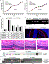
Figure 1.
Figure 1.
MCMV disseminates to and becomes latent in the eye following systemic infection of neonatal VEGF-Ahyper or wild-type 129 mice. (A) Postnatal changes in body weight in male (left) and female (right) neonatally-infected VEGF-Ahyper or WT mice, compared with uninfected control male and female mice of the same age (n = 20–30). (B) Titer of MCMV (upper) and MCMV positivity rate (lower) at day 14 p.i. in eyes, salivary glands, lungs, and kidneys determined by plaque assay (n = 5–8; *p < 0.05). (C) PCR assay for MCMV DNA in eyes of VEGF-Ahyper and WT mice at day 14 p.i. All mice tested positive. (D) Merged photomicrographs of ocular staining for RPE65 (red), MCMV EA (green), and DAPI (blue) from an infected WT mouse (left) and an infected VEGF-Ahyper mouse (right) at day 14 p.i. The majority of MCMV EA-positive cells are located in the choroid (scale bars: 50 μm). (E) Photomicrographs of ocular H&E staining of an infected WT mouse; an infected VEGF-Ahyper mouse at day 14 p.i.; an age-matched, uninfected control WT mouse; and an age-matched uninfected VEGF-Ahyper mouse (scale bars: 100 μm). (F) PCR assay for MCMV DNA in eyes of VEGF-Ahyper and WT mice at 6 months p.i. All eyes tested positive. (G) RT-PCR assay for MCMV IE1 mRNA in MCMV-infected VEGF-Ahyper and WT mice at 6 months p.i. IE1 mRNA was detected in 6 of 13 eyes from VEGF-Ahyper mice, while all 15 eyes from MCMV-infected WT mice tested negative.
Figure 2.
Figure 2.
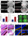
MCMV infection exacerbates CNV in VEGF-Ahyper but not in WT mice. (A) Representative images of SD-OCT from infected WT (lower left) and VEGF-Ahyper mice (lower right) at 6 months p.i.; also age-matched, uninfected WT (upper left) and VEGF-Ahyper mice (upper right). CNV lesions were found in MCMV-infected and uninfected VEGF-Ahyper mice (indicated by red squares). (B) The numbers (left) and volumes (right) of CNV lesions in MCMV-infected and uninfected VEGF-Ahyper mice (*p < 0.05). (C) Fluorescein angiography images of MCMV-latently-infected and uninfected VEGF-Ahyper and WT mice. (D) Photomicrographs of ocular H&E staining of MCMV-infected WT (lower left) and VEGF-Ahyper mice (lower right) at 6 months p.i., as well as from age-matched, uninfected WT (upper left) and VEGF-Ahyper mice (upper right). CNV lesions were observed in the subretinal space (upper right) and inside the inner retina (lower right) (scale bars: 100 μm). (E) Merged photomicrographs of ocular staining for CD31 (red) and DAPI (blue) from infected WT (lower left) and VEGF-Ahyper mice (lower right) at 6 months p.i., as well as from age-matched, uninfected WT (upper left) and VEGF-Ahyper mice (upper right). CD31-stained neovessels are present in the inner retina (lower right) (scale bars: 100 μm).
Figure 3.
Figure 3.
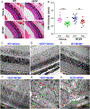
Features of photoreceptor degeneration and CNV lesions in VEGF-Ahyper mice. (A, B) Photomicrographs of ocular H&E staining (A) and analysis of ONL thickness (B) among infected and uninfected WT and VEGF-Ahyper mice 6 months p.i. A significantly thinner outer nuclear layer (ONL) was observed in MCMV-infected and uninfected VEGF-Ahyper mice, compared with infected and uninfected WT mice (scale bars: 400 μm). **p < 0.01; ****p < 0.0001. (C–H) Representative electron micrographs of ocular ultrastructure from infected WT (E) and VEGF-Ahyper mice (F, G, H) at 6 months p.i.; also age-matched, uninfected WT (C) and VEGF-Ahyper mice (D). No remarkable pathological changes were observed in the RPE layer or inner retina of non-CNV areas of both infected (F) and uninfected (D) VEGF-Ahyper mice. Some CNV lesions (indicated by aster isks) were observed in the RPE layer, surrounded by pigmented RPE-like cells (G). In addition, the morphology of nearby photoreceptors was disturbed, with shortening and loss of outer segments (G, indicated by arrowheads). Some CNV lesions (indicated by asterisks) grew into the inner retina (H) and many pigmented RPE-like cells were observed adjacent to new vessels in the inner retina (H, indicated by arrows). No remarkable pathological changes were observed in the RPE layer or inner retina of infected (E) or uninfected (C) WT mice.
Figure 4.
Figure 4.
MCMV-infected VEGF-Ahyper mice contain increased ocular VEGF-A compared with uninfected VEGF-Ahyper mice at 6 months p.i. (A) Protein levels of VEGF in plasma assayed by ELISA (n = 6; *p < 0.05; ***p < 0.001; ****p < 0.0001). (B) Protein levels of ocular VEGF assayed by ELISA (n = 6; **p < 0.01; ***p < 0.001; ****p < 0.0001). (C) Merged photomicrographs of ocular staining for F4/80 (red), VEGF (green), and DAPI (blue) of MCMV-infected WT (lower left) and VEGF-Ahyper mice (lower middle and right) at 6 months p.i., as well as from age-matched, uninfected WT (upper left) and VEGF-Ahyper mice (upper middle and right). A few F4/80-stained cells were observed in CNV lesions, some of which co-stained with VEGF (yellow arrows), while some were VEGF-negative (red arrows). The majority of VEGF-stained cells were not F4/80 macrophages (white arrows) (scale bars: 100 μm). (D) The number of F4/80-stained cells per CNV lesion in MCMV-infected and uninfected VEGF-Ahyper mice at 6 months p.i.
Figure 5.
Figure 5.
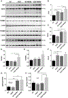
Protein levels of several inflammatory/angiogenic factors were increased in eyes of infected VEGF-Ahyper mice at 6 months p.i. (A–F) Western blots of RIP3, TGF-β1, TGF-β-R1, TGF-β-R2, and GFAP in eyes of MCMV-infected or uninfected WT and VEGF-Ahyper mice at 6 months p.i. Ratio of RIP3 to β-actin(B), TGF-β1 to β-actin(C), TGF-β-R1 to β-actin(D), TGF-β-R2 to β-actin(E), and GFAP to β-actin(F). (G, H) Protein levels of CCL5 (G) and IL-6(H) in eyes of MCMV-infected or uninfected WT and VEGF-Ahyper mice at 6 months p.i. Data are shown as mean ± SEM (for western blot, n = 3; for ELISA, n = 4) and compared by t-test. ***p < 0.001; **p < 0.01; *p < 0.05.

Similar articles

Cited by

References

    1. Friedman E Update of the vascular model of AMD. Br J Ophthalmol 2004; 88: 161–163. - PMC - PubMed
    1. Bowes Rickman C, Farsiu S, Toth CA, et al. Dry age-related macular degeneration: mechanisms, therapeutic targets, and imaging. Invest Ophthalmol Vis Sci 2013; 54: ORSF68–ORSF80. - PMC - PubMed
    1. Bird AC. Therapeutic targets in age-related macular disease. J Clin Invest 2010; 120: 3033–3041. - PMC - PubMed
    1. Gorin MB. Genetic insights into age-related macular degeneration: controversies addressing risk, causality, and therapeutics. Mol Aspects Med 2012; 33: 467–486. - PMC - PubMed
    1. Pennesi ME, Neuringer M, Courtney RJ. Animal models of age related macular degeneration. Mol Aspects Med 2012; 33: 487–509. - PMC - PubMed

Publication types

MeSH terms