. 2020 May 18;30(10):1821-1833.e8.
doi: 10.1016/j.cub.2020.03.006. Epub 2020 Apr 2.

Stress Disrupts Human Hippocampal-Prefrontal Function during Prospective Spatial Navigation and Hinders Flexible Behavior

Affiliations

Stress Disrupts Human Hippocampal-Prefrontal Function during Prospective Spatial Navigation and Hinders Flexible Behavior

Thackery I Brown et al. Curr Biol. .

Abstract

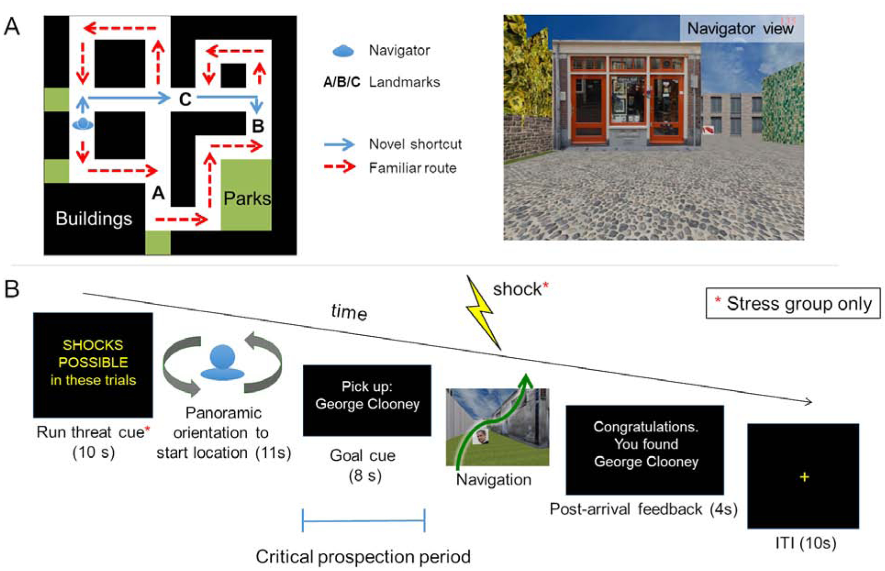
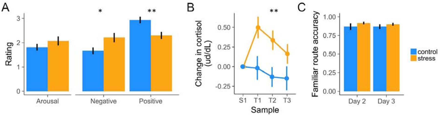
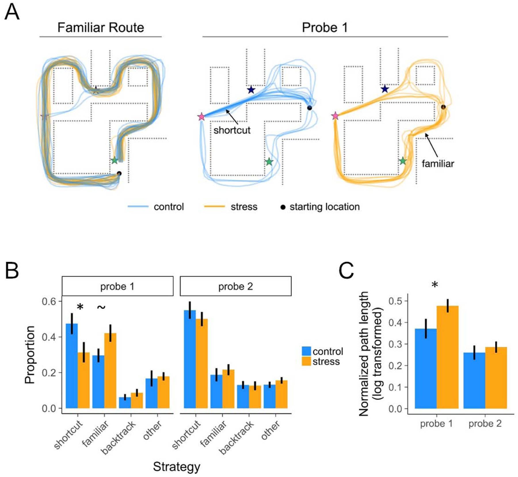
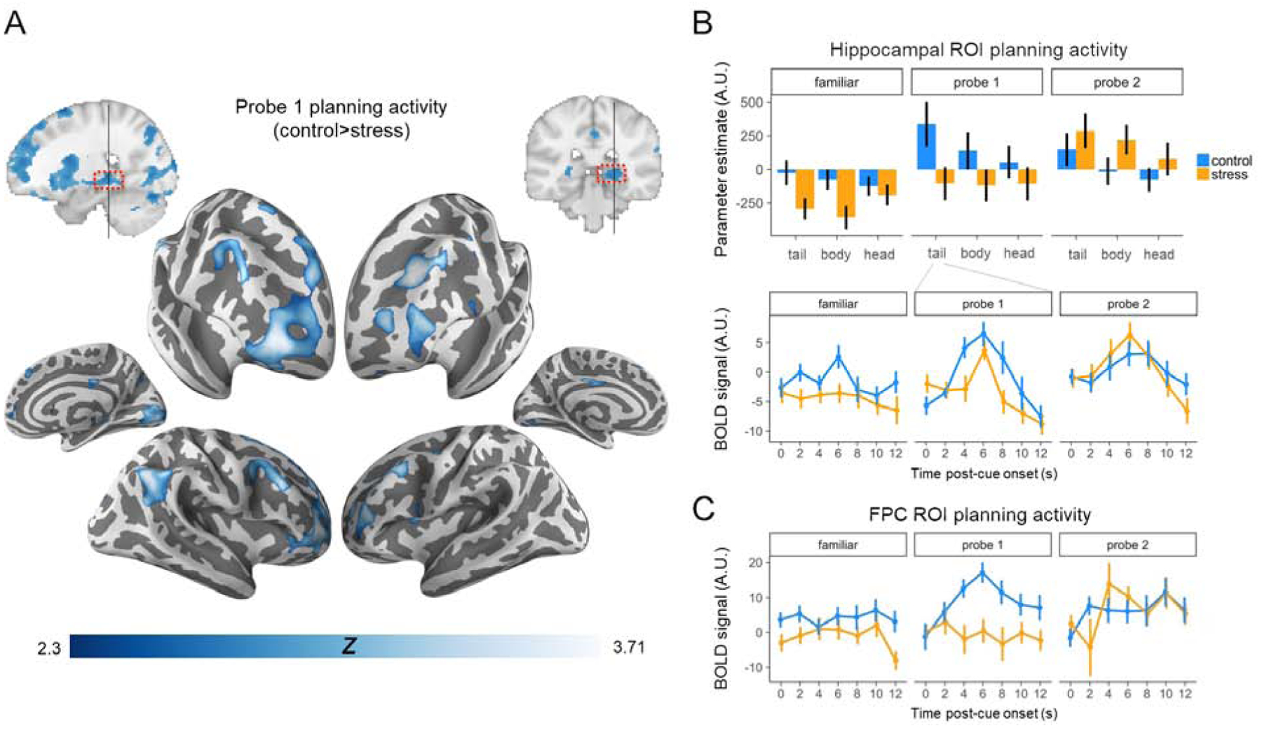
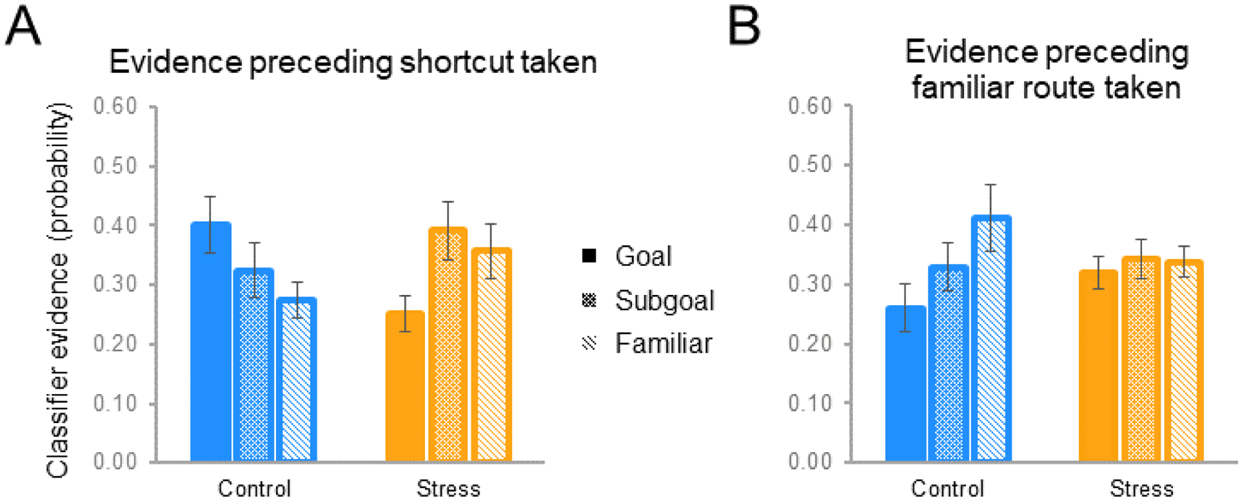
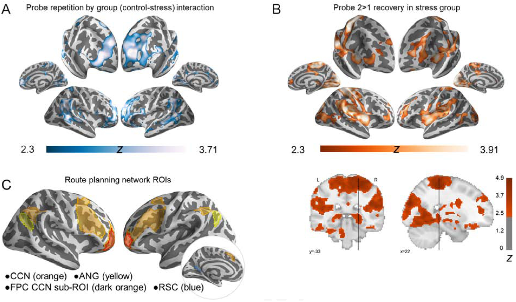
The ability to anticipate and flexibly plan for the future is critical for achieving goal-directed outcomes. Extant data suggest that neural and cognitive stress mechanisms may disrupt memory retrieval and restrict prospective planning, with deleterious impacts on behavior. Here, we examined whether and how acute psychological stress influences goal-directed navigational planning and efficient, flexible behavior. Our methods combined fMRI, neuroendocrinology, and machine learning with a virtual navigation planning task. Human participants were trained to navigate familiar paths in virtual environments and then (concurrent with fMRI) performed a planning and navigation task that could be most efficiently solved by taking novel shortcut paths. Strikingly, relative to non-stressed control participants, participants who performed the planning task under experimentally induced acute psychological stress demonstrated (1) disrupted neural activity critical for mnemonic retrieval and mental simulation and (2) reduced traversal of shortcuts and greater reliance on familiar paths. These neural and behavioral changes under psychological stress were tied to evidence for disrupted neural replay of memory for future locations in the spatial environment, providing mechanistic insight into why and how stress can alter planning and foster inefficient behavior.

Keywords: Cognitive Control; Hippocampus; Memory; Navigation; Stress; fMRI.


Conflict of interest statement

Declaration of Interests The authors declare no competing interests.

Figures

Figure 1.
Figure 1.. Task overview.
(A) Topographical view of one of 12 virtual city environments. On Days 1 and 2, participants over-learned a familiar route (red line) through each environment. Each environment contained buildings, parks, fences, trees, and three unique goal landmark objects (images of famous faces, fruits/vegetables, animals, or tools displayed on boxes along the familiar route). On Day 3, participants were first asked to repeat navigation of each familiar route once more during fMRI (Familiar route trials, beginning and ending at a pseudo-randomly placed finish line). Then, during Probe trials they were placed along the familiar route (blue character), and asked to navigate as quickly as possible to a goal [B] within that environment. Critically, while participants were not informed that shortcuts exist, each environment was designed such that a novel shortcut (blue line) provided the most efficient route to the goal from the Probe-trial start location. As such, participants could navigate to the goal by taking the familiar route (which would take longer) or by flexibly drawing on memory to plan and take a shortcut. Participants were not informed of these alternative strategies. (B) Day 3 fMRI Probe trial structure. At the outset of each scanning run, stress group participants were given a reminder that they would be under threat of shock throughout the run (Day 3 Familiar route trials were also performed under threat). On each Probe trial, they were oriented to their start location, then presented the name of a landmark object as a goal and held in place for 8s, with the environment hidden from view. During this period, participants could plan how to get to the goal, if they freely chose/had the cognitive bandwidth to do so. This prospective planning period was the target of our fMRI analyses. Participants were then allowed to freely navigate to the goal to end the trial. Stress participants performed the entirety of these trials under anticipatory stress (see STAR Methods).
Figure 2.
Figure 2.. Manipulation Checks.
(A) Stress increased subjective ratings of negative valence and decreased ratings of positive valence. (B) Stress increased cortisol levels throughout the Day 3 scan session. (C) Control and stress participants navigated the familiar routes with similar accuracy by the end of the last day of training (Day 2) and during fMRI task performance (Day 3). For subsequent Probe-trial behavioral analyses, we excluded environments on which participants did not accurately follow the familiar route by the end of training. Error bars indicate within-participant standard error of the mean. See also Table S1 and Table S2.
Figure 3.
Figure 3.. Behavior.
(A) Illustration of all participants’ paths, by group, in a representative environment. Stars indicate categorically distinct landmark objects. (B) Proportion of well-learned environments navigated using various strategies. During the first Probe round (Probe 1), the probability of taking a precise shortcut was higher in the control vs. stress group, and vice versa for taking a familiar route. By the second Probe round (Probe 2), the stress group took an equivalent proportion of shortcuts. (C) Normalized path length as a function of group and Probe round. During Probe 1, the stress group had significantly longer path lengths than the control group, indicating that stress restricted flexible access to environmental knowledge during planning thereby impairing efficient navigation. Error bars indicate within-participant standard error of the mean. See also Table S3 and Figure S1.
Figure 4.
Figure 4.. Prospective planning activity across groups.
(A) The control group recruited hippocampus and lateral prefrontal and parietal CCN regions more than the stress group when planning navigation to novel goals (Probe 1). p<0.01, voxel-wise threshold; cluster-corrected to a false positive rate of p<0.05[17]. (B) Hippocampal ROI planning activity was reduced under stress, particularly in the tail (timecourse in middle panel). Hippocampal activity, along with shortcut behavior, recovered (increased) in the stress group during Probe 2 planning. (C) Bilateral FPC ROI activity as a function of task and group. FPC activity increased during Probe 1 planning in the control group, but decreased with goal familiarity (Probe 2). In contrast, activity increased in the stress group selectively during Probe 2 planning. Statistics were conducted on parameter estimates; time courses are for visualization purposes only. See also Table S4, Figure S3, and Figure S4.
Figure 5.
Figure 5.. Landmark evidence during the Probe 1 planning period as a function of ultimate route taken.
(A) When ultimately taking a shortcut, controls exhibited the strongest planning-period evidence for long-term goals (Goal), followed by shortcut subgoals and familiar route landmarks. By contrast, stress participants showed weaker long-term goal evidence (than control participants, and than subgoals). (B) This pattern in controls reversed during trials on which the familiar route was ultimately taken, whereas stress participants failed to exhibit differential reinstatement evidence. See also Table S5.
Figure 6.
Figure 6.. Interaction of novel planning (Probe 1) relative to repeated planning (Probe 2) for controls vs. stress groups.
(A) Regions showing a group × Probe round interaction were more active during novel than repeated Probe planning in the control group, but greater activity during repeated than novel Probe planning in the stress group. (B) Recovery in the stress group (Probe 2>Probe 1) underlying interaction in A. Frontoparietal control regions (particularly anterior PFC and lateral IPS) and the hippocampal tail were notable a priori loci of this recovery based on our predictions. (C) Visualization of a priori cortical ROIs: cognitive control network – CCN; frontopolar cortex – FPC; angular gyrus – ANG; retrosplenial cortex – RSC. p<0.01, voxel-wise threshold; cluster-corrected p<0.05. See also Table S6, Figure S5, and Figure S6.

Comment in

References

    1. Buckner RL, and Carroll DC (2007). Self-projection and the brain. Trends Cogn. Sci 11, 49–57. - PubMed
    1. Addis DR, Wong AT, and Schacter DL (2007). Remembering the past and imagining the future: common and distinct neural substrates during event construction and elaboration. Neuropsycholgia 7, 1363–1377. - PMC - PubMed
    1. Race E, Keane MM, and Verfaellie M (2011). Medial temporal lobe damage causes deficits in episodic memory and episodic future thinking not attributable to deficits in narrative construction. J. Neurosci 31, 10262–9. - PMC - PubMed
    1. Race E, Keane MM, and Verfaellie M (2013). Losing sight of the future: Impaired semantic prospection following medial temporal lobe lesions. Hippocampus 23, 268–77. - PMC - PubMed
    1. Hassabis D, Kumaran D, and Maguire EA (2007). Using imagination to understand the neural basis of episodic memory. J. Neurosci 27, 14365–74. - PMC - PubMed

Publication types

LinkOut - more resources