. 2020 Jun 30;30(8):4361-4380.
doi: 10.1093/cercor/bhaa023.

A Domain-General Cognitive Core Defined in Multimodally Parcellated Human Cortex

Affiliations

A Domain-General Cognitive Core Defined in Multimodally Parcellated Human Cortex

Moataz Assem et al. Cereb Cortex. .

Abstract

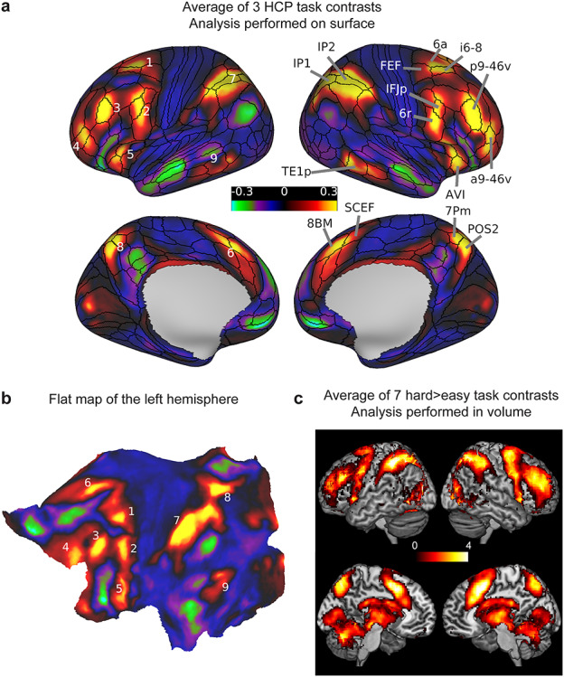
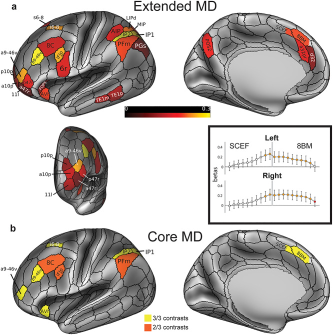
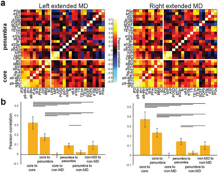
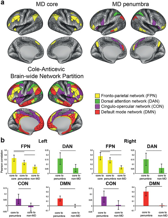
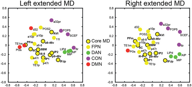
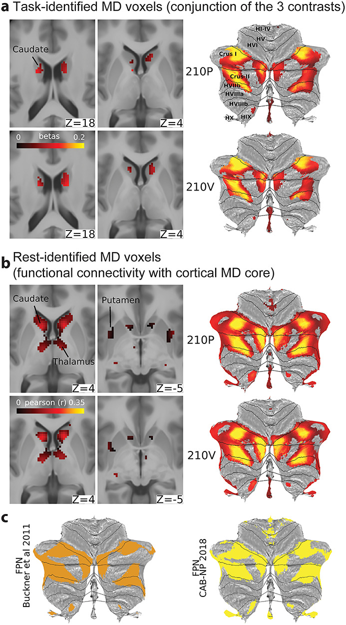
Numerous brain imaging studies identified a domain-general or "multiple-demand" (MD) activation pattern accompanying many tasks and may play a core role in cognitive control. Though this finding is well established, the limited spatial localization provided by traditional imaging methods precluded a consensus regarding the precise anatomy, functional differentiation, and connectivity of the MD system. To address these limitations, we used data from 449 subjects from the Human Connectome Project, with the cortex of each individual parcellated using neurobiologically grounded multimodal MRI features. The conjunction of three cognitive contrasts reveals a core of 10 widely distributed MD parcels per hemisphere that are most strongly activated and functionally interconnected, surrounded by a penumbra of 17 additional areas. Outside cerebral cortex, MD activation is most prominent in the caudate and cerebellum. Comparison with canonical resting-state networks shows MD regions concentrated in the fronto-parietal network but also engaging three other networks. MD activations show modest relative task preferences accompanying strong co-recruitment. With distributed anatomical organization, mosaic functional preferences, and strong interconnectivity, we suggest MD regions are well positioned to integrate and assemble the diverse components of cognitive operations. Our precise delineation of MD regions provides a basis for refined analyses of their functions.

Keywords: cognitive control; domain-general; fronto-parietal; multimodal cortical parcellation; multiple-demand.


Figures

Figure 1
Figure 1
(a) Average of the 3 HCP group average task contrasts (WM 2bk > 0bk, Relational H > E, Math > Story). Values are beta estimates. Black contours correspond to the HCP multimodal parcellation MMP_1.0 (210V) areal borders. Numbers on the left hemisphere correspond to visually separable patches of activity distributed across the cortex. (b) The same activity of the left hemisphere projected on a flattened cortical sheet. Numbers correspond to the same patches labeled in (a). (c) Volumetric MD map from Fedorenko et al. (2013) computed by averaging seven hard > easy task contrasts (2-mm smoothed) displayed on a volume rendering lateral surface (above) and medial slice (below) of the MNI template. Values are t-statistics. Data available at http://balsa.wustl.edu/lL9nj.
Figure 2
Figure 2
(a) The extended MD system: conjunction of significant areas across three functional contrasts. Areal colors reflect average beta values across the three contrasts analyzed in relation to subject-specific parcellations. Data are averaged across hemispheres and for illustration projected here onto the left lateral and medial surfaces (top) and an anterior view of frontal pole parcels (bottom left). Box (bottom right) displays pattern of activity in regions SCEF (posterior) and 8BM (anterior), divided into posterior to anterior segments in relation to subject-specific parcellations. Gray bar indicates 8BM/SCEF border. Orange indicates segments that are part of the extended MD system when activity from both hemispheres is combined (i.e., segments with activity significantly above zero in all three behavioral contrasts). Red indicates one additional segment that survives as part of the extended MD system when activity from each hemisphere is tested separately. (b) The core MD system: areas with activity estimates that were significantly higher than the mean activity of all extended MD areas in all three contrasts (yellow) and two out of three contrasts (orange). Data available at http://balsa.wustl.edu/qNLq8.
Figure 3
Figure 3
FC of the MD system. (a) FC (Pearson correlation) across the MD system. Regions of the extended MD system are separated into core and penumbra, with regions within each set ordered by mean activation (beta) across our three functional contrasts. Note the strength of core MD connectivity (lower left box) versus penumbra connectivity (upper right box). (b) Statistical comparison (paired sample t-test) between different groups of cortical connections. Lines highlight a statistically significant difference (P < 0.05, Bonferroni corrected for 30 comparisons). Data available at http://balsa.wustl.edu/jjL1x.
Figure 4
Figure 4
MD system and resting-state networks. (a) Resting-state network assignments from the CAB-NP (Ji et al. 2019) for the core (top left) and penumbra (top right) MD areas, compared with the whole CAB-NP FPN (bottom left). (b) Statistical comparison (paired sample t-test) of cortical connection types for each CAB-NP network. Data available at http://balsa.wustl.edu/wNGV6.
Figure 5
Figure 5
Multidimensional scaling plot of the connectivities between extended MD regions. Axis units are arbitrary. Data available at http://balsa.wustl.edu/jjL1x.
Figure 6
Figure 6
Task profiles across the MD system. (a) Raw activation estimates (betas) for each contrast. Areas are sorted from left to right according to the strength of their MD response (average across the three contrasts). Error bars represent SEM. Core MD areal labels are colored in orange (survived in all three contrasts) and red (survived in two out of three contrasts). (b) Task profiles for two independent groups of subjects (210P and 210V). (c) Correlation of task profiles between groups. (d) Normalized task profiles across the MD system as line plots. Bar heights represent between-task standard deviation, separately calculated for each subject and averaged over subjects. Bar colors indicate relative preferences between tasks. Color wheel indicates red for WM, green for relational reasoning (Rel), and blue for math. Intermediate colors show mixed preferences. Brighter and darker colors reflect stronger and weaker MD activation, respectively. (e) Cortical projection of the RGB color weighted normalized task profiles. Data available at http://balsa.wustl.edu/4m747.
Figure 7
Figure 7
Group average beta maps for the WM 0bk > fix contrast (upper) and Relational E > fix contrast (lower). The borders of extended MD regions are colored in green. Data available at http://balsa.wustl.edu/mDkgN.
Figure 8
Figure 8
Subcortical and cerebellar MD components. (a) Left: conjunction of significant voxels across the three tasks for the 210P (top) and 210V (bottom). Right: cerebellar activity is displayed on a flat cerebellum with lines representing anatomical borders (Diedrichsen and Zotow 2015). Data available at http://balsa.wustl.edu/Z4NXp. (b) Left: subcortical voxels with significant connections to the cortical core MD areas. Right: cerebellar MD connectivity displayed on a flat map. Data available at http://balsa.wustl.edu/VjwZg. (c) FPN from Buckner et al. (2011) (left) and Ji et al. (2018) (right). Data available at http://balsa.wustl.edu/3g7wv.

References

    1. Alexander GE, DeLong MR, Strick PL. 1986. Parallel organization of functionally segregated circuits linking basal ganglia and cortex. Annu Rev Neurosci. 9:357–381. - PubMed
    1. Amalric M, Dehaene S. 2016. Origins of the brain networks for advanced mathematics in expert mathematicians. Proc Natl Acad Sci. 113:4909–4917. - PMC - PubMed
    1. Amalric M, Dehaene S. 2017. Cortical circuits for mathematical knowledge: evidence for a major subdivision within the brain’s semantic networks. Philos Trans R Soc B Biol Sci. 373:20160515. - PMC - PubMed
    1. Amiez C, Wutte MG, Faillenot I, Petrides M, Burle B, Procyk E. 2016. Single subject analyses reveal consistent recruitment of frontal operculum in performance monitoring. Neuroimage. 133:266–278. - PubMed
    1. Averbeck BB, Lehman J, Jacobson M, Haber SN. 2014. Estimates of projection overlap and zones of convergence within frontal-striatal circuits. J Neurosci. 34:9497–9505. - PMC - PubMed

Publication types