. 2020 Mar 31;21(7):2394.
doi: 10.3390/ijms21072394.

Comparative Proteomic Analysis by iTRAQ Reveals that Plastid Pigment Metabolism Contributes to Leaf Color Changes in Tobacco (Nicotiana tabacum) during Curing

Affiliations

Comparative Proteomic Analysis by iTRAQ Reveals that Plastid Pigment Metabolism Contributes to Leaf Color Changes in Tobacco (Nicotiana tabacum) during Curing

Shengjiang Wu et al. Int J Mol Sci. .

Abstract

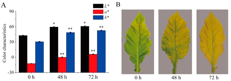
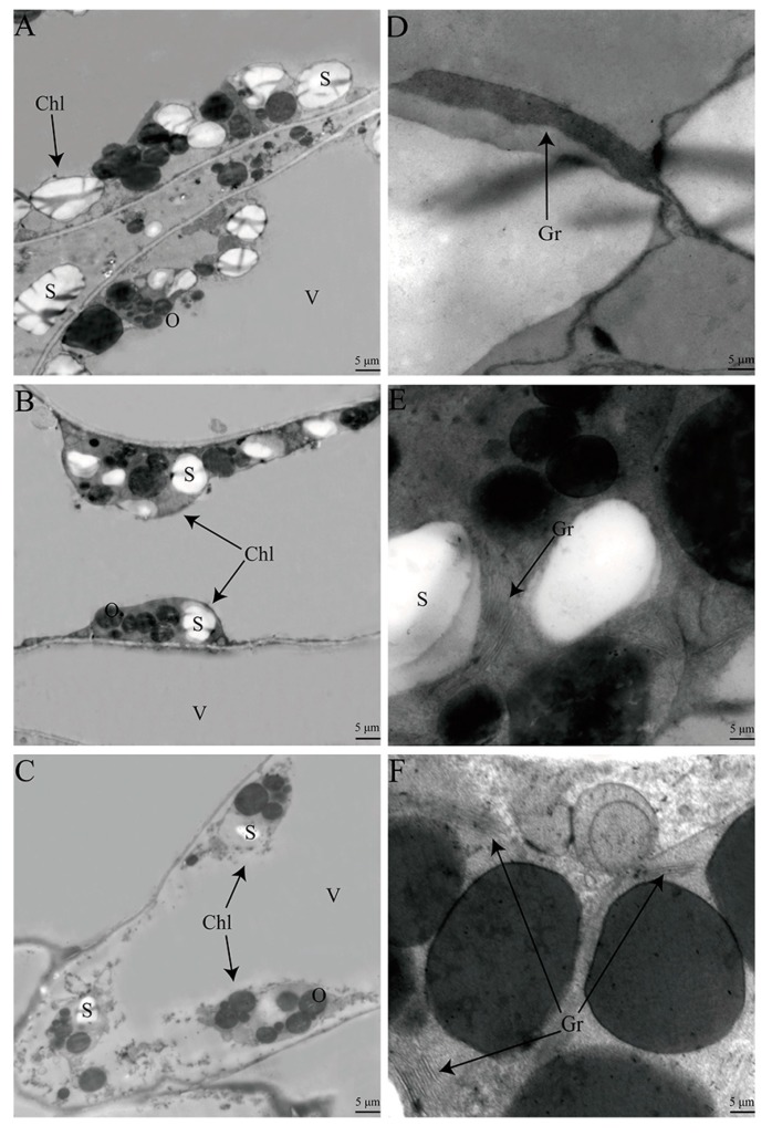
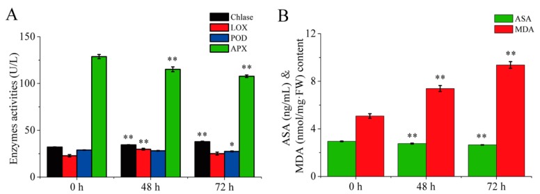
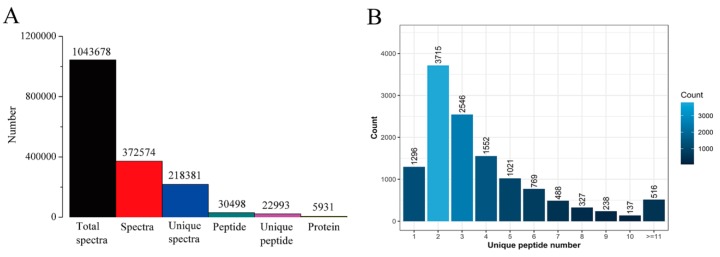
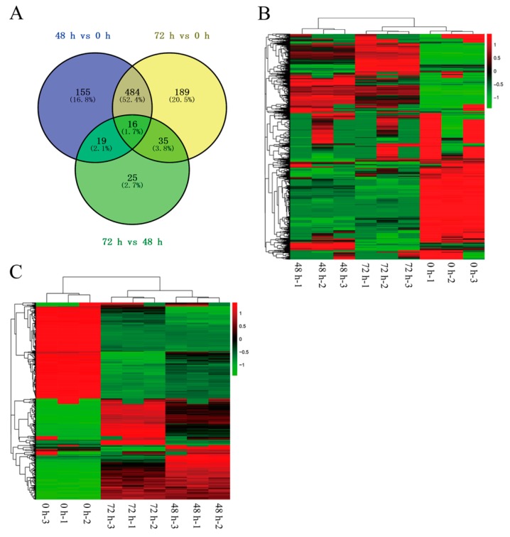
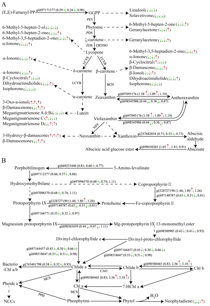
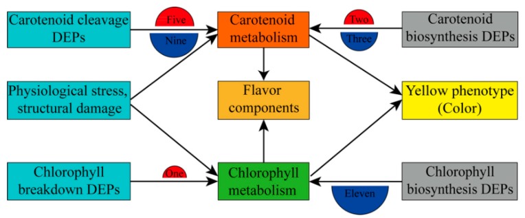
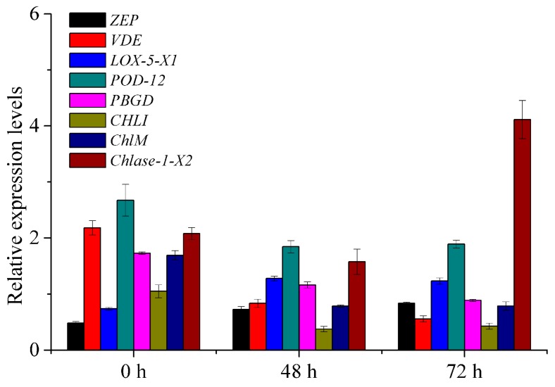
Tobacco (Nicotiana tabacum), is a world's major non-food agricultural crop widely cultivated for its economic value. Among several color change associated biological processes, plastid pigment metabolism is of trivial importance in postharvest plant organs during curing and storage. However, the molecular mechanisms involved in carotenoid and chlorophyll metabolism, as well as color change in tobacco leaves during curing, need further elaboration. Here, proteomic analysis at different curing stages (0 h, 48 h, 72 h) was performed in tobacco cv. Bi'na1 with an aim to investigate the molecular mechanisms of pigment metabolism in tobacco leaves as revealed by the iTRAQ proteomic approach. Our results displayed significant differences in leaf color parameters and ultrastructural fingerprints that indicate an acceleration of chloroplast disintegration and promotion of pigment degradation in tobacco leaves due to curing. In total, 5931 proteins were identified, of which 923 (450 up-regulated, 452 down-regulated, and 21 common) differentially expressed proteins (DEPs) were obtained from tobacco leaves. To elucidate the molecular mechanisms of pigment metabolism and color change, 19 DEPs involved in carotenoid metabolism and 12 DEPs related to chlorophyll metabolism were screened. The results exhibited the complex regulation of DEPs in carotenoid metabolism, a negative regulation in chlorophyll biosynthesis, and a positive regulation in chlorophyll breakdown, which delayed the degradation of xanthophylls and accelerated the breakdown of chlorophylls, promoting the formation of yellow color during curing. Particularly, the up-regulation of the chlorophyllase-1-like isoform X2 was the key protein regulatory mechanism responsible for chlorophyll metabolism and color change. The expression pattern of 8 genes was consistent with the iTRAQ data. These results not only provide new insights into pigment metabolism and color change underlying the postharvest physiological regulatory networks in plants, but also a broader perspective, which prompts us to pay attention to further screen key proteins in tobacco leaves during curing.

Keywords: Nicotiana tabacum; iTRAQ; pigment metabolism; postharvest physiology; ultrastructure.


Conflict of interest statement

The authors declare no conflict of interest.

Figures

Figure 1
Figure 1
Color and phenotypic changes in tobacco leaves during curing. (A) The leaf color values of L*, a*, and b* were determined during curing. Data are shown as the means ± SE, n = 30. Asterisks indicate significant differences between the values at 0 h and 48 h or 72 h based on Duncan’s multiple range test in SPSS (* p < 0.05, ** p < 0.01). (B) Representative tobacco leaf phenotypes were documented for each flue-curing stage.
Figure 2
Figure 2
Ultrastructural changes in tobacco leaves during curing. Chloroplasts were gradually disrupted in tobacco leaves cells during curing (A, 0 h; B, 48 h and C, 72 h). Grana thylakoid lamellae were disrupted in tobacco leaves during curing (D, 0 h; E, 48 h and F, 72 h). Chl, chloroplast; Gr, grana thylakoid lamellae; S, starch granule; O, osmiophilic granule; V, vacuole.
Figure 3
Figure 3
Changes of enzyme activities (A, Chlase, LOX, POD, APX) and chemical components (B, ASA and MDA) in tobacco leaves during curing. Data are shown as the means ± SE (n = 6). Asterisks indicate significant differences between the values at 0 h and 48 h or 72 h based on Duncan’s multiple range test in SPSS (* p < 0.05, ** p < 0.01). Chlase, chlorophyllase; LOX, lipoxygenase; POD, peroxidase; APX, ascorbate peroxidase; ASA, ascorbic acid; MDA, malondialdehyde.
Figure 4
Figure 4
Basic information of iTRAQ output. (A) Spectra, peptides, and proteins identified in tobacco leaves. (B) Number of peptides that matched proteins.
Figure 5
Figure 5
Venn diagram and heatmap representing identified proteins and DEPs from different comparison groups in tobacco leaf samples. (A) Total DEPs identified in all tobacco leaf samples; (B) Heatmap/hierarchical clustering of all identified proteins; and (C) Heatmap/hierarchical clustering of DEPs identified in all tobacco leaf samples. The numbers of DEPs identified from three biological replicates are shown in the different segments (Figure 5A). Red and green indicate higher expression and lower expression, respectively (Figure 5B,C).
Figure 6
Figure 6
The carotenoid (A) and chlorophyll (B) metabolic pathway in tobacco leaves during curing. Up-regulated proteins or increased metabolites are marked with upward red arrows, while down-regulated proteins or decreased metabolites are marked with downward green arrows. The numbers represent the fold change. The left arrows or numbers represented the difference of proteins or metabolites during 0–48 h, the middle arrows or numbers indicated the difference during 0–72 h, and the right indicated the difference during 48–72 h. Gi numbers and ratios of the DEPs are shown in Supplementary Table S4. (A) GGPP, geranylgeranyl diphosphate; PSY, phytoene synthase; PDS, phytoene desaturase; ZDS, ζ-carotene desaturase; CRTISO, carotenoid isomerase; LCYB, lycopene β-cyclase; LCYE, lycopeneε-cyclase; BCH, β-carotene hydroxylase; NSY, neoxanthin synthase. (B) Chlide a/b, chlorophyllide a/b; MCS, metal-chelating substance; CAO, chlorophyllide-a oxygenase; Pheide a, pheophorbide a; Chl a/b, chlorophyll a/b; 7HChl a, 7-Hydroxy-chlorophyll a; NCCs, non-fluorescent chlorophyll catabolites.
Figure 7
Figure 7
Branching program related to pigment metabolism and color change in tobacco leaves during curing. The semicircle filled with the red color indicated that the DEPs were positive regulators, and the blue color suggested that the DEPs were negative regulators in the pigment biosynthetic and breakdown pathway. The numbers represented the numbers of the total up- and down-regulated proteins in different pathways.
Figure 8
Figure 8
Verification of iTRAQ results by qRT-PCR. Values represent the means ± SE (n = 3). ZEP, zeaxanthin epoxidase, chloroplastic-like; VDE, violaxanthin de-epoxidase, chloroplastic; LOX-5-X1, probable linoleate 9S-lipoxygenase 5 isoform X1; POD-12, peroxidase 12-like; PBGD, porphobilinogen deaminase, chloroplastic-like; CHLI, magnesium-chelatase subunit ChlI, chloroplastic; ChlM, magnesium protoporphyrin IX methyltransferase, chloroplastic; Chlase-1-X2, chlorophyllase-1-like isoform X2.

Similar articles

Cited by

References

    1. Ma C., Liang B., Chang B., Liu L., Yan J., Yang Y., Zhao Z. Transcriptome Profiling Reveals Transcriptional Regulation by DNA Methyltransferase Inhibitor 5-Aza-2′-Deoxycytidine Enhancing Red Pigmentation in Bagged “Granny Smith” Apples (Malus domestica) Int. J. Mol. Sci. 2018;19:3133. doi: 10.3390/ijms19103133. - DOI - PMC - PubMed
    1. Chen J., Funnell K.A., Lewis D.H., Eason J.R., Woolley D.J. Relationship between changes in colour and pigment content during spathe regreening of Zantedeschia ‘Best Gold’. Postharvest Biol. Technol. 2012;67:124–129. doi: 10.1016/j.postharvbio.2011.12.019. - DOI
    1. Wei X., Deng X., Cai D., Ji Z., Wang C., Yu J., Li J., Chen S. Decreased tobacco-specific nitrosamines by microbial treatment with Bacillus amyloliquefaciens DA9 during the air-curing process of burley tobacco. J. Agric. Food Chem. 2014;62:12701–12706. doi: 10.1021/jf504084z. - DOI - PubMed
    1. Li D., Zhang X., Li L., Aghdam M.S., Wei X., Liu J., Xu Y., Luo Z. Elevated CO2 delayed the chlorophyll degradation and anthocyanin accumulation in postharvest strawberry fruit. Food Chem. 2019;285:163–170. doi: 10.1016/j.foodchem.2019.01.150. - DOI - PubMed
    1. Yuan Z., Deng L., Yin B., Yao S., Zeng K. Effects of blue LED light irradiation on pigment metabolism of ethephon-degreened mandarin fruit. Postharvest Biol. Technol. 2017;134:45–54. doi: 10.1016/j.postharvbio.2017.08.005. - DOI

LinkOut - more resources