Review
. 2020 Mar 31;9(4):837.
doi: 10.3390/cells9040837.

Role and Mechanisms of Mitophagy in Liver Diseases

Affiliations
Review

Role and Mechanisms of Mitophagy in Liver Diseases

Xiaowen Ma et al. Cells. .

Abstract

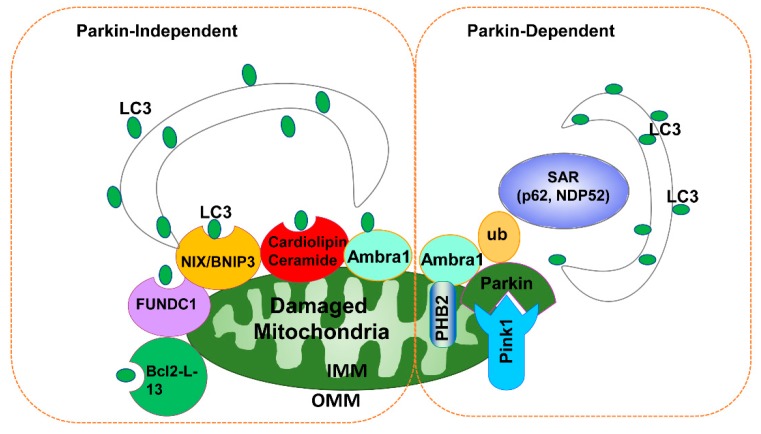
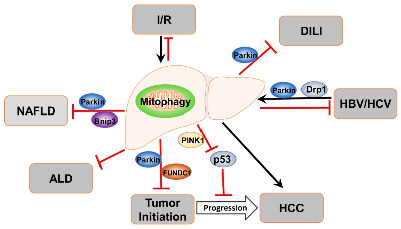
The mitochondrion is an organelle that plays a vital role in the regulation of hepatic cellular redox, lipid metabolism, and cell death. Mitochondrial dysfunction is associated with both acute and chronic liver diseases with emerging evidence indicating that mitophagy, a selective form of autophagy for damaged/excessive mitochondria, plays a key role in the liver's physiology and pathophysiology. This review will focus on mitochondrial dynamics, mitophagy regulation, and their roles in various liver diseases (alcoholic liver disease, non-alcoholic fatty liver disease, drug-induced liver injury, hepatic ischemia-reperfusion injury, viral hepatitis, and cancer) with the hope that a better understanding of the molecular events and signaling pathways in mitophagy regulation will help identify promising targets for the future treatment of liver diseases.

Keywords: NAFLD; Parkin; Pink1; alcohol; autophagy; mitochondria.


Conflict of interest statement

The authors declare no conflict of interest.

Figures

Figure 1
Figure 1
Parkin-dependent and Parkin-independent mitophagy. In the presence of Parkin, damaged/depolarized mitochondria (e.g., following CCCP treatment) stabilize PINK1 that recruits Parkin to the mitochondria. Once on the mitochondria, Parkin promotes ubiquitination of outer mitochondrial membrane proteins, which serve as binding partners for SAR, such as p62 or NDP52. SAR acts as an adaptor molecule through direct interaction with LC3 to recruit autophagosomal membranes to the mitochondria. The mitochondrial inner membrane protein PHB2 also binds to LC3 through its LIR domain upon mitochondrial depolarization after proteasome-dependent outer mitochondrial membrane rupture in a Parkin-dependent manner. For the Parkin-independent pathway, damaged mitochondria (particularly under hypoxia conditions) increase the expression of FUNDC1, NIX, and BNIP3, which may in turn recruit autophagosomes to mitochondria by direct interaction with LC3 through their LIR domains. Upon mitochondrial depolarization, Bcl-2-L13 also promotes mitophagy independent parkin. Upon toxin or drug-induced mitochondrial damage, mitochondrial lipid Cardiolipin and ceramide also bind to LC3 and promote mitophagy independent of Parkin. Notably, Ambra1 may promote mitophagy in both Parkin-dependent and Parkin-independent manners. IMM, inner mitochondrial membrane; OMM, outer mitochondrial membrane; SAR, soluble autophagy receptor; ub, ubiquitin.
Figure 2
Figure 2
The structure of the mitochondrial spheroids under electron microscopy (EM). Wild-type mouse embryonic fibroblasts (MEF) were treated with CCCP (20 µM) for 16 h. Cells were fixed and further processed for EM analysis. Representative EM images of a MEF (A), typical mitochondrial spheroid structures (B,C). Black arrows denote mitochondrial spheroids and the red arrow denotes mitochondria spheroid-enwrapped mitochondria.
Figure 3
Figure 3
Monitoring mitophagy in primary hepatocytes using Cox8-GFP-mCherry. Primary mouse hepatocytes were infected with adenovirus-Cox8-GFP-mCherry (10 MOI) for 72 h followed by confocal microscopy. Arrows denote red-only autolysosome-enwrapped mitochondria.
Figure 4
Figure 4
Megamitochondria in mouse livers after chronic plus binge alcohol feeding. Two three-month-old C57BL/6 mice were subjected to the chronic-plus-binge (Gao-binge) alcohol model. Liver sections were fixed and electron microscopy followed (A and B from Gao-binge alcohol-fed mouse livers). Arrows denote megamitochondria. L, lysosome; LD, lipid droplet; M, mitochondria; N, nucleus. Bar: 50 nm.
Figure 5
Figure 5
Summary of the role of mitophagy in liver disease. Mitophagy plays protective roles against drug-induced liver injury, the pathogenesis of ALD and NAFLD, as well as ischemia/reperfusion. In contrast, mitophagy may play dual roles in liver tumorigenesis and its progression. In viral hepatitis, mitophagy is highly activated, serving as a survival mechanism by inhibiting cell death in HBV/HCV infected cells.

Similar articles

Cited by

References

    1. Mizushima N., Levine B., Cuervo A.M., Klionsky D.J. Autophagy fights disease through cellular self-digestion. Nature. 2008;451:1069–1075. doi: 10.1038/nature06639. - DOI - PMC - PubMed
    1. Mizushima N., Komatsu M. Autophagy: Renovation of Cells and Tissues. Cell. 2011;147:728–741. doi: 10.1016/j.cell.2011.10.026. - DOI - PubMed
    1. Li Y., Ding W.-X. Adipose tissue autophagy and homeostasis in alcohol-induced liver injury. Liver Res. 2017;1:54–62. doi: 10.1016/j.livres.2017.03.004. - DOI - PMC - PubMed
    1. Czaja M.J., Ding W.-X., Donohue T.M., Friedman S.L., Kim J.-S., Komatsu M., Lemasters J.J., Lemoine A., Lin J.D., Ou J.-H.J., et al. Functions of autophagy in normal and diseased liver. Autophagy. 2013;9:1131–1158. doi: 10.4161/auto.25063. - DOI - PMC - PubMed
    1. Yin X.-M., Ding W.-X., Gao W. Autophagy in the liver. Hepatology. 2008;47:1773–1785. doi: 10.1002/hep.22146. - DOI - PubMed

Publication types

Substances