. 2020 Mar 31;9(4):838.
doi: 10.3390/cells9040838.

Involvement of Enteric Glia in Small Intestine Neuromuscular Dysfunction of Toll-Like Receptor 4-Deficient Mice

Affiliations

Involvement of Enteric Glia in Small Intestine Neuromuscular Dysfunction of Toll-Like Receptor 4-Deficient Mice

Silvia Cerantola et al. Cells. .

Abstract

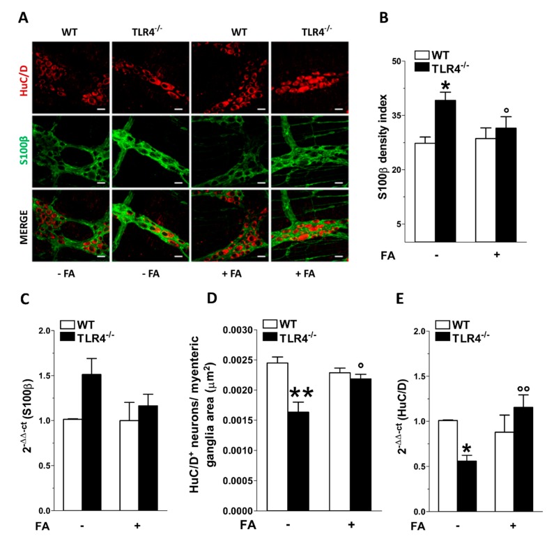
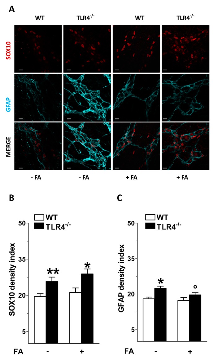
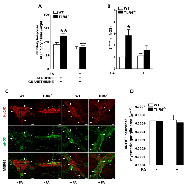
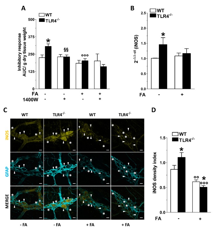
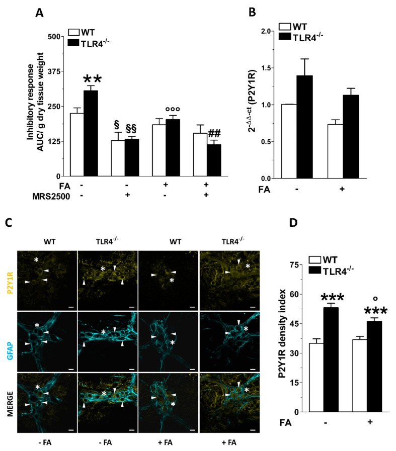
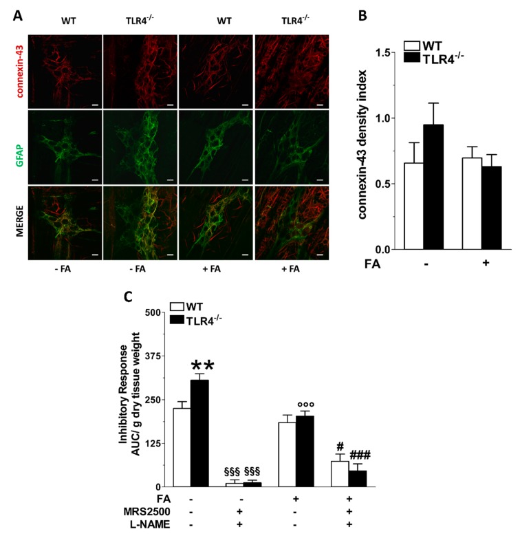
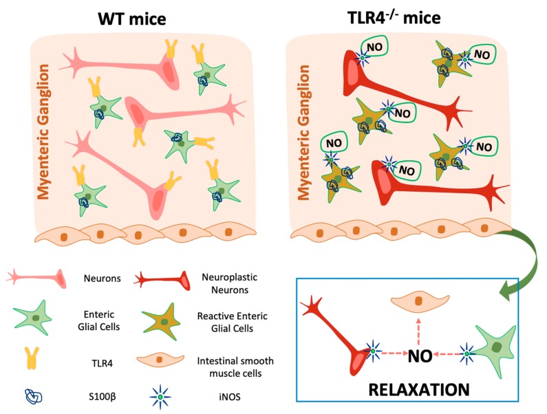
Enteric glial cells (EGCs) influence nitric oxide (NO)- and adenosine diphosphate (ADP)- mediated signaling in the enteric nervous system (ENS). Since Toll-like receptor 4 (TLR4) participates to EGC homoeostasis, this study aimed to evaluate the possible involvement of EGCs in the alterations of the inhibitory neurotransmission in TLR4-/- mice. Ileal segments from male TLR4-/- and wild-type (WT) C57BL/6J mice were incubated with the gliotoxin fluoroacetate (FA). Alterations in ENS morphology and neurochemical coding were investigated by immunohistochemistry whereas neuromuscular responses were determined by recording non-adrenergic non-cholinergic (NANC) relaxations in isometrically suspended isolated ileal preparations. TLR4-/- ileal segments showed increased iNOS immunoreactivity associated with enhanced NANC relaxation, mediated by iNOS-derived NO and sensitive to P2Y1 inhibition. Treatment with FA diminished iNOS immunoreactivity and partially abolished NO- and ADP- mediated relaxation in the TLR4-/- mouse ileum, with no changes of P2Y1 and connexin-43 immunofluorescence distribution in the ENS. After FA treatment, S100β and GFAP immunoreactivity in TLR4-/- myenteric plexus was reduced to levels comparable to those observed in WT. Our findings show the involvement of EGCs in the alterations of ENS architecture and in the increased purinergic and nitrergic-mediated relaxation, determining gut dysmotility in TLR4-/- mice.

Keywords: enteric glial cells; enteric nervous system; fluoroacetate; innate immunity; intestinal motility; knockout mice; small intestine; toll-like receptor 4.


Conflict of interest statement

The authors declare no conflict of interest.

Figures

Figure 1
Figure 1
Effect of fluoroacetate (FA) treatment on myenteric ganglia neural and glial cells. (A) Representative confocal microphotographs showing the distribution of S100β (green) and HuC/D (red) in small intestine LMMPs of WT and TLR4−/− mice in the presence or absence of 10 μM FA (bars = 22 μm). (B,C) Analysis of S100β density index in ileal myenteric ganglia (B) and S100β mRNA levels in small intestine segments (C) of WT and TLR4−/− mice in the presence or absence of 10 μM FA. (D,E) Total number of HuC/D+ neurons in ileal myenteric ganglia (D) and HuC/D mRNA levels in small intestine segments (E) of WT and TLR4−/− mice in the presence or absence of 10 μM FA. Data are reported as mean ± SEM for all panels. * P < 0.05, ** P < 0.01 vs. WT; ° P < 0.05, °° P < 0.01 vs. respective control without FA; N = 6 mice/group.
Figure 2
Figure 2
Effect of fluoroacetate (FA) treatment on enteric glial phenotype. (A) Representative confocal microphotographs showing the distribution of SOX10 (red) and GFAP (cyan) in ileal LMMPs of WT and TLR4−/− mice in the presence or absence of 10 μM FA (bars = 22 μm). (B,C) Analysis of SOX10 and GFAP density index in ileal LMMPs of WT and TLR4−/− mice in the presence or absence of 10 μM FA. Data are reported as mean ± SEM for all panels. * P < 0.05, ** P < 0.01 vs. WT; ° P < 0.05 vs. respective control without FA; N = 6 mice/group.
Figure 3
Figure 3
Involvement of EGCs in the modulation of nitrergic neurotransmission of small intestine in TLR4−/− mice. (A) 10 Hz EFS-evoked NANC relaxation responses in ileal segments of WT and TLR4−/− mice in the presence or absence of 10 μM FA. (B) qRT-PCR quantification of nNOS mRNA levels in small intestine segments of WT and TLR4−/− mice in the presence or absence of 10 μM FA. (C) Representative confocal microphotographs showing the distribution of nNOS+ (green) and HuC/D+ (red) neurons in ileal LMMPs of WT and TLR4−/− mice in the presence or absence of 10 μM FA (bars = 22 μm). White arrowhead point to HuC/D+ nNOS+ neurons. (D) Number of nNOS+ neurons in ileal LMMPs of WT and TLR4−/− mice in the presence or absence of 10 μM FA. Data are reported as mean ± SEM for all panels. * P < 0.05, ** P < 0.01 vs. WT; °°° P < 0.005 vs. respective control without FA; N = 6 mice/group.
Figure 4
Figure 4
TLR4 signaling and EGC activity influence NO-mediated relaxation. (A) 10 Hz EFS-evoked relaxation in NANC conditions with or without 10 μM FA or 10 μM 1400 W (iNOS inhibitor) in ileal segments of WT and TLR4−/− mice. (B) qRT-PCR quantification of iNOS mRNA levels in small intestine segments of WT and TLR4−/− mice in the presence or absence of 10 μM FA. (C) Representative confocal microphotographs showing the distribution of iNOS (yellow) and GFAP (cyan) and (D) analysis of iNOS density index in ileal LMMPs of WT and TLR4−/− mice in the presence or absence of 10 μM FA (bars = 22 μm). White arrowheads indicate iNOS+ neurons, stars indicate iNOS+ glial cell bodies, respectively. Data are reported as mean ± SEM for all panels. * P < 0.05 vs. WT; °° P < 0.01, °°° P < 0.001 vs. respective control without FA; §§ P < 0.01 vs. respective control without 1400 W; N = 6 mice/group.
Figure 5
Figure 5
TLR4 signaling and EGC activity affect P2Y1 receptor-mediated small intestinal relaxation. (A) 10 Hz EFS-evoked relaxation in NANC conditions with or without 10 μM FA or 10 μM MRS2500 (P2Y1R inhibitor) in ileal segments of WT and TLR4−/− mice. (B) qRT-PCR quantification of P2Y1R mRNA levels in small intestine segments of WT and TLR4−/− mice in the presence or absence of 10 μM FA. (C) Representative confocal microphotographs showing the distribution of P2Y1R (yellow) and GFAP (cyan) and (D) analysis of P2Y1R density index in ileal LMMPs of WT and TLR4-/- mice in the presence or absence of 10 μM FA (bars = 22 μm). White arrowheads indicate P2Y1+ neurons, stars indicate P2Y1+ glial cell bodies, respectively. Data are reported as mean ± SEM for all panels. ** P < 0.01, *** P < 0.001 vs. WT; ° P < 0.05, °°° P < 0.001 vs. respective control without FA; § P < 0.05, §§ P < 0.01 vs. respective control without MRS2500; ## P < 0.01 vs. respective control with FA and without MRS2500; N = 6 mice/group.
Figure 6
Figure 6
TLR4 signaling and EGC activity regulate nitrergic- and purinergic-mediated inhibitory response of the small intestine. (A) Representative confocal microphotographs showing the distribution of connexin-43 (red) and GFAP (green) and (B) analysis of connexin-43 density index in ileal LMMPs of WT and TLR4−/− mice in the presence or absence of 10 μM FA (bars = 22 μm). (C) 10 Hz EFS-evoked relaxation in NANC conditions with or without 10 μM FA or 10 μM MRS2500 (P2Y1R inhibitor) or 1 μM L-NAME (pan-NOS inhibitor) in WT and TLR4−/− mice. Data are reported as mean ± SEM for all panels. ** P < 0.01 vs. WT; °°° P < 0.001 vs. respective control without FA; §§§ P < 0.001 vs. respective control without MRS2500 and L-NAME. # P < 0.05, ### P < 0.001 vs. respective control with FA and without MRS2500 and L-NAME; N = 6 mice/group.
Figure 7
Figure 7
Graphical diagram depicting the influence of enteric glia on small intestine motility of TLR4−/− mice.

Similar articles

Cited by

References

    1. Neunlist M., Rolli-Derkinderen M., Latorre R., Van Landeghem L., Coron E., Derkinderen P., De Giorgio R. Enteric glial cells: Recent developments and future directions. Gastroenterology. 2014;147:1230–1237. doi: 10.1053/j.gastro.2014.09.040. - DOI - PubMed
    1. Capoccia E., Cirillo C., Gigli S., Pesce M., D’Alessandro A., Cuomo R., Sarnelli G., Steardo L., Esposito G. Enteric glia: A new player in inflammatory bowel diseases. Int. J. Immunopathol. Pharmacol. 2015;28:443–451. doi: 10.1177/0394632015599707. - DOI - PubMed
    1. Gulbransen B.D., Sharkey K.A. Novel functional roles for enteric glia in the gastrointestinal tract. Nat. Rev. Gastroenterol. Hepatol. 2012;9:625–632. doi: 10.1038/nrgastro.2012.138. - DOI - PubMed
    1. Yu Y.B., Li Y.Q. Enteric glial cells and their role in the intestinal epithelial barrier. World J. Gastroenterol. 2014;20:11273–11280. doi: 10.3748/wjg.v20.i32.11273. - DOI - PMC - PubMed
    1. Ochoa-Cortes F., Turco F., Linan-Rico A., Soghomonyan S., Whitaker E., Wehner S., Cuomo R., Christofi F.L. Enteric Glial Cells: A New Frontier in Neurogastroenterology and Clinical Target for Inflammatory Bowel Diseases. Inflamm. Bowel Dis. 2016;22:433–449. doi: 10.1097/MIB.0000000000000667. - DOI - PMC - PubMed

Publication types

MeSH terms

LinkOut - more resources