. 2020 Mar 31;12(4):385.
doi: 10.3390/v12040385.

A Heterologous Viral Protein Scaffold for Chimeric Antigen Design: An Example PCV2 Virus Vaccine Candidate

Affiliations

A Heterologous Viral Protein Scaffold for Chimeric Antigen Design: An Example PCV2 Virus Vaccine Candidate

Emilio Lamazares et al. Viruses. .

Abstract

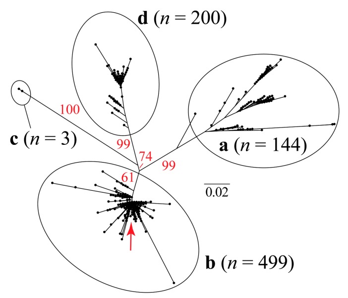
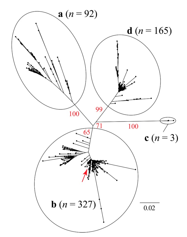
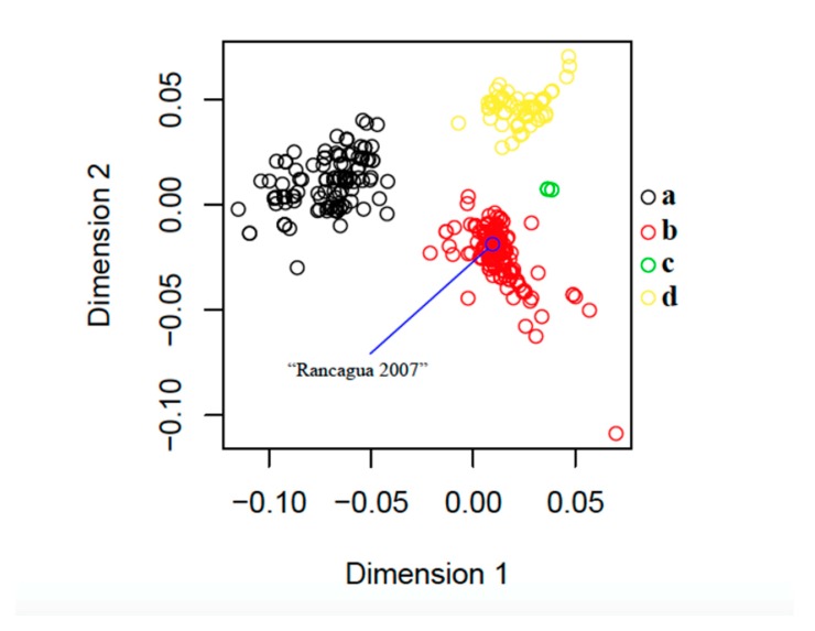
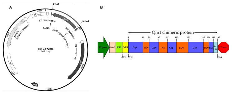
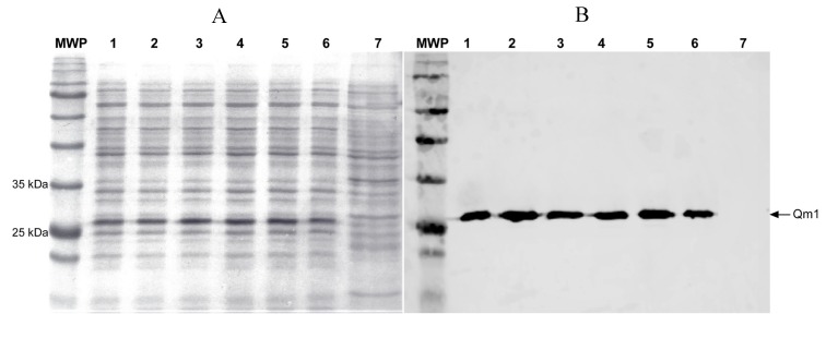
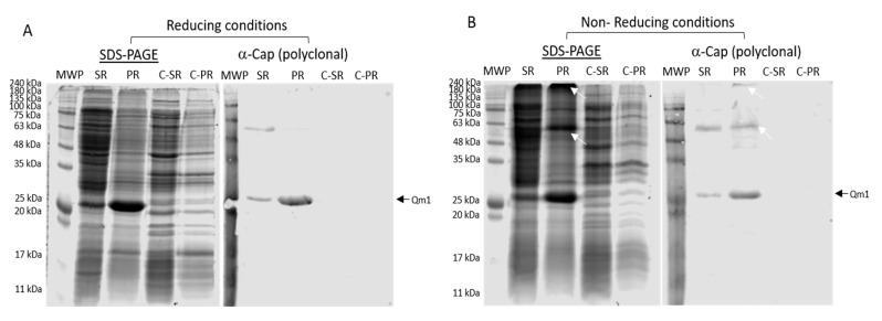
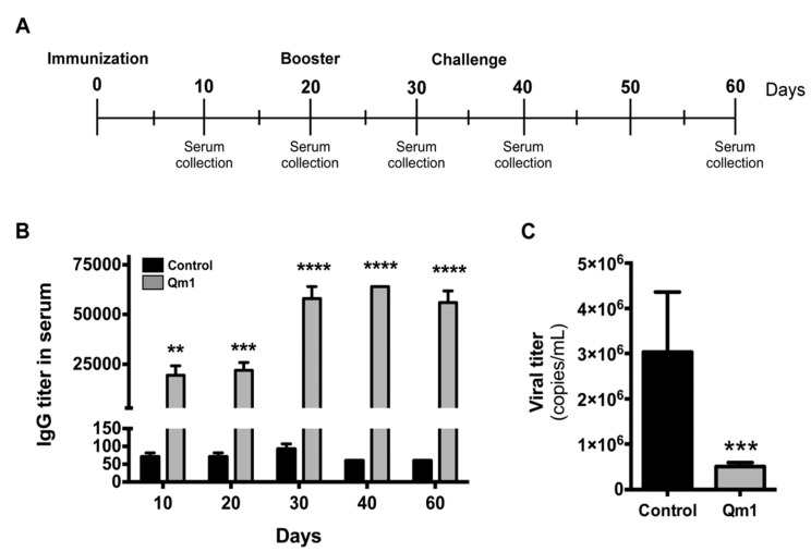
Recombinant vaccines have low-cost manufacturing, regulatory requirements, and reduced side effects compared to attenuated or inactivated vaccines. In the porcine industry, post-weaning multisystemic disease syndrome generates economic losses, characterized by progressive weight loss and weakness in piglets, and it is caused by porcine circovirus type 2 (PCV2). We designed a chimeric antigen (Qm1) to assemble the main exposed epitopes of the Cap-PCV2 protein on the capsid protein of the tobacco necrosis virus (TNV). This design was based on the Cap-N-terminal of an isolated PCV2 virus obtained in Chile. The virus was characterized, and the sequence was clustered within the PCV2 genotype b clade. This chimeric protein was expressed as inclusion bodies in both monomeric and multimeric forms, suggesting a high-molecular-weight aggregate formation. Pigs immunized with Qm1 elicited a strong and specific antibody response, which reduced the viral loads after the PCV2 challenge. In conclusion, the implemented design allowed for the generation of an effective vaccine candidate. Our proposal could be used to express the domains or fragments of antigenic proteins, whose structural complexity does not allow for low-cost production in Escherichia coli. Hence, other antigen domains could be integrated into the TNV backbone for suitable antigenicity and immunogenicity. This work represents new biotechnological strategies, with a reduction in the costs associated with vaccine development.

Keywords: PCV2 virus; biotechnology strategies; recombinant antigens production; vaccines.


Conflict of interest statement

The authors declare no conflicts of interest.

Figures

Figure 1
Figure 1
Unrooted maximum likelihood tree based on 705 bp of the porcine circovirus type 2 (PCV2) capsid gene. The red arrow shows the approximate phylogenetic position of the sequence that was recovered in Rancangua in 2007. The genotype classification (a, b, c, and d) follows Franzo et al. (2015) [37] (Supplementary Material, Table S1). In red, the bootstrap values based on 1000 replicates are indicated for the main internal branches.
Figure 2
Figure 2
Unrooted maximum likelihood tree based on 1650 bp of the PCV2 capsid + concatenated replicase genes. The red arrow shows approximately the phylogenetic position of the sequence that was recovered in Rancangua in 2007. The genotype classification (a, b, c, and d) follows Franzo et al. (2015) [37] (Supplementary Material, Table S1). In red, the bootstrap values based on 1000 replicates are indicated for the main internal branches.
Figure 3
Figure 3
Classical multidimensional scaling (CMDS) based on 846 protein sequences of the PCV2 capsid gene (235 amino acid residues). The blue circle indicates the exact position of the sequence that was recovered in Rancagua in 2007. Following Franzo et al. (2015) [37], genotypes a (n = 144), b (n = 499), c (n = 3), and d (n = 200) are shown in black, red, green, and yellow, respectively (Supplementary Material, Table S1).
Figure 4
Figure 4
The sequence and structure comparison of the chimera 1 (Qm1) and PCV2 capsid protein (Cap). (A) The EMBOSS Needle alignment of the Cap (PDB: 3r0r) and Qm1 protein sequences. The sequences have 59.4% of identity and 67.5% of similarity according to the analysis. (B) The structural superposition the crystal structures of PCV2 (pink) and Qm1 (yellow).
Figure 5
Figure 5
pET22-Qm1 expression plasmid and the genetic engineering design of the chimeric Qm1. (A) The Qm1 gene synthesized by GeneScript and inserted between the XhoI-NdeI restriction sites in pET-22b to obtain the pET22-Qm1 final expression vector. (B) The genetic engineering design of the chimeric Qm1 protein coded into a bacterial expression cassette. The expression cassette into pET-22b+ shows the T7 promoter in green, and the T7 terminator in red. After the promoter, the genetic elements are the lac operator in pink and the ribosome binding site (RBS) in light green. The pel B leader sequence in yellow is fused to the chimeric protein (Qm1) with the tobacco necrosis virus (TNV) backbone in orange that possess Cap epitopes in blue.
Figure 6
Figure 6
The expression analysis of Chimera 1 (Qm1) in Escherichia coli (SHuffle® T7 Express) under denaturant conditions. SDS-PAGE (A) and Western blot assay (B) using monoclonal anti-histidine antibodies (Clontech). MWP: molecular weight pattern, 1–6: six transformant colonies. 7: untransformed SHuffle® T7 Express E. coli strain used as a negative control. All samples were processed after 6 h of induction.
Figure 7
Figure 7
The identification of Qm1 expressed in the SHuffle® T7 Express E. coli strain. (A) SDS-PAGE Coomassie blue staining in the reducing condition (left) and Western blot (right) using a polyclonal anti-Cap antibody, previously released against proteins of the SHuffle® T7 Express strain of E. coli. (B) The same as (A), but SDS-PAGE under non-reducing conditions. MWP: molecular weight pattern; SR: supernatants of cell rupture; PR: pellet of cell rupture of the Qm1 clone 1. C-SR and C-PR correspond to the negative control sample of the SHuffle® T7 Express. The white arrows indicate protein aggregates detected by the antibody. A volume of 20 µL per sample was used in each case in both conditions.
Figure 8
Figure 8
SDS-PAGE of the Qm1 expression in fermentation. SDS-PAGE with Coomassie blue staining under reducing conditions after 6 h of induction. MWP: molecular weight pattern; SRF1, SRF2, and SRF3: rupture supernatants from each of the three fermentations of the Qm1 with SHuffle® T7 Express clone; PRF1, PRF2, and PRF3: the pellet of rupture from each of the three fermentations of Qm1 SHuffle® T7 Express clone; C-SR and C-PR: the rupture supernatants and pellet of rupture from the fermentation negative control sample. PRC +: the previous sample obtained positive detected by Western blot with an anti-his antibody. We used 20 µL per sample.
Figure 9
Figure 9
Analysis of the antibody and antiviral response to PCV2 in pigs. (A) The immunization and viral challenge schedule. (B) IgG titer in serum measured in pigs vaccinated with Qm1 (grey) or the vehicle PBS (black) by indirect ELISA at the indicated days. (C) The pig serum was collected at day 60 and the viral titer was quantified in challenged pigs by qRT-PCR using SensiMixTM SYBR Hi-ROX kit (Bioline). The values are the mean ± standard error mean (SEM). n = 6, two-way ANOVA and the Sidak test for IgG titer and the Student’s t-test for viral titer ** p < 0.01; *** p < 0.001; **** p < 0.0001 vs. the control group.

Similar articles

Cited by

References

    1. Oliveira C., Domingues L. Guidelines to reach high-quality purified recombinant proteins. Appl. Microbiol. Biotechnol. 2018;102:81–92. doi: 10.1007/s00253-017-8623-8. - DOI - PubMed
    1. Jozala A.F., Geraldes D.C., Tundisi L.L., Feitosa V.A., Breyer C.A., Cardoso S.L., Mazzola P.G., Oliveira-Nascimento L., Rangel-Yagui C.O., Magalhaes P.O., et al. Biopharmaceuticals from microorganisms: From production to purification. Braz. J. Microbiol. 2016;47(Suppl. 1):51–63. doi: 10.1016/j.bjm.2016.10.007. - DOI - PMC - PubMed
    1. Owczarek B., Gerszberg A., Hnatuszko-Konka K. A brief reminder of systems of production and chromatography-based recovery of recombinant protein biopharmaceuticals. Biomed. Res. Int. 2019;2019:4216060. doi: 10.1155/2019/4216060. - DOI - PMC - PubMed
    1. Sanchez-Garcia L., Martin L., Mangues R., Ferrer-Miralles N., Vazquez E., Villaverde A. Recombinant pharmaceuticals from microbial cells: A 2015 update. Microb. Cell Fact. 2016;15:33. doi: 10.1186/s12934-016-0437-3. - DOI - PMC - PubMed
    1. Karch C.P., Burkhard P. Vaccine technologies: From whole organisms to rationally designed protein assemblies. Biochem. Pharm. 2016;120:1–14. doi: 10.1016/j.bcp.2016.05.001. - DOI - PMC - PubMed

Publication types

MeSH terms

LinkOut - more resources