. 2020 Mar 31;10(4):530.
doi: 10.3390/biom10040530.

Antiproliferative and Antimigration Activities of Fluoro-Neplanocin A via Inhibition of Histone H3 Methylation in Triple-Negative Breast Cancer

Affiliations

Antiproliferative and Antimigration Activities of Fluoro-Neplanocin A via Inhibition of Histone H3 Methylation in Triple-Negative Breast Cancer

Woong Sub Byun et al. Biomolecules. .

Abstract

Triple-negative breast cancer (TNBC) is among the most aggressive and potentially metastatic malignancies. Most affected patients have poor clinical outcomes due to the lack of specific molecular targets on tumor cells. The upregulated expression of disruptor of telomeric silencing 1-like (DOT1L), a histone methyltransferase specific for the histone H3 lysine 79 residue (H3K79), is strongly correlated with TNBC cell aggressiveness. Therefore, DOT1L is considered a potential molecular target in TNBC. Fluoro-neplanocin A (F-NepA), an inhibitor of S-adenosylhomocysteine hydrolase, exhibited potent antiproliferative activity against various types of cancer cells, including breast cancers. However, the molecular mechanism underlying the anticancer activity of F-NepA in TNBC cells remains to be elucidated. We determined that F-NepA exhibited a higher growth-inhibitory activity against TNBC cells relative to non-TNBC breast cancer and normal breast epithelial cells. Moreover, F-NepA effectively downregulated the level of H3K79me2 in MDA-MB-231 TNBC cells by inhibiting DOT1L activity. F-NepA also significantly inhibited TNBC cell migration and invasion. These activities of F-NepA might be associated with the upregulation of E-cadherin and downregulation of N-cadherin and Vimentin in TNBC cells. Taken together, these data highlight F-NepA as a strong potential candidate for the targeted treatment of high-DOT1L-expressing TNBC.

Keywords: DOT1L; H3K79me2; fluoro-neplanocin A; histone H3 lysine methylation; metastasis; triple-negative breast cancer.


Conflict of interest statement

The authors declare no conflict of interest.

Figures

Figure 1
Figure 1
DOT1L expression levels in BCs and correlation between DOT1L expression and relapse-free survival in patients with TNBC. (A) Structures of NepA analogs: NepA, 1; F-NepA, 2; N6-methyl-F-NepA, 3. (B) DOT1L, H3K79me2, and E-cadherin levels in various BC cell lines were detected by Western blotting. β-Actin and Histone H3 were used as an internal control. (C) The Kaplan–Meier survival curve represents the relapse-free survival durations of patients with TNBC according to the DOT1L (Affy ID: 226201_at) expression level.
Figure 2
Figure 2
Suppressive effects of NepA analogs on H3K79me2 via the inhibition of DOT1L. (A) MDA-MB-231 and HCC1937 cells were treated with NepA analogs (200 nM) for 48 h, and H3K79me2 levels were determined by Western blotting. Histone H3 was used as an internal control. The relative intensities of the indicated protein levels were analyzed semi-quantitatively using NIH ImageJ software. (B) DOT1L (500 ng/well) enzyme activity levels were analyzed after incubation with NepA analogs (0.5–2 μM) for 2 h. All data are expressed as mean values ± SD (n = 3) and are representative of three separate experiments. **p < 0.01 indicates significant differences relative to the vehicle-treated control group.
Figure 3
Figure 3
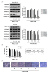
Effects of F-NepA on the methylation of various H3 lysine residues and viability in human TNBC cells. (A) MDA-MB-231 and HCC1937 cells were treated with the indicated concentrations of F-NepA for 48 h, and the methylation levels of various H3 lysine residues were determined by Western blotting. Histone H3 was used as an internal control. The relative intensities of the indicated proteins were analyzed semi-quantitatively using NIH ImageJ software. (B) MDA-MB-231 cells were incubated with the indicated concentrations of F-NepA for 24–72 h, after which cell viability was measured using an MTT assay. Data are expressed as means ± SD from three independent experiments. The IC50 values were calculated via a nonlinear regression analysis using TableCurve 2D v5.01 software. (C) Morphological changes in MDA-MB-231 cells induced by F-NepA (0–2 μM) were viewed under an inverted phase-contrast microscope (100× magnification). All data are expressed as mean values ± SD (n = 3) and are representative of three separate experiments. *p < 0.05, **p < 0.01, and ***p < 0.001 indicate significant differences relative to the vehicle-treated control group.
Figure 4
Figure 4
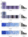
Regulatory effects of F-NepA on the migration and invasion potential of human TNBC cells. (A) Monolayers of MDA-MB-231 and HCC1937 cells were scratched mechanically and treated with F-NepA for 24 h. Representative images of wound closure obtained under a light microscope are shown (left). The areas of the wounds were quantified using ImageJ (right). (B) MDA-MB-231 and HCC1937 cells were pretreated with F-NepA at the indicated concentrations for 24 h, reseeded into the upper chambers of Transwell inserts, and incubated for 24 h. The cells that invaded the lower chambers were fixed, stained, imaged (left), and counted (right). *p < 0.05 and **p < 0.01 indicate significant differences relative to the vehicle-treated control group.
Figure 5
Figure 5
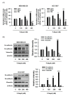
F-NepA regulates EMT gene expression in human TNBC cells. (A) MDA-MB-231 and HCC1937 cells were treated with F-NepA at the indicated concentrations for 24 h, and the mRNA levels of CDH1, CDH2, and VIM were determined using real-time PCR. (B) The protein levels of E-cadherin, N-cadherin, and Vimentin in MDA-MB-231 and HCC1937 cells treated with F-NepA for 48 h were determined by Western blotting. β-Actin was used as an internal control. The relative intensities of the indicated proteins were analyzed semi-quantitatively using NIH ImageJ software. The data are expressed as the mean values ± SD (n = 3) and are representative of three separate experiments. *p < 0.05, **p < 0.01, and ***p < 0.001 indicate significant differences relative to the vehicle-treated control group.
Figure 6
Figure 6
F-NepA reverses TGF-β induced EMT biomarker expressions. (A) MCF10A cells were treated with TGF-β1 (5 ng/mL) for indicated time periods, and the protein expression of E-cadherin, N-cadherin, and Vimentin was analyzed by Western blotting. β-Actin was used as an internal control. (B) MCF10A cells were treated in the presence or absence of TGF-β1 (5 ng/mL) for 8 days, and then fresh media without TGF-β1 were replaced and treated with indicated concentrations of F-NepA for an additional 2 days. The expression of E-cadherin was analyzed by Western blotting. β-Actin was used as an internal control. (C) MDA-MB-231 and HCC1937 cells were treated in the presence or absence of TGF-β1 (5 ng/mL) for 8 days, and then fresh media were replaced without TGF-β1 and treated with indicated concentrations of F-NepA for an additional 2 days. The expression of E-cadherin, N-cadherin, and Vimentin was analyzed by Western blotting. β-Actin was used as an internal control.

Similar articles

Cited by

References

    1. Siegel R.L., Miller K.D., Jemal A. Cancer statistics, 2019. CA Cancer J. Clin. 2019;69:7–34. doi: 10.3322/caac.21551. - DOI - PubMed
    1. Byun W.S., Kim W.K., Han H.J., Chung H.-J., Jang K., Kim H.S., Kim S., Kim D., Bae E.S., Park S., et al. Targeting histone methyltransferase DOT1L by a novel psammaplin A analog inhibits growth and metastasis of triple-negative breast cancer. Mol. Ther. Oncolytics. 2019;15:140–152. doi: 10.1016/j.omto.2019.09.005. - DOI - PMC - PubMed
    1. Colleoni M., Sun Z., Price K.N., Karlsson P., Forbes J.F., Thürlimann B., Gianni L., Castiglione M., Gelber R.D., Coates A.S., et al. Annual hazard rates of recurrence for breast cancer during 24 years of follow-up: Results from the international breast cancer study group trials I to V. J. Clin. Oncol. 2016;34:927–935. doi: 10.1200/JCO.2015.62.3504. - DOI - PMC - PubMed
    1. Zhu J., Wang F., Shi L., Cai H., Zheng Y., Zheng W., Bao P., Shu X.O. Accelerated aging in breast cancer survivors and its association with mortality and cancer recurrence. Breast Cancer Res. Treat. 2020;180:449–459. doi: 10.1007/s10549-020-05541-5. - DOI - PMC - PubMed
    1. Itani N., Grogan N., Mott S., Phadke S. Metastatic presentations of previously treated early-stage breast cancer patients and association with survival. Clin. Breast Cancer. 2019 doi: 10.1016/j.clbc.2019.11.004. - DOI - PubMed

Publication types

MeSH terms