

Systemic Blood Immune Cell Populations as Biomarkers for the Outcome of Immune Checkpoint Inhibitor Therapies
- PMID: 32244396
- PMCID: PMC7177687
- DOI: 10.3390/ijms21072411
Systemic Blood Immune Cell Populations as Biomarkers for the Outcome of Immune Checkpoint Inhibitor Therapies
Abstract
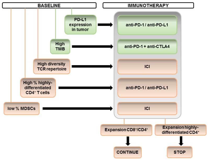
The development of cancer immunotherapy in the last decade has followed a vertiginous rhythm. Nowadays, immune checkpoint inhibitors (ICI) which include anti-CTLA4, anti-PD-1 and anti-PD-L1 antibodies are in clinical use for the treatment of numerous cancers. However, approximately only a third of the patients benefit from ICI therapies. Many efforts have been made for the identification of biomarkers allowing patient stratification into potential responders and progressors before the start of ICI therapies or for monitoring responses during treatment. While much attention is centered on biomarkers from the tumor microenvironment, in many cases biopsies are not available. The identification of systemic immune cell subsets that correlate with responses could provide promising biomarkers. Some of them have been reported to influence the response to ICI therapies, such as proliferation and activation status of CD8 and CD4 T cells, the expression of immune checkpoints in peripheral blood cells and the relative numbers of immunosuppressive cells such as regulatory T cells and myeloid-derived suppressor cells. In addition, the profile of soluble factors in plasma samples could be associated to response or tumor progression. Here we will review the cellular subsets associated to response or progression in different studies and discuss their accuracy in diagnosis.
Keywords: CD4+; CD8+; MDSCs; immune checkpoint inhibitors; immunotherapy; systemic blood subsets.
Conflict of interest statement
The authors declare no conflict of interest.
Figures
References
-
- Nomi T., Sho M., Akahori T., Hamada K., Kubo A., Kanehiro H., Nakamura S., Enomoto K., Yagita H., Azuma M., et al. Clinical Significance and Therapeutic Potential of the Programmed Death-1 Ligand/Programmed Death-1 Pathway in Human Pancreatic Cancer. Clin. Cancer Res. 2007;13:2151–2157. doi: 10.1158/1078-0432.CCR-06-2746. - DOI - PubMed
-
- Jiang J., Zhu Y., Jiang J., Zhao J., Zhang X., Xu N. Immunohistochemical localization of programmed death-1 ligand-1 (PD-L1) in gastric carcinoma and its clinical significance. Acta Histochem. 2006;108:19–24. - PubMed
-
- Hamanishi J., Mandai M., Iwasaki M., Okazaki T., Tanaka Y., Yamaguchi K., Higuchi T., Yagi H., Takakura K., Minato N., et al. Programmed cell death 1 ligand 1 and tumor-infiltrating CD8+ T lymphocytes are prognostic factors of human ovarian cancer. Proc. Natl. Acad. Sci. USA. 2007;104:3360–3365. doi: 10.1073/pnas.0611533104. - DOI - PMC - PubMed
-
- Gato-Cañas M., Zuazo M., Arasanz H., Ibáñez-Vea M., Lorenzo L., Fernández-Hinojal G., Vera R., Smerdou C., Martisova E., Arozarena I., et al. PDL1 Signals through Conserved Sequence Motifs to Overcome Interferon-Mediated Cytotoxicity. Cell Rep. 2017;20:1818–1829. doi: 10.1016/j.celrep.2017.07.075. - DOI - PubMed
Publication types
MeSH terms
Substances
Grants and funding
LinkOut - more resources
Full Text Sources
Research Materials
