. 2020 Mar 31;21(7):2418.
doi: 10.3390/ijms21072418.

Chronic Regulation of miR-124-3p in the Perilesional Cortex after Experimental and Human TBI

Affiliations

Chronic Regulation of miR-124-3p in the Perilesional Cortex after Experimental and Human TBI

Niina Vuokila et al. Int J Mol Sci. .

Abstract

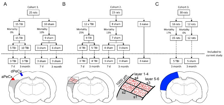
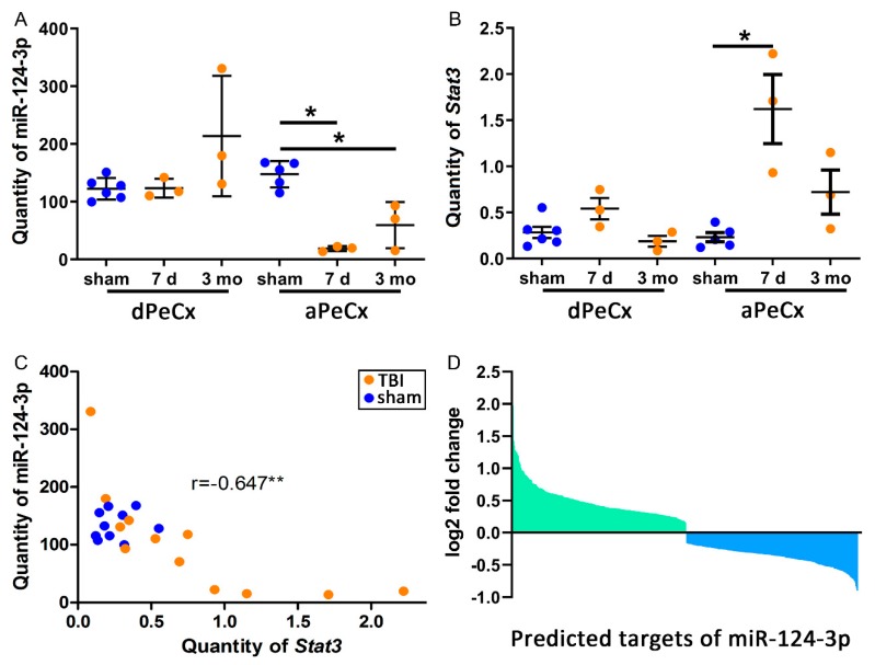
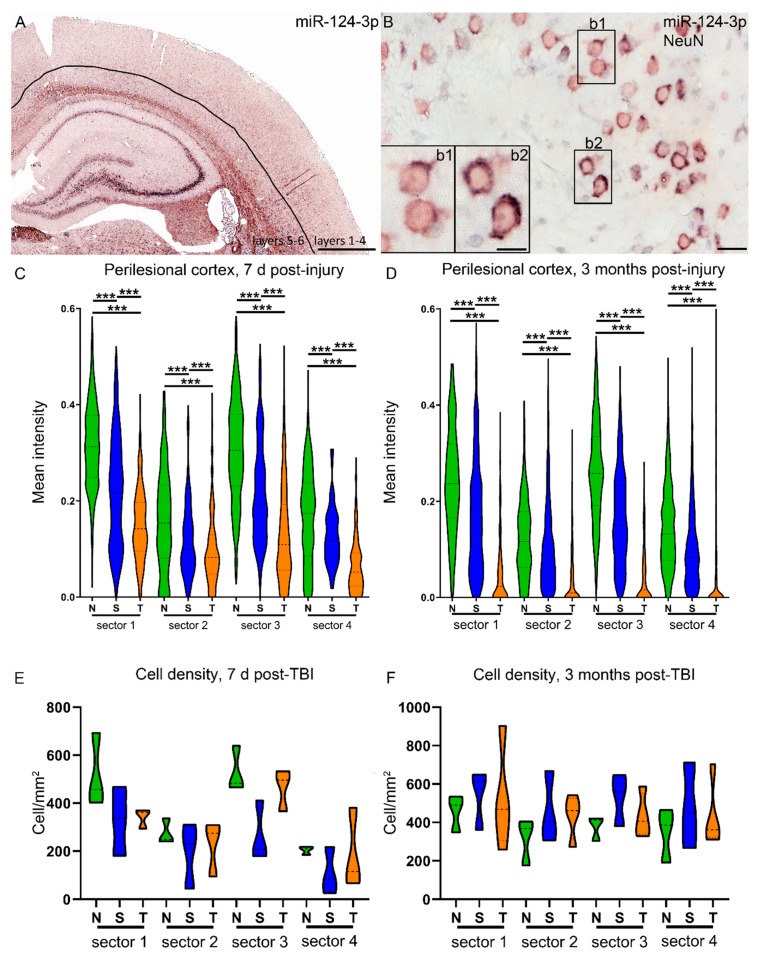
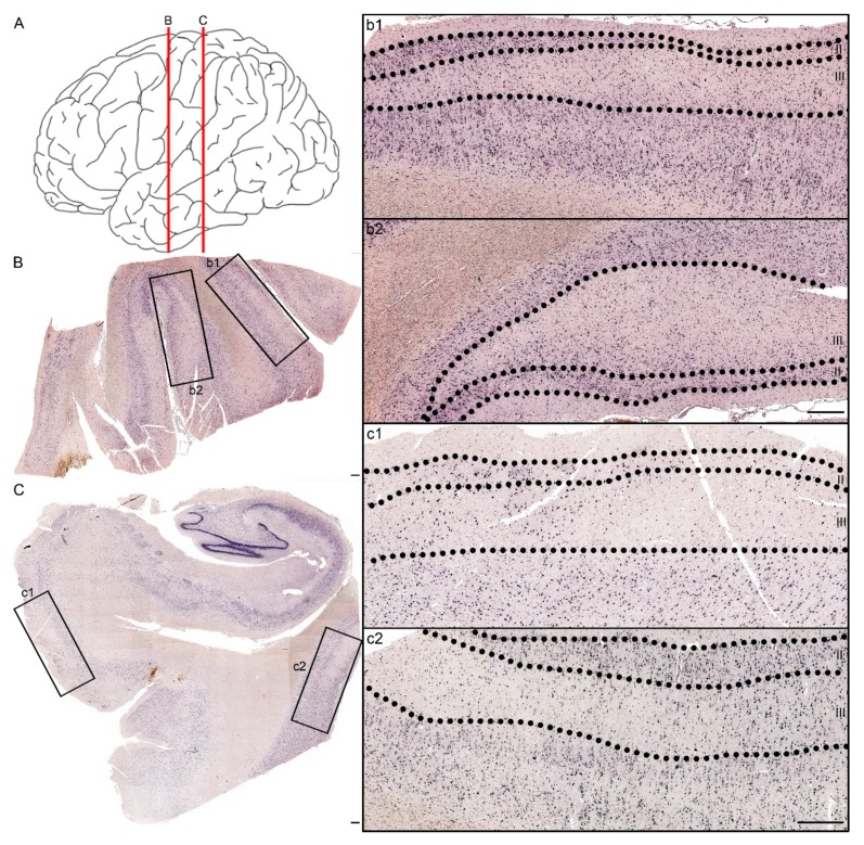
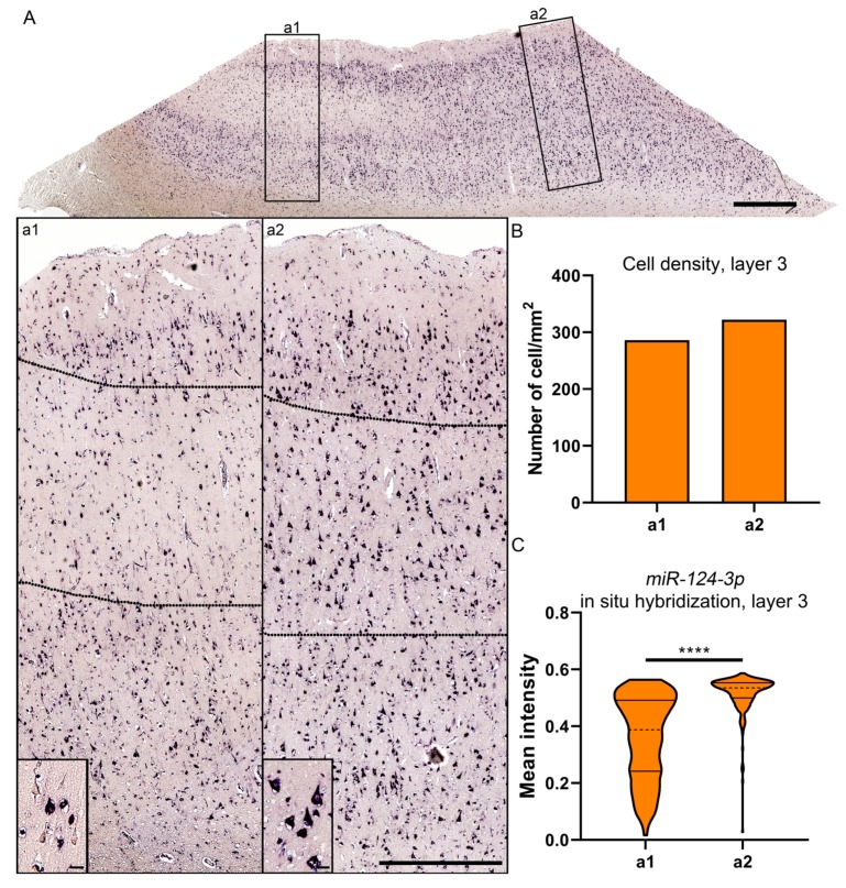
Traumatic brain injury (TBI) dysregulates microRNAs, which are the master regulators of gene expression. Here we investigated the changes in a brain-enriched miR-124-3p, which is known to associate with major post-injury pathologies, such as neuroinflammation. RT-qPCR of the rat tissue sampled at 7 d and 3 months in the perilesional cortex adjacent to the necrotic lesion core (aPeCx) revealed downregulation of miR-124-3p at 7 d (fold-change (FC) 0.13, p < 0.05 compared with control) and 3 months (FC 0.40, p < 0.05) post-TBI. In situ hybridization confirmed the downregulation of miR-124-3p at 7 d and 3 months post-TBI in the aPeCx (both p < 0.01). RT-qPCR confirmed the upregulation of the miR-124-3p target Stat3 in the aPeCx at 7 d post-TBI (7-fold, p < 0.05). mRNA-Seq revealed 312 downregulated and 311 upregulated miR-124 targets (p < 0.05). To investigate whether experimental findings translated to humans, we performed in situ hybridization of miR-124-3p in temporal lobe autopsy samples of TBI patients. Our data revealed downregulation of miR-124-3p in individual neurons of cortical layer III. These findings indicate a persistent downregulation of miR-124-3p in the perilesional cortex that might contribute to post-injury neurodegeneration and inflammation.

Keywords: bioinformatics; biomarker; epileptogenesis; miR-124-3p; microRNA; traumatic brain injury.


Conflict of interest statement

The authors declare no conflict of interest.

Figures

Figure 1
Figure 1
Study design and tissue sampling. (A) Cohort 1: a total of 25 animals underwent either traumatic brain injury (TBI; n = 15) or sham (n = 10) operation. Of the surviving 24 animals, 8 (5 TBI, 3 sham) were killed at 7 d post-TBI, and the rest (10 TBI, 6 sham) at 3 months post-TBI. Six TBI and 6 sham animals were randomly chosen for RT-qPCR analysis. Two samples were dissected for analysis: the perilesional cortex adjacent to the lesion core (aPeCx, red) and the somatosensory cortex distal to the lesion (dPeCx, blue). (B) Cohort 2: a total of 23 animals underwent either TBI (n = 12) or sham (n = 8) operation. From the surviving 16 animals 8 (4 TBI, 4 sham) were killed at 7 d post-TBI, and the remaining animals (5 TBI, 3 sham) at 3 months post-TBI. Altogether 7 TBI and 6 sham rats were used for miR-124-3p in situ hybridization. The remaining 3 animals did not undergo any surgical procedures (naïve controls). Analysis of miR-124-3p expression in in situ-processed sections was conducted in individual cells (red dots) within a 1000-µm thick sector adjacent to the lesion core. A corresponding area was analyzed in the control samples. (C) Cohort 3: a total of 30 animals underwent either TBI (n = 18) or sham (n = 12) operation. At 3 months post-TBI, animals were killed and the brain was cut into 2-mm thick coronal slices (bregma levels from −2.2 to −6.2) and tissue from the perilesional cortex (blue area in A) was sampled for RNA-sequencing.
Figure 2
Figure 2
TBI downregulates miR-124-3p and upregulates Stat3 in the perilesional cortex. (A) A RT-qPCR analysis of laser-capture microdissected cortex indicated a downregulation of miR-124-3p in the perilesional cortex adjacent to the lesion core (aPeCx, FC 0.13 at 7 d, FC 0.40 at 3 months; both p < 0.05) but not in the more distal perilesional cortex (dPeCx, approximately 600 µm more dorsal) at both 7 d and 3 months post-TBI. (B) Expression of Stat3, one of the targets of miR-124-3p, was upregulated in aPeCx at 7 d post-TBI (p < 0.05, FC = 6.97) and tended to be upregulated at 3 months post-TBI (p > 0.05, FC = 3.11). (C) A correlation analysis revealed that the higher the miR-124-3p levels, the lower the Stat3 levels (r = −0.647, p < 0.01). (D) RNA sequencing revealed 312 upregulated and 311 downregulated targets of miR-124-3p. The gene set enrichment analysis (GSEA) of these targets indicated that only the downregulated targets were enriched in the dataset (ES = −0.33, FDR < 0.01). dPeCx, distal part of cortex; d, day; ES, enrichment score; FC, fold change; FDR, false discovery rate; LCM, Laser capture microdissection; mo, month; aPeCx, perilesional cortex; RT-qPCR, quantitative reverse transcriptase polymerase chain reaction. Statistical significance: *p < 0.05, **p < 0.01 (Mann–Whitney U).
Figure 3
Figure 3
Perilesional expression of miR-124-3p is downregulated by lateral fluid-percussion injury as well as by craniectomy. (A) In situ hybridization was used to detect the distribution and intensity of cellular expression of miR-124-3p in different layers of the cortex. (B) Double-labeling with NeuN revealed that the expression level of miR-124-3p varied between the neurons in the perilesional cortex (inserts b1, b2). Our analysis focused on a 1-mm wide area of the dorsal perilesional cortex, which was further divided into four sectors (see Figure 1). The intensity of in situ hybridization signal was measured in the individual neurons within each sector (mean number of cells counted: 7 d, naïve 123; sham 89; TBI 94; 3 months, naïve 125; sham 151; TBI 63). (C,D) Naïve animals (N) showed a clear expression pattern at both 7 d and 3 months post-TBI, with a higher level of miR-124-3p expression in the deeper (s1, s3) than superficial layers (s2, s4). The proximal and distal sectors had similar expression levels (s1 vs. s3; s2 vs. s4). In sham-operated experimental controls (S) with a craniotomy, the overall pattern of miR-124-3p expression was preserved. At both time-points and within each of the four sectors, however, the miR-124-3p levels were downregulated compared with those in naïve animals (p < 0.001). In rats with TBI (T), the miR-124-3p expression levels were even lower than those in sham-operated animals (p < 0.001) in all sectors and at both time-points. (E,F) The cell density in the sectors did not differ between time-points or groups. N, naïve; S, sham; s1–s4, sector as described in Figure 1; T/TBI, traumatic brain injury. Statistical significance: *** p < 0.001 (Mann–Whitney U). Scale bars: A 1000 µm, B 20 µm, insert in B 10 µm.
Figure 4
Figure 4
Patchy loss of miR-124-3p expression in the temporal cortex of a patient with severe TBI 5 days earlier. (A) Red lines indicate the coronal levels of sections illustrated in panels B,C. (B) miR-124-3p in situ hybridization from a section of the lateral temporal cortex. Note the neuronal loss in higher magnification images, particularly in layer III (b1, b2 correspond to boxed areas in panel B). (C) Hippocampus, entorhinal cortex (higher magnification in panel c1), and more lateral temporal cortex (c2). Note the preservation of the miR-124-3p hybridization signal in layer II and its loss in layer III. Scale bar: 500 µm.
Figure 5
Figure 5
Expression of miR-124-3p in layer III cells of the lesioned cortex in a patient with severe TBI 5 days earlier. (A) Expression of miR-124-3p inside (a1) and outside (a2) the area with a visible loss of miR-124-3p. (B) Cellular density was comparable in the investigated areas. (C) Violin plots showing the greater variability in the distribution of the mean miR-124-3p signal intensity inside (a1) than outside (a2) the patchy neuronal loss region. The mean intensity of neuronal miR-124-3p in situ hybridization signal was lower inside than outside the lesion area (p < 0.01). Statistical significance: **** p < 0.001. Scale bars: A 1000 µm; a1 and a2 1000 µm; insert in a1 and a2 20 µm.

Similar articles

Cited by

References

    1. Feigin V.L., Theadom A., Barker-Collo S., Starkey N.J., McPherson K., Kahan M., Jones K. Incidence of traumatic brain injury in New Zealand: A population-based study. Lancet Neurol. 2013;12:53–64. doi: 10.1016/S1474-4422(12)70262-4. - DOI - PubMed
    1. McGraw J., Hiebert G.W., Steeves J.D. Modulating astrogliosis after neurotrauma. J. Neurosci. Res. 2001;63:109–115. doi: 10.1002/1097-4547(20010115)63:2<109::AID-JNR1002>3.0.CO;2-J. - DOI - PubMed
    1. Raghupathi R. Cell Death Mechanisms Following Traumatic Brain Injury. Brain Pathol. 2004;14:215–222. doi: 10.1111/j.1750-3639.2004.tb00056.x. - DOI - PMC - PubMed
    1. Wong V.S., Langley B. Epigenetic changes following traumatic brain injury and their implications for outcome, recovery and therapy. Neurosci. Lett. 2016;625:26–33. doi: 10.1016/j.neulet.2016.04.009. - DOI - PMC - PubMed
    1. Rola R., Mizumatsu S., Otsuka S., Morhardt D., Noblehaeusslein L., Fishman K., Fike J.R. Alterations in hippocampal neurogenesis following traumatic brain injury in mice. Exp. Neurol. 2006;202:189–199. doi: 10.1016/j.expneurol.2006.05.034. - DOI - PubMed