. 2020 Mar 31;21(7):2419.
doi: 10.3390/ijms21072419.

Targeting PKCθ Promotes Satellite Cell Self-Renewal

Affiliations

Targeting PKCθ Promotes Satellite Cell Self-Renewal

Anna Benedetti et al. Int J Mol Sci. .

Abstract

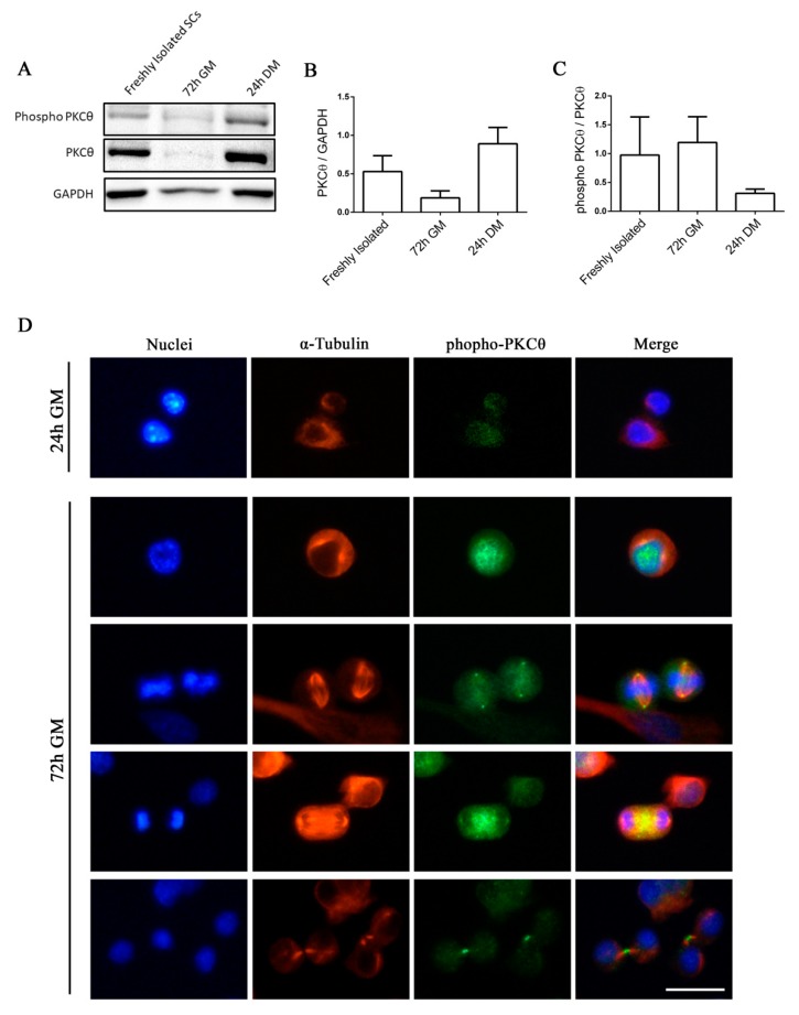
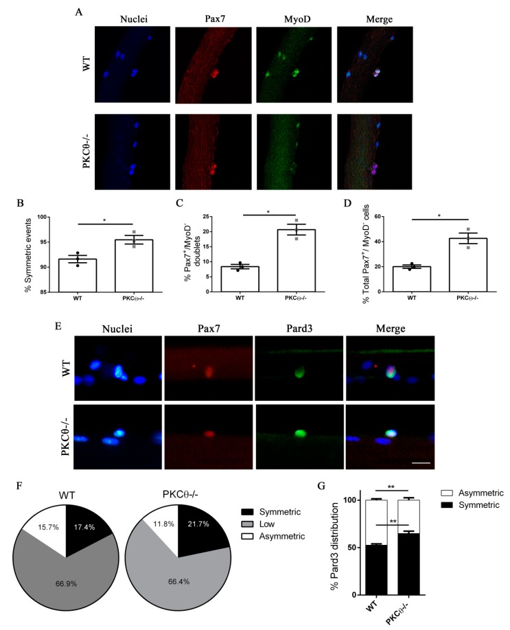
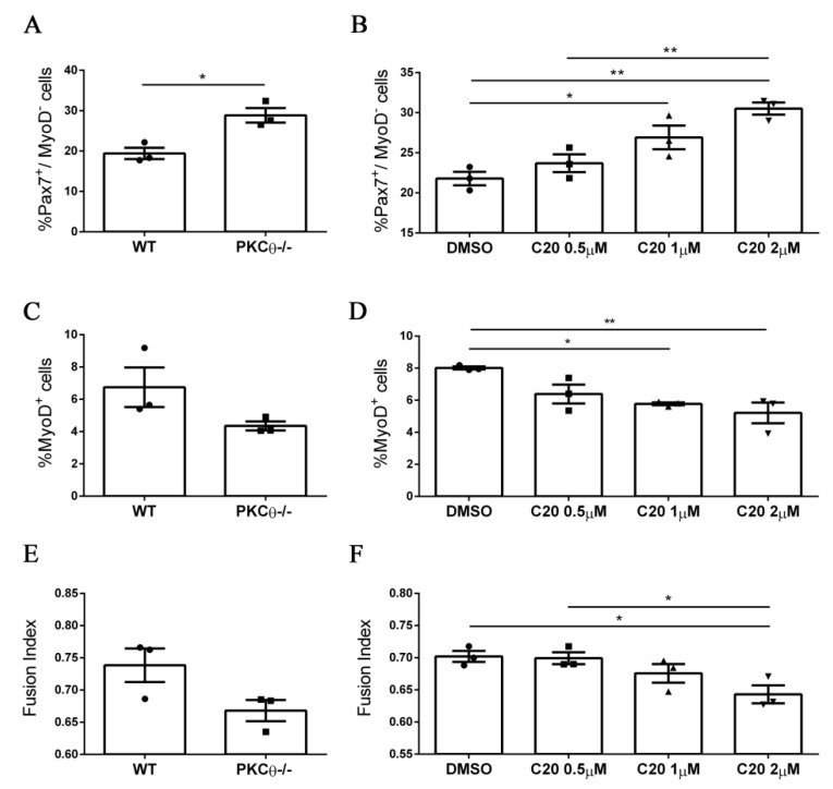
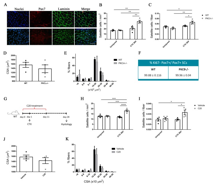
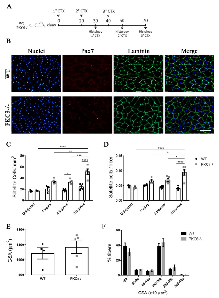
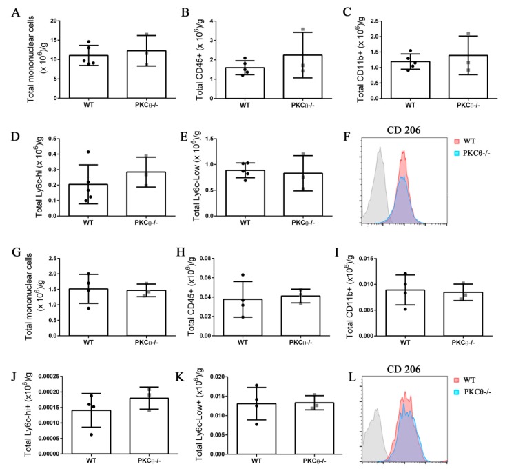
Skeletal muscle regeneration following injury depends on the ability of satellite cells (SCs) to proliferate, self-renew, and eventually differentiate. The factors that regulate the process of self-renewal are poorly understood. In this study we examined the role of PKCθ in SC self-renewal and differentiation. We show that PKCθ is expressed in SCs, and its active form is localized to the chromosomes, centrosomes, and midbody during mitosis. Lack of PKCθ promotes SC symmetric self-renewal division by regulating Pard3 polarity protein localization, without affecting the overall proliferation rate. Genetic ablation of PKCθ or its pharmacological inhibition in vivo did not affect SC number in healthy muscle. By contrast, after induction of muscle injury, lack or inhibition of PKCθ resulted in a significant expansion of the quiescent SC pool. Finally, we show that lack of PKCθ does not alter the inflammatory milieu after acute injury in muscle, suggesting that the enhanced self-renewal ability of SCs in PKCθ-/- mice is not due to an alteration in the inflammatory milieu. Together, these results suggest that PKCθ plays an important role in SC self-renewal by stimulating their expansion through symmetric division, and it may represent a promising target to manipulate satellite cell self-renewal in pathological conditions.

Keywords: Protein kinase C θ; muscle regeneration; satellite cells; self-renewal.


Conflict of interest statement

The authors declare no conflict of interest.

Figures

Figure 1
Figure 1
Phospho-PKCθ is localized to the nucleus, centrosomes, mitotic spindle and midbody in satellite cells during mitosis. (A): WB analysis showing PKCθ and phospho-PKCθ expression in activated SCs (freshly isolated), proliferating SCs (72h GM), differentiated myotubes (24h DM) (n= 3 individual experiments). (B): WB densitometric analysis of the level of PKCθ expression, normalized on the level of GAPDH protein. (C): WB Densitometric analysis of the phospho-PKCθ/PKCθ ratio. (D): Representative immunofluorescence images of satellite cells after 24h and 72h in culture, stained for α-Tubulin (red) and phospho-PKCθ (green). Nuclei were counterstained with Hoechst. Scale bar 20 µm. Error bars represent mean ± sem.
Figure 2
Figure 2
Knock out of Protein kinase C Theta (PKCθ) does not affect satellite cell (SC) proliferation. (A): Flow-cytometry analysis of CFSE stained WT and PKCθ-/- SCs, cultured for 72h in GM. (B): Percentage of proliferating SCs, identified by immunofluorescence asPax7+/Ki67+ cells on WT and PKCθ-/- GA muscle sections, 3 and 7 days after CTX injury, over the total Pax7+ cells. (C): Representative immunofluorescence images of WT and PKCθ-/- GA muscles, stained for Pax7 and Ki67, 3 days after the induction of CTX injury, scale bar: 100 µm.
Figure 3
Figure 3
Lack of PKCθ stimulates symmetric self-renewal by regulating Pard3 polarization. (A): Representative pictures of single myofibers isolated from EDL muscles of WT and PKCθ-/- mice, after 48h in culture. Myofibers were stained for Pax7 (red) and MyoD (green), nuclei were counterstained with Topro3. (B): Quantification of symmetric division events. (C): Quantification of Pax7+/MyoD cell doublets, and (D): quantification of total Pax7+/MyoD cells in WT and PKCθ-/- single myofibers. (E): Representative pictures of single myofibers isolated from EDL muscles of WT and PKCθ-/- mice, after 36h in culture. Myofibers were stained for Pax7 (red) and Pard3 (green), nuclei were counterstained with Topro3. (F): Percentage ofSCs showing symmetric, low or asymmetric Pard3 distribution in WT and PKCθ-/- myofibers. (G): Percentage of SCs showing symmetric and asymmetric Pard3 distribution (WT, n = 3 mice, PKCθ-/-, n = 3 mice, n > 20 myofibers analyzed per mouse). Error bars represent mean ± sem, * p < 0.05 calculated by Student’s t-test.
Figure 4
Figure 4
Pharmacological inhibition of PKCθ increases the fraction of reserve cell population in cultured primary myoblasts. (A): Percentage of Pax7+/MyoD SCs in WT and PKCθ-/- cultures, or in WT cultures treated with increasing concentration of C20 (B); cells were cultured for 4 days in GM followed by2 days in DM. (C): Percentage of total MyoD+ SCs in WT and PKCθ-/- cultures, or in WT cultures treated with C20, as in B (D). (E): Fusion index of WT and PKCθ-/- myotubes, or WT myotubes treated with C20 (F) as in A (n = 3 replicate dishes per group). Error bars represent mean ± sem, * p < 0.05, ** p < 0.01 calculated by one-way ANOVAwith adjustment for multiple comparison test.
Figure 5
Figure 5
PKCθ absence/inhibition increases the quiescent satellite cell pool after induction of acute injury. (A): Representative immunofluorescence pictures of WT and PKCθ-/- GA sections, 28 days after CTX injury. Sections were stained for Pax7 (red) and Laminin (green). Nuclei were counterstained with Hoechst. Scale bar: 100 µm. (B): Number of SCs per mm2 and (C): number of SCs per fiber in uninjured and 28 day-injured GA muscle, in WT and PKCθ-/- mice. (D): Mean CSA and (E): CSA distribution of muscle fibers in WT and PKCθ-/- GA sections, 28 days after injury. (F): Quantification of non-proliferating SCs 28 days after CTX injury, in WT and PKCθ-/- GA, identified by immunofluorescence co-staining for Pax7 and Ki67. (WT, n = 4 mice, PKCθ-/-, n = 4 mice). (G): experimental plan for in vivo C20 treatment in injured muscle. (H): Number of SCs per mm2 and (I): number of SCs per fiber in uninjured and 28 day-injured GA muscle, in WT mice treated with C20 or vehicle. (J): mean CSA and (K): CSA distribution of muscle fibers in WT mice treated with C20 or vehicle, 28 days after injury. (C20 treated WT, n = 4 mice, Vehicle treated WT n = 4 mice). Error bars represent mean ± sem, * p < 0.05, ** p < 0.01 *** p < 0.001, **** p < 0.0001 calculated by Two-way Anova with adjustment for multiple comparison test.
Figure 6
Figure 6
The number of quiescent satellite cells increases in PKCθ-/- mice after repeated injuries. (A): Experimental plan. (B): representative immunofluorescence pictures of WT and PKCθ-/- GA sections 30 days after 3 CTX injury, scale bar: 100 µm. Sections were stained for Pax7 (red) and Laminin (green). Nuclei were counterstained with Hoechst. (C): Quantification of the number of SCs/mm2 (D): and number of SCs per fiber in uninjured GA from WT and PKCθ-/- mice, or after 1, 2 and 3 injuries. (E): mean CSA of regenerated fibers 30 days after the third injury in WT and PKCθ-/- mice. (F): Frequency of myofiber CSA from GA muscles of WT and PKCθ-/- mice, 30 days after the third injury. Error bars represent mean ± sem, * p < 0.05, ** p < 0.01, *** p < 0.001, **** p < 0.0001 calculated by two-way ANOVA with adjustment for multiple comparison test.
Figure 7
Figure 7
Knock-out of PKCθ does not alter the inflammatory milieu after induction of acute injury. (A): Total number of mononuclear cells, (B): CD45+ cells, (C): CD11b+ cells, (D): Ly6c-hi+ cells, and (E): Ly6c-low+ cells, normalized on muscle mass 3 days after CTX injury in WT and PKCθ-/- GA. (F): histogram showing CD206 mean fluorescence in WT and PKCθ-/- GA 3d after CTX. (G): Total number of mononuclear cells, (H): CD45+ cells, (I): CD11b+ cells, (J): Ly6c-hi+ cells, and (K): Ly6c-low+ cells, normalized on muscle mass 10 days after CTX injury in WT and PKCθ-/- GA. (L): histogram showing CD206 mean fluorescence in WT and PKCθ-/- GA 10d after CTX. Error bars represent mean ± sem.

Similar articles

Cited by

References

    1. Mauro A. Satellite Cell of Skeletal Muscle Fibers. J. Cell Biol. 1961;9:493–495. doi: 10.1083/jcb.9.2.493. - DOI - PMC - PubMed
    1. Cheung T.H., Rando T.A. Molecular regulation of stem cell quiescence. Nat. Rev. Mol. Cell Biol. 2013;14:329–340. doi: 10.1038/nrm3591. - DOI - PMC - PubMed
    1. Kuang S., Kuroda K., Le Grand F., Rudnicki M.A. Asymmetric Self-Renewal and Commitment of Satellite Stem Cells in Muscle. Cell. 2007;129:999–1010. doi: 10.1016/j.cell.2007.03.044. - DOI - PMC - PubMed
    1. Troy A., Cadwallader A.B., Fedorov Y., Tyner K., Tanaka K.K., Olwin B.B. Coordination of Satellite Cell Activation and Self-Renewal by Par-Complex-Dependent Asymmetric Activation of p38α/β MAPK. Cell Stem Cell. 2012;11:541–553. doi: 10.1016/j.stem.2012.05.025. - DOI - PMC - PubMed
    1. Knoblich J.A. Asymmetric cell division: Recent developments and their implications for tumour biology. Nat. Rev. Mol. Cell Biol. 2010;11:849–860. doi: 10.1038/nrm3010. - DOI - PMC - PubMed

Substances

LinkOut - more resources