

Characterisation of Peste Des Petits Ruminants Disease in Pastoralist Flocks in Ngorongoro District of Northern Tanzania and Bluetongue Virus Co-Infection
- PMID: 32244509
- PMCID: PMC7232183
- DOI: 10.3390/v12040389
Characterisation of Peste Des Petits Ruminants Disease in Pastoralist Flocks in Ngorongoro District of Northern Tanzania and Bluetongue Virus Co-Infection
Abstract
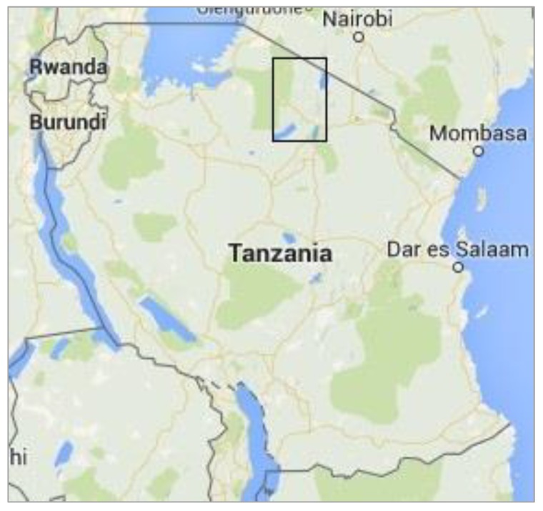
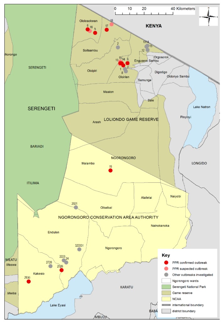
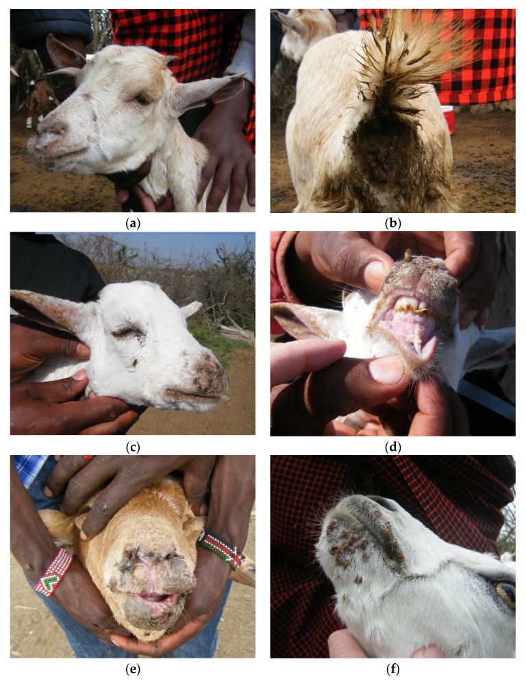
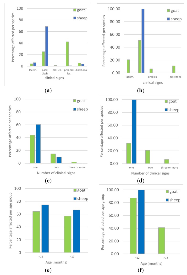
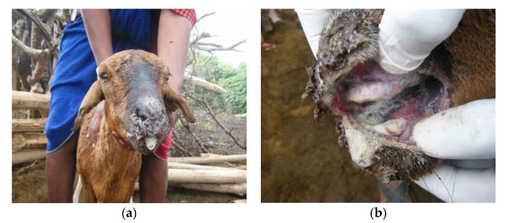
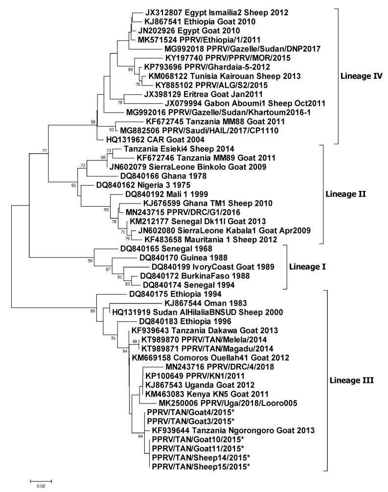
Peste des petits ruminants (PPR) disease was first confirmed in Tanzania in 2008 in sheep and goats in Ngorongoro District, northern Tanzania, and is now endemic in this area. This study aimed to characterise PPR disease in pastoralist small ruminant flocks in Ngorongoro District. During June 2015, 33 PPR-like disease reports were investigated in different parts of the district, using semi-structured interviews, clinical examinations, PPR virus rapid detection test (PPRV-RDT), and laboratory analysis. Ten flocks were confirmed as PPRV infected by PPRV-RDT and/or real-time reverse transcription-polymerase chain reaction (RT-qPCR), and two flocks were co-infected with bluetongue virus (BTV), confirmed by RT-qPCR. Phylogenetic analysis of six partial N gene sequences showed that the PPR viruses clustered with recent lineage III Tanzanian viruses, and grouped with Ugandan, Kenyan and Democratic Republic of Congo isolates. No PPR-like disease was reported in wildlife. There was considerable variation in clinical syndromes between flocks: some showed a full range of PPR signs, while others were predominantly respiratory, diarrhoea, or oro-nasal syndromes, which were associated with different local disease names (olodua-a term for rinderpest, olkipiei-lung disease, oloirobi-fever, enkorotik-diarrhoea). BTV co-infection was associated with severe oro-nasal lesions. This clinical variability makes the field diagnosis of PPR challenging, highlighting the importance of access to pen-side antigen tests and multiplex assays to support improved surveillance and targeting of control activities for PPR eradication.
Keywords: PPR; differential diagnosis; ethno-veterinary knowledge; goats; outbreak investigation; sheep; surveillance.
Conflict of interest statement
The authors declare no conflict of interest. The funders had no role in the design of the study; in the collection, analyses, or interpretation of data; in the writing of the manuscript, or in the decision to publish the results.
Figures

Similar articles
-
Concurrent infection of Bluetongue and Peste-des-petits-ruminants virus in small ruminants in Haryana State of India.Transbound Emerg Dis. 2018 Feb;65(1):235-239. doi: 10.1111/tbed.12610. Epub 2017 Jan 24. Transbound Emerg Dis. 2018. PMID: 28116836
-
Partial genetic characterization of peste des petits ruminants virus from goats in northern and eastern Tanzania.Transbound Emerg Dis. 2014 Aug;61 Suppl 1(Suppl 1):56-62. doi: 10.1111/tbed.12229. Transbound Emerg Dis. 2014. PMID: 25135464 Free PMC article.
-
Retrospective Characterization of Initial Peste des petits ruminants Outbreaks (2008-2012) in the Democratic Republic of the Congo.Viruses. 2021 Nov 26;13(12):2373. doi: 10.3390/v13122373. Viruses. 2021. PMID: 34960642 Free PMC article.
-
Review of Peste des Petits Ruminants Occurrence and Spread in Tanzania.Animals (Basel). 2021 Jun 7;11(6):1698. doi: 10.3390/ani11061698. Animals (Basel). 2021. PMID: 34200290 Free PMC article. Review.
-
Peste des petits ruminants in Africa: a review of currently available molecular epidemiological data, 2020.Arch Virol. 2020 Oct;165(10):2147-2163. doi: 10.1007/s00705-020-04732-1. Epub 2020 Jul 11. Arch Virol. 2020. PMID: 32653984 Free PMC article. Review.
Cited by
-
GCRV NS38 counteracts SVCV proliferation by intracellular antagonization during co-infection.Virol Sin. 2023 Feb;38(1):142-156. doi: 10.1016/j.virs.2022.12.003. Epub 2022 Dec 13. Virol Sin. 2023. PMID: 36526167 Free PMC article.
-
Unveiling of the Co-Infection of Peste des Petits Ruminants Virus and Caprine Enterovirus in Goat Herds with Severe Diarrhea in China.Viruses. 2024 Jun 19;16(6):986. doi: 10.3390/v16060986. Viruses. 2024. PMID: 38932277 Free PMC article.
-
Development and Evaluation of a Nested PCR for Improved Diagnosis and Genetic Analysis of Peste des Petits Ruminants Virus (PPRV) for Future Use in Nascent PPR Eradication Programme.Animals (Basel). 2021 Nov 5;11(11):3170. doi: 10.3390/ani11113170. Animals (Basel). 2021. PMID: 34827902 Free PMC article.
-
The evaluation of five serological assays in determining seroconversion to peste des petits ruminants virus in typical and atypical hosts.Sci Rep. 2023 Sep 8;13(1):14787. doi: 10.1038/s41598-023-41630-3. Sci Rep. 2023. PMID: 37684280 Free PMC article.
-
Peste Des Petits Ruminants in Atypical Hosts and Wildlife: Systematic Review and Meta-Analysis of the Prevalence between 2001 and 2021.Arch Razi Inst. 2021 Dec 30;76(6):1589-1606. doi: 10.22092/ari.2021.356900.1939. eCollection 2021 Dec. Arch Razi Inst. 2021. PMID: 35546985 Free PMC article.
References
Publication types
MeSH terms
Substances
Grants and funding
- BBS/E/I/00007034/BB_/Biotechnology and Biological Sciences Research Council/United Kingdom
- BBS/E/I/00007037/BB_/Biotechnology and Biological Sciences Research Council/United Kingdom
- BBS/E/I/00007036/BB_/Biotechnology and Biological Sciences Research Council/United Kingdom
- BB/L013657/1/BB_/Biotechnology and Biological Sciences Research Council/United Kingdom
- BB/L013592/1/BB_/Biotechnology and Biological Sciences Research Council/United Kingdom
LinkOut - more resources
Full Text Sources
