Review
. 2020 Mar 31;25(7):1605.
doi: 10.3390/molecules25071605.

Encapsulation for Cancer Therapy

Affiliations
Review

Encapsulation for Cancer Therapy

Xavier Montané et al. Molecules. .

Abstract

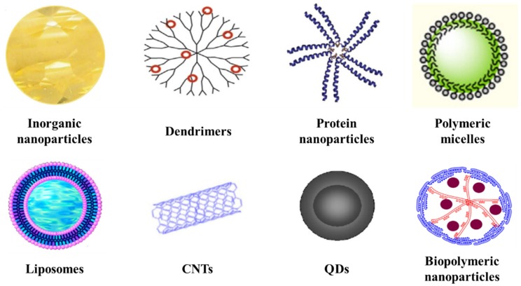
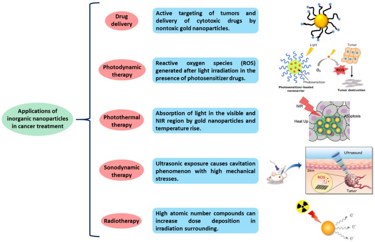
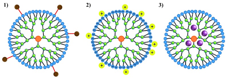
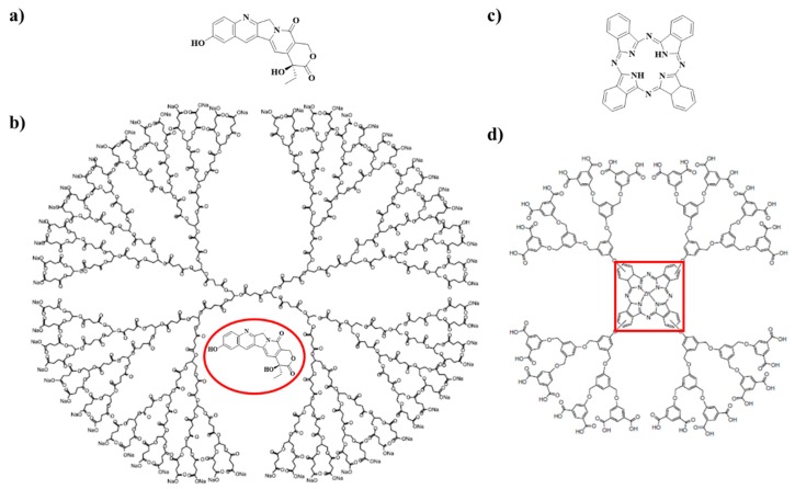
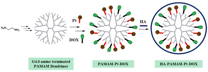
The current rapid advancement of numerous nanotechnology tools is being employed in treatment of many terminal diseases such as cancer. Nanocapsules (NCs) containing an anti-cancer drug offer a very promising alternative to conventional treatments, mostly due to their targeted delivery and precise action, and thereby they can be used in distinct applications: as biosensors or in medical imaging, allowing for cancer detection as well as agents/carriers in targeted drug delivery. The possibility of using different systems-inorganic nanoparticles, dendrimers, proteins, polymeric micelles, liposomes, carbon nanotubes (CNTs), quantum dots (QDs), biopolymeric nanoparticles and their combinations-offers multiple benefits to early cancer detection as well as controlled drug delivery to specific locations. This review focused on the key and recent progress in the encapsulation of anticancer drugs that include methods of preparation, drug loading and drug release mechanism on the presented nanosystems. Furthermore, the future directions in applications of various nanoparticles are highlighted.

Keywords: anticancer therapy; biocompatibility; biodegradability; bioimaging; cancer; drug delivery system; nanocapsules (NCs); nanomedicine; nanoparticles; nanotechnology.


Conflict of interest statement

The authors declare no conflict of interest.

Figures

Figure 1
Figure 1
Number of peer-reviewed publications in the field of nanomedicine in Web of Science divided in periods of 5 years.
Figure 2
Figure 2
Examples of different nanoparticle drug delivery systems: inorganic nanoparticles, dendrimers, protein nanoparticles, polymeric micelles, liposomes, CNTs, QDs and biopolymeric nanoparticles.
Figure 3
Figure 3
Applications of inorganic nanoparticles in cancer treatment.
Figure 4
Figure 4
TEM images of different types of gold nanoparticles: (a) gold nanospheres; reprinted with permission from [30]; copyright (2005) American Chemical Society; (b) gold nanorods; reprinted with permission from [31]; copyright (2013) American Chemical Society; (c) gold nanocages; reprinted with permission from [31]; (d) gold nanohexapods; reprinted with permission from [31]; (e) gold bipyramids; reprinted with permission from [32]; copyright (2005) American Chemical Society; (f) “nanorice” (gold-coated Fe2O3 nanorods); reprinted with permission from [33]; copyright (2006) American Chemical Society; (g) SiO2/Au nanoshells (the inset shows a hollow nanoshell); reprinted with permission from [29]; copyright (2010) Elsevier; (h) nanobowls with bottom cores; reprinted with permission from [34]; copyright (2009) American Chemical Society; (i) spiky SiO2/Au nanoshells (reprinted with permission from [35]; copyright (2010) American Chemical Society; the inset shows a gold nanostar); reprinted with permission from [36]; copyright (2006) American Chemical Society.
Figure 5
Figure 5
Schematic representation of the 3 mechanisms used to load drugs in dendrimers: (1) through a covalent interaction between the drug to the periphery of the dendrimer; (2) by coordination of the drug to the outer functional groups of the dendrimer via ionic interactions; (3) by simple encapsulation of the drugs into the dendrimer cavities.
Figure 6
Figure 6
(a) Chemical structure of 10-hydroxycamptothecin (10HCPT), (b) Chemical structure of a poly(glycerol-succinic acid) (PGLSA)-COONa dendrimer with encapsulated 10HCPT, (c) phthalocyanin, a photosensitizer for non-invasive cancer treatment and (d) zinc phthalocyanin covalently conjugated to a dendrimer structure. Not drawn to scale.
Figure 7
Figure 7
Scheme of the synthesis of PAMAM dendrimers with two drugs (Pt and DOX) covalently linked to the amine groups of the outer generation of the dendrimer. The addition of an external shell of HA, which is coordinated through electrostatic interactions with PAMAM, improve the biocompatibility of the dendrimers.
Figure 8
Figure 8
Scheme of the electrospray deposition system to synthesize gliadin-based nanoparticles. Reprinted with permission from [57]; copyright (2012) American Chemical Society.
Figure 9
Figure 9
Schematic representation of silk nanoparticles and pH-dependent release of DOX. Diagram not drawn to scale. Reprinted with permission from [58]; copyright (2013) Wiley Online Library.
Figure 10
Figure 10
(a) Solubilization of PTX in a polymeric micelle made of amphiphilic block copolymers (Physical loading of anticancer drug into the micelles). Adapted with permission from [63]; copyright (2007) American Chemical Society. (b) Inner structure of micelles with encapsulated Epirubicin covalently linked to the polyaspartate chain of PEG-polyaspartate block copolymer by an acid-labile hydrazone bond. The amphiphilic block copolymers (PEG and polyaspartate) forms spontaneously micellar structures in an aqueous media.
Figure 11
Figure 11
Cancer drug delivery via the injection of drug-loaded polymeric micelles. Adapted with permission from [61]; copyright (2016) Springer.
Figure 12
Figure 12
Schematic representation of the inner structure of a liposome drug delivery system with encapsulated CUR.
Figure 13
Figure 13
Schematic illustration of drug delivery systems comprising SWNTs. (a) A scheme showing non-covalent supramolecular π–π stacking of DOX with SWNTs. (b) Representative photos of mice from different groups were taken after treatments with DOX. (c) Gastrointestinal toxicity was observed in mice treated with free DOX but not in mice treated with SWNT-DOX. Histological sections of intestinal epithelium showed damage of the intestinal epithelium in the free DOX treated group. The arrows show the area of loss of columnar epithelial cells in tips of villi. (Scale bars: 100 micrometers). Reprinted with permission from [88]; copyright (2009) Wiley-VCH.
Figure 14
Figure 14
(a) Synthetic pathway for the preparation of QD705-RGD. (b) In vivo NIR fluorescence imaging of U87MG tumor-bearing mice (left shoulder, pointed by white arrows) injected with 200 pmol of QD705-RGD (left) and QD705 (right), respectively. All images were acquired under the same instrumental conditions. The mice autofluorescence is color coded green while the unmixed QD signal is color coded red. Prominent uptake in the liver, bone marrow, and lymph nodes was also visible. (c) Tumor-to-background ratios of mice injected with QD705 or QD705-RGD. The data were represented as mean (standard deviation (SD)). Using one-tailed paired Student’s t-test (n) 3),”*” denotes where p < 0.05 as compared to the mice injected with QD705. (d) Serum stability of QD705 and QD705-RGD in complete mouse serum over the course of 24 h. Reprinted with permission from [95]; copyright (2006) American Chemical Society.
Figure 15
Figure 15
(a) Synthetic pathway and assay of working protocol of the Hyaluronic Acid (HA)-ZnO QDs-Dicarboxyl-Terminated PEG drug delivery system. (b) TEM images of: (A) ZnO QDs, (B) NH2-ZnO QDs, and (C) PEG-ZnO QDs. The scale bar represents 20 nm. Reprinted with permission from [99]; copyright (2016) American Chemical Society.
Figure 16
Figure 16
Synthesis pathway for the preparation of cellulose nanocrystals complexes with encapsulated CUR. Adapted with permission from [104]; copyright (2016) Elsevier.

Similar articles

Cited by

References

    1. Wiwanitkit V. Cancer nanotherapy: Concept for design of new drug. J. Med. Hypotheses Ideas. 2013;7:3–4. doi: 10.1016/j.jmhi.2012.10.002. - DOI
    1. Costa J. Cancer. [(accessed on 2 January 2020)]; Available online: https://www.britannica.com/science/cancer-disease.
    1. Afshar M., Madani S., Tarazoj A.A., Papi S.H., Otroshi O., Gandomani H.S., Rahimi A., Salehiniya H. Physical Activity and Types of Cancer. WCRJ. 2018;5:1–11.
    1. Anand P., Kunnumakara A.B., Sundaram C., Harikumar K.B., Tharakan S.T., Lai O.S., Sung B., Aggarwal B.B. Cancer is a preventable disease that requires major lifestyle changes. Pharm. Res. 2008;25:2097–2116. doi: 10.1007/s11095-008-9661-9. - DOI - PMC - PubMed
    1. International Agency for Research on Cancer . World Cancer Report 2014. IARC; Lyon, France: 2014.

LinkOut - more resources