. 2020 Mar 31;11(4):378.
doi: 10.3390/genes11040378.

Increasing the Genetic Diagnosis Yield in Inherited Retinal Dystrophies: Assigning Pathogenicity to Novel Non-canonical Splice Site Variants

Affiliations

Increasing the Genetic Diagnosis Yield in Inherited Retinal Dystrophies: Assigning Pathogenicity to Novel Non-canonical Splice Site Variants

Vasileios Toulis et al. Genes (Basel). .

Abstract

Aims: We aimed to validate the pathogenicity of genetic variants identified in inherited retinal dystrophy (IRD) patients, which were located in non-canonical splice sites (NCSS).

Methods: After next generation sequencing (NGS) analysis (target gene panels or whole exome sequencing (WES)), NCSS variants were prioritized according to in silico predictions. In vivo and in vitro functional tests were used to validate their pathogenicity.

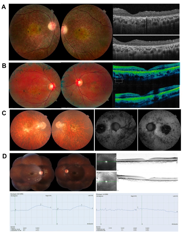
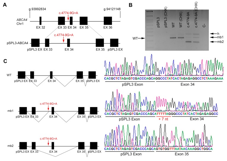
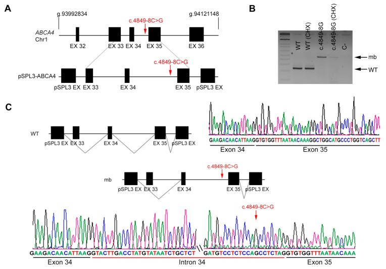
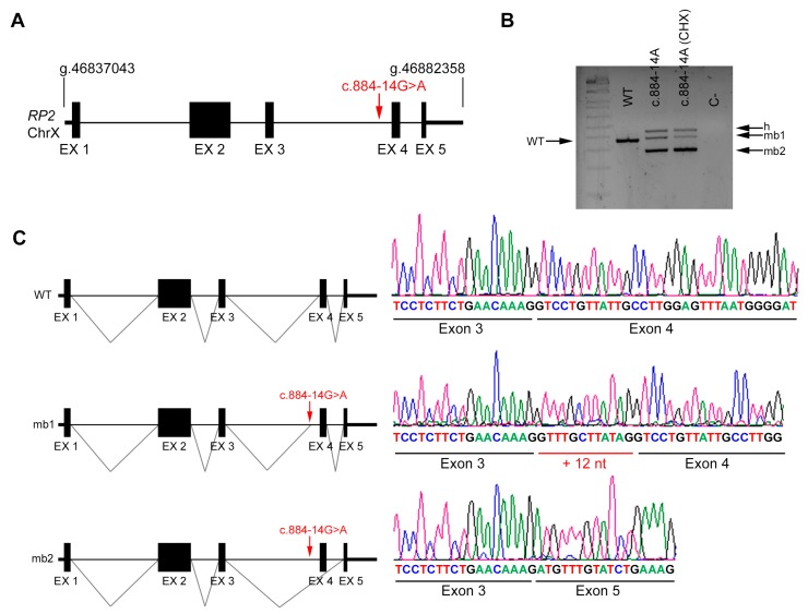
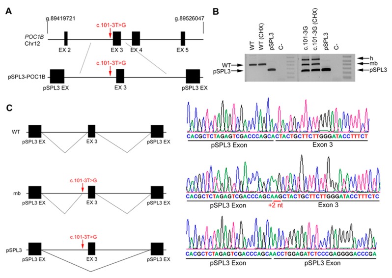
Results: Four novel NCSS variants have been identified. They are located in intron 33 and 34 of ABCA4 (c.4774-9G>A and c.4849-8C>G, respectively), intron 2 of POC1B (c.101-3T>G) and intron 3 of RP2 (c.884-14G>A). Functional analysis detected different aberrant splicing events, including intron retention, exon skipping and intronic nucleotide addition, whose molecular effect was either the disruption or the elongation of the open reading frame of the corresponding gene.

Conclusions: Our data increase the genetic diagnostic yield of IRD patients and expand the landscape of pathogenic variants, which will have an impact on the genotype-phenotype correlations and allow patients to opt for the emerging gene and cell therapies.

Keywords: aberrant splicing; inherited retinal dystrophies; minigenes; non-canonical splice sites.


Conflict of interest statement

The authors hereby declare that there is no competing interest.

Figures

Figure 1
Figure 1
Ocular phenotypes of patients analyzed in this study, with fundus examination, autofluorescence imaging, OCT imaging and ERGs (if available). (A) DBG1, clinically diagnosed of RP; Fundus image showing retinal vascular attenuation, central EPR and retinal atrophy, with peripheral retinal bony spicule hyperpigmentation. OCT shows diffuse retinal thinning, and severe alteration of the external layers. (B) DBG2, clinically diagnosed of RP with macular affectation; Fundus showing the central macular area exhibiting hypoautofluorescence corresponding to foveal area. Macular area of granular aspect with points of hyperautofluorescence. There are several scattered parches of hypoautofluorescence located out of vascular arcades at temporal and nasal side; OCT shows preserved foveal morphology and thinning of the macula in both eyes. (C) DBG3, clinically diagnosed of RP; Fundus showing tilted disc, peripapilary atrophy, atrophic RPE at the macula with a geographical shape, some vitreous opacities and some bony spicule pigmentation at the fundus equator. The atrophic macular area presents hypoautofluorescence with a small sparing area at the fovea. (D) DBG4 clinically diagnosed of Cone-Rod Dystrophy. Both eyes with retinal pigmentary changes and mild central hyperautofluorescence, with normal OCT; ERGs with very reduced scototpic (left) and photopic (right) response.
Figure 2
Figure 2
In vitro splicing analysis of the non-canonical splice site (NCSS) variant identified in ABCA4 in patient DBG1. (A) Genomic position of the identified NCSS variant in intron 33 of the ABCA4 gene (c.4774 -9G>A). Diagram showing the genomic region amplified from patient’s DNA, cloned into the pSPL3 vector. Note that exon 33 was not included in full. (B) Analysis of ABCA4 mRNAs from HEK293T cells transfected with either empty vector, WT or mutant genomic sequences (treated or untreated with cycloheximide, CHX). The band of 247 bp corresponds to the wild-type transcript (WT), whereas cells transfected with pSPL3 carrying the NCSS variant produced two different aberrant transcripts (mb1 and mb2). h indicates the heteroduplex band of mb1 and mb2. C- indicates the PCR negative control. (C) Subsequent Sanger sequencing of cloned individual bands (indicated in the left diagrams) and comparison to the wild-type transcript confirmed the insertion of 7 bp from intron 33 (mb1) due to acceptor splice site shift as well as exon skipping of exon 34 (mb2).
Figure 3
Figure 3
In vitro splicing analysis of the non-canonical splice site (NCSS) variant identified in ABCA4 in patient DBG2. (A) Genomic position of the identified NCSS variant in intron 34 of the ABCA4 gene (c.4849-8C>G). Diagram showing the genomic region amplified from patient’s DNA, cloned into the pSPL3 vector. Note that exon 33 was not included in full. (B) Analysis of ABCA4 mRNAs from HEK293T cells transfected with either WT or mutant genomic sequences (treated or untreated with cycloheximide, CHX). The band of 247 bp corresponds to the wild-type transcript (WT), whereas cells transfected with pSPL3 carrying the NCSS variant produced one aberrant transcript of 477 bp (indicated as mb). C- indicates the PCR negative control. (C) Subsequent Sanger sequencing of cloned individual bands confirmed intron 34 retention due to the NCSS variant.
Figure 4
Figure 4
In vivo splicing analysis of the non-canonical splice site (NCSS) variant identified in RP2. (A) Diagram showing the genomic position of the NCSS variant in intron 3 of the RP2 gene in chromosome X (c.884 -14G>A) from patient R1. (B) In vivo analysis of RP2 mRNAs from control (WT) and patient’s blood samples (treated or untreated with cycloheximide, CHX). The band of 370 bp corresponds to the wild-type transcript (WT), whereas blood from the hemizygous patient carrying the NCSS variant resulted into two different aberrant transcripts (mb1 and mb2). h indicates the heteroduplex band of mb1 and mb2. C- indicates the PCR negative control. (C) Subsequent Sanger sequencing of cloned individual bands, indicated in the left diagrams, confirmed the in-frame insertion of 12 bp (mb1) and exon skipping of exon 4 (mb2) compared to the wild-type transcript.
Figure 5
Figure 5
In vitro splicing analysis of the non-canonical splice site (NCSS) variant identified in POC1B. (A) Genomic position of the NCSS variant in intron 2 of the POC1B gene (c.101-3T>G) from patient DBG4. Diagram showing the genomic region between introns 2 and 3 amplified from patient’s DNA, cloned into the pSPL3 vector. (B) Analysis of POC1B mRNAs from HEK293T cells transfected with either empty vector, WT or mutant genomic sequences (treated or untreated with cycloheximide, CHX). The band of 429 bp corresponds to the wild-type transcript (WT), whereas cells transfected with pSPL3 carrying the NCSS variant produced one aberrant transcript (mb) plus the same transcript band produced by the pSPL3 control vector, indicating an exon skipping event. h indicates the heteroduplex band of the mb and skipped transcript products. C- indicates the PCR negative control. (C) Subsequent Sanger sequencing of cloned individual bands, indicated in the left diagrams, confirmed the insertion of +2 nt (mb) and the skipping of exon 2 compared to the wild-type transcript.

References

    1. Ellingford J.M., Barton S., Bhaskar S., O’Sullivan J., Williams S.G., Lamb J.A., Panda B., Sergouniotis P.I., Gillespie R.L., Daiger S.P., et al. Molecular findings from 537 individuals with inherited retinal disease. J. Med. Genet. 2016;53:761–767. doi: 10.1136/jmedgenet-2016-103837. - DOI - PMC - PubMed
    1. Zernant J., Xie Y.A., Ayuso C., Riveiro-Alvarez R., Lopez-Martinez M.-A., Simonelli F., Testa F., Gorin M.B., Strom S.P., Bertelsen M., et al. Analysis of the ABCA4 genomic locus in Stargardt disease. Hum. Mol. Genet. 2014;23:6797–6806. doi: 10.1093/hmg/ddu396. - DOI - PMC - PubMed
    1. Sangermano R., Khan M., Cornelis S.S., Richelle V., Albert S., Garanto A., Elmelik D., Qamar R., Lugtenberg D., van den Born L.I., et al. ABCA4 midigenes reveal the full splice spectrum of all reported noncanonical splice site variants in Stargardt disease. Genome Res. 2018;28:100–110. doi: 10.1101/gr.226621.117. - DOI - PMC - PubMed
    1. Carss K.J., Arno G., Erwood M., Stephens J., Sanchis-Juan A., Hull S., Megy K., Grozeva D., Dewhurst E., Malka S., et al. Comprehensive rare variant analysis via whole-genome sequencing to determine the molecular pathology of inherited retinal disease. Am. J. Hum. Genet. 2017;100:75–90. doi: 10.1016/j.ajhg.2016.12.003. - DOI - PMC - PubMed
    1. Simunovic M.P., Jolly J.K., Xue K., Edwards T.L., Groppe M., Downes S.M., MacLaren R.E. The spectrum of CHM gene mutations in choroideremia and their relationship to clinical phenotype. Investig. Opthalmol. Vis. Sci. 2016;57:6033–6039. doi: 10.1167/iovs.16-20230. - DOI - PMC - PubMed

Publication types