Review
. 2020 Mar 31;12(4):848.
doi: 10.3390/cancers12040848.

Morphologic and Genomic Heterogeneity in the Evolution and Progression of Breast Cancer

Affiliations
Review

Morphologic and Genomic Heterogeneity in the Evolution and Progression of Breast Cancer

Jamie R Kutasovic et al. Cancers (Basel). .

Abstract

: Breast cancer is a remarkably complex and diverse disease. Subtyping based on morphology, genomics, biomarkers and/or clinical parameters seeks to stratify optimal approaches for management, but it is clear that every breast cancer is fundamentally unique. Intra-tumour heterogeneity adds further complexity and impacts a patient's response to neoadjuvant or adjuvant therapy. Here, we review some established and more recent evidence related to the complex nature of breast cancer evolution. We describe morphologic and genomic diversity as it arises spontaneously during the early stages of tumour evolution, and also in the context of treatment where the changing subclonal architecture of a tumour is driven by the inherent adaptability of tumour cells to evolve and resist the selective pressures of therapy.

Keywords: Breast cancer; genomics; intra-tumour heterogeneity; metastasis; subclonal diversity; treatment resistance.


Conflict of interest statement

The authors declare no conflict of interest

Figures
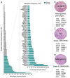
Figure 1
Figure 1
Genomic alterations across breast cancers. (A) Frequency of genomic alteration (mutation and copy number variation) in the 147 breast cancer driver genes across the TCGA pancancer breast cancer dataset (n = 1033); and stratified by oestrogen receptor (ER) status (in magnified plot): ER positive, n = 795; ER negative, n = 238. Top ten most frequently mutated genes in (B) Invasive Carcinoma-No Special Type (IC-NST) [15]; (C) Invasive Lobular Carcinoma (ILC) [15]; and, (D) Metaplastic breast cancer [36,37,38].
Figure 2
Figure 2
The morphological and molecular evolution of breast cancer. (A) Hypothetical schematic showing how the mutation of cancer genes drives the clonal and subclonal evolution of cancer (adapted from [55]). Key early driver genes impact the subsequent lineage and tumour type that arises, including mutations in PI3KCA in ER+ tumours, CDH1 in lobular lineage, TP53 in high grade ER- tumours, ETV6-NTRK3 and MYB-NFIB translocations in secretory and adenoid cystic carcinomas respectively. (B) the multistep model of breast cancer showing morphological stages of development from normal epithelium. This simplified model is based on the evolution of ER positive and ER negative breast cancer, as portrayed in more detail elsewhere [49,50,51]; evidence derived from morphological evaluation and the frequency with which lesions are co-localized, as well as molecular evidence showing co-localized lesions share identical mutations indicating clonal relatedness. (C) Cartoon to illustrate how this might arise in a ‘sick lobe’, that is a clonal outgrowth of apparently morphologically normal-looking epithelial cells (green), which harbour early genetic changes. In some areas of the lobe, the earliest morphologically abnormal changes may appear in some terminal duct-lobular units (lobule) as columnar cell lesions. These lesions are considered precursors of ADH (light blue cells) and DCIS (purple cells), which arise in lobules and may travel down ducts. The mutation or loss of CDH1 (E-cadherin) triggers the evolution of the ’lobular lineage’ (sky blue cells) as ALH then LCIS (lobular neoplasia); these cells may travel down ducts underneath the normal epithelial lining (pageotoid spread). Both LCIS and DCIS are genetically advanced lesions and so likely exhibit sub-clonal mutations. As these neoplastic cells can travel along ductal structures then this means invasion can occur at multiple sites giving rise to multifocal invasive disease (ILC, IC NST), which continues to undergo subclonal change. CCL: columnar cell lesion; ADH: atypical ductal hyperplasia. ALH: atypical lobular hyperplasia, APH: atypical apocrine hyperplasia; DCIS: ductal carcinoma in situ; LCIS: lobular carcinoma in situ; IC NST: invasive carcinoma no special type; ILC: invasive lobular carcinoma; MDL: mixed ductal lobular carcinoma; MGA: microglandular adenosis.
Figure 3
Figure 3
Illustrating morphological and molecular heterogeneity. Low power, haematoxylin and eosin stained sections of two tissue blocks from the same surgical specimen (scale bar = 2 mm). Both blocks are widely affected by breast disease with cystically dilated ducts, in situ carcinoma and invasive carcinoma. The three images in the lower panel are high power views of the same sections stained for E-cadherin (scale bar in middle image = 0.5 mm). Left picture shows cells of LCIS (E-cadherin negative) that have grown and then expanded underneath normal epithelial cells lining a duct (pagetoid spread), adjacent to E-cadherin positive, invasive cells of IC NST. Middle picture shows adjacent ducts in a complex branching network, one duct populated by DCIS (E-cadherin positive), two smaller ducts by LCIS and two other ducts co-involved by cells of DCIS and LCIS (DCIS/LCIS). Right picture showing ducts separately involved by DCIS or LCIS, plus an area of invasive cancer (ILC, E-cadherin negative). The individual components of this case were previously analysed by whole exome sequencing and all lesions were shown to be clonally related with early, common diver mutations identified in BRCA2 and TBX3, ‘lobular’ lineage-specific mutations including in CDH1 and ‘ductal’ lineage-specific mutations including in NF1 (see [42]). DCIS: ductal carcinoma in situ; LCIS: lobular carcinoma in situ; IC NST: invasive carcinoma no special type; ILC: invasive lobular carcinoma.

Similar articles

Cited by

References

    1. WHO Classification of Tumours Editorial Board . World Health Organisation Classification of Tumours: Breast Tumours. 5th ed. International Agency for Research on Cancer (IARC); Lyon, France: 2019.
    1. Perou C.M., Sorlie T., Eisen M.B., van de Rijn M., Jeffrey S.S., Rees C.A., Pollack J.R., Ross D.T., Johnsen H., Akslen L.A., et al. Molecular portraits of human breast tumours. Nature. 2000;406:747–752. doi: 10.1038/35021093. - DOI - PubMed
    1. Sorlie T., Perou C.M., Tibshirani R., Aas T., Geisler S., Johnsen H., Hastie T., Eisen M.B., van de Rijn M., Jeffrey S.S., et al. Gene expression patterns of breast carcinomas distinguish tumor subclasses with clinical implications. Proc. Natl. Acad. Sci. USA. 2001;98:10869–10874. doi: 10.1073/pnas.191367098. - DOI - PMC - PubMed
    1. Prat A., Parker J.S., Karginova O., Fan C., Livasy C., Herschkowitz J.I., He X., Perou C.M. Phenotypic and molecular characterization of the claudin-low intrinsic subtype of breast cancer. Breast Cancer Res. 2010;12:R68. doi: 10.1186/bcr2635. - DOI - PMC - PubMed
    1. Mathews J.C., Nadeem S., Levine A.J., Pouryahya M., Deasy J.O., Tannenbaum A. Robust and interpretable PAM50 reclassification exhibits survival advantage for myoepithelial and immune phenotypes. NPJ Breast Cancer. 2019;5:30. doi: 10.1038/s41523-019-0124-8. - DOI - PMC - PubMed

LinkOut - more resources