. 2020 Mar 31;21(7):2439.
doi: 10.3390/ijms21072439.

Identification and Regulation of Interleukin-17 (IL-17) Family Ligands in the Teleost Fish European Sea Bass

Affiliations

Identification and Regulation of Interleukin-17 (IL-17) Family Ligands in the Teleost Fish European Sea Bass

Carmen González-Fernández et al. Int J Mol Sci. .

Abstract

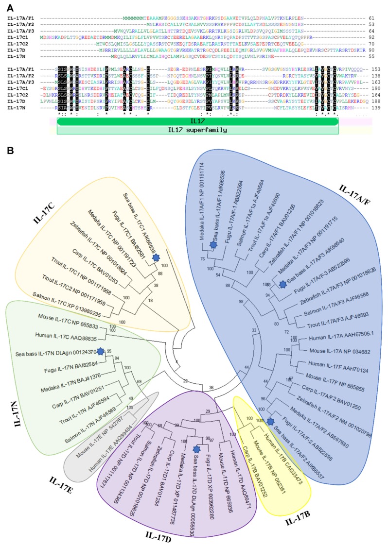
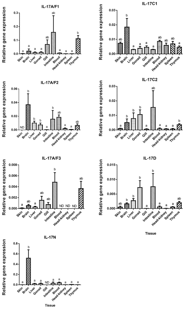
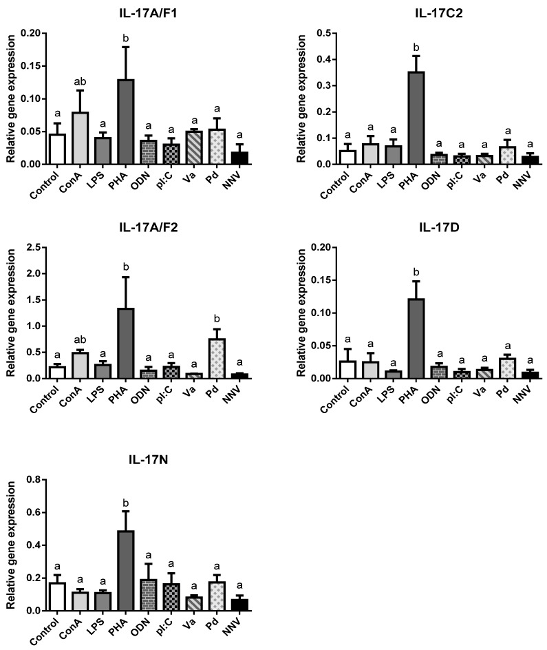
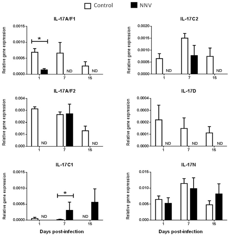
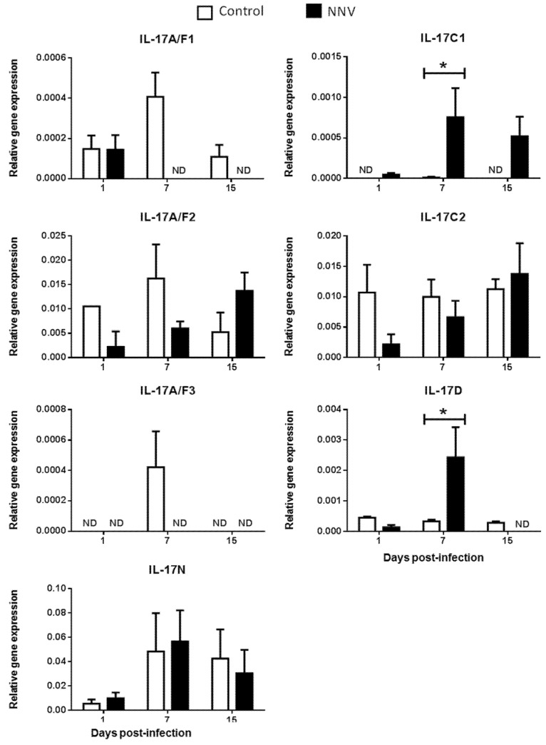
Interleukin-17 (IL-17) cytokine comprises a family of six ligands in mammals with proinflammatory functions, having an important role in autoimmune disorders and against bacterial, viral, and fungal pathogens. While IL-17A and IL-17F ligands are mainly produced by Th cells (Th17 cells), the rest of the ligands are expressed by other immune and non-immune cells and have different functions. The identification of IL-17 ligands in fish has revealed the presence of six members, counterparts to mammalian ones, and a teleost-specific form, the fish IL-17N. However, tissue distribution, the regulation of gene expression, and scarce bioactivity assays point to similar functions compared to mammalian ones, though this yet to be investigated and confirmed. Thus, we have identified seven IL-17 ligands in the teleost European sea bass (Dicentrarchus labrax), for the first time, corresponding to IL-17A/F1, IL-17A/F2, IL-17A/F3, IL-17C1, IL-17C2, IL-17D, and IL-17N, according to the predicted protein sequences and phylogenetic analysis. They are constitutively and widely transcribed in sea bass tissues, with some of them being mainly expressed in the thymus, brain or intestine. Upon in vitro stimulation of head-kidney leucocytes, the mRNA levels of all sea bass IL-17 ligands were up-regulated by phytohemagglutinin treatment, a well-known T cell mitogen, suggesting a major expression in T lymphocytes. By contrast, the infection of sea bass juveniles with nodavirus (NNV), a very pathogenic virus for this fish species, resulted in the up-regulation of the transcription of IL-17C1 in the head-kidney and of IL-17C1 and IL-17D in the brain, the target tissue for NNV replication. By contrast, NNV infection led to a down-regulated transcription of IL-17A/F1, IL-17A/F2, IL-17C1, IL-17C2, and IL-17D in the head-kidney and of IL-17A/F1 and IL-17A/F3 in the brain. The data are discussed accordingly with the IL-17 ligand expression and the immune response under the different situations tested.

Keywords: PAMPs; Th17 cells; fish; immunity; interleukin 17 (IL-17); nodavirus (NNV).


Conflict of interest statement

The authors declare no conflict of interest. The funders had no role in the design of the study; in the collection, analyses, or interpretation of data; in the writing of the manuscript, or in the decision to publish the results.

Figures

Figure 1
Figure 1
Identification of seven IL-17 ligands in European sea bass. (A) The predicted amino acid sequences of European sea bass IL-17 were aligned by ClustalW. The dashes denote gaps introduced to maximize alignments. Identical and similar residues are indicated by asterisks and colons, respectively. The position of the IL-17 domain is indicated. (B) The neighbour-joining tree of the European sea bass IL-17 deduced proteins with fish and mammalian orthologues. The genetic distances were calculated based on protein differences (p-distance) with complete deletion of gaps. The number at each node indicates the percentage of bootstrapping after 1000 replications. Accession numbers are indicated.
Figure 2
Figure 2
European sea bass Interleukin-17 (IL-17) transcripts are widely and constitutively expressed in the tissues. The relative gene expression of IL-17 ligands in the tissues of naïve European sea bass specimens. The data are presented as the mean (n= 3) ± SEM relative to the expression of the endogenous controls. Different letters indicate differences in expression between tissues (ANOVA, p < 0.05). ND, undetected.
Figure 3
Figure 3
Transcription of European sea bass IL-17 ligands is induced in head-kidney leucocytes by T cell mitogens. The relative gene expression of IL-17 ligands in the European sea bass head-kidney leucocytes incubated for 4 h with culture medium (Control), 5 μg/mL concanavalin A (ConA), 5 μg/mL lipopolysaccharide (LPS), 10 μg/mL phytohemagglutinin (PHA), 50 μg/mL synthetic unmethylated cytosine-phosphodiester-guanosine oligodeoxynucleotide 1668 (ODN), 25 μg/mL PolyI:C (pI:C), 108 heat-killed Vibrio anguillarum (Va) or Photobacterium damselae (Pd) bacteria/mL, and 106 TCID50 nodavirus (NNV)/mL. The data are presented as the mean (n = 5) ± SEM relative to the expression of the endogenous controls. Different letters indicate differences in gene expression (ANOVA, p < 0.05).
Figure 4
Figure 4
Nodavirus regulates the transcription of European sea bass IL-17 genes in the head-kidney. The relative gene expression of IL-17 ligands in the head-kidney of European sea bass specimens intramuscularly injected with 100 µL of culture medium alone (control) or containing 106 TCID50 of nodavirus (NNV)/fish. The data are presented as the mean (n= 4–6) ± SEM relative to the expression of the endogenous controls. Asterisks denote significant differences with the control group at each sampling time (Student-t-test, p < 0.05). ND, undetected.
Figure 5
Figure 5
Nodavirus regulates the transcription of European sea bass IL-17 genes in the brain. The relative gene expression of IL-17 ligands in the brain of European sea bass specimens intramuscularly injected with 100 µL of culture medium alone (control) or containing 106 TCID50 of nodavirus (NNV)/fish. The data are presented as the mean (n= 4–6) ± SEM relative to the expression of the endogenous controls. Asterisks denote significant differences with the control group at each sampling time (Student-t-test, p < 0.05). ND, undetected.

Similar articles

Cited by

References

    1. Akira S., Uematsu S., Takeuchi O. Pathogen recognition and innate immunity. Cell. 2006;124:783–801. doi: 10.1016/j.cell.2006.02.015. - DOI - PubMed
    1. Luckheeram R.V., Zhou R., Verma A.D., Xia B. CD4 +T cells: Differentiation and functions. Clin. Dev. Immunol. 2012;1-12 doi: 10.1155/2012/925135. - DOI - PMC - PubMed
    1. Saravia J., Chapman N.M., Chi H. Helper T cell differentiation. Cell. Mol. Immunol. 2019;16:634–643. doi: 10.1038/s41423-019-0220-6. - DOI - PMC - PubMed
    1. Park H., Li Z., Yang X.O., Chang S.H., Nurieva R., Wang Y.-H., Wang Y., Hood L., Zhu Z., Tian Q., et al. A distinct lineage of CD4 T cells regulates tissue inflammation by producing interleukin 17. Nat. Immunol. 2005;6:1133–1141. doi: 10.1038/ni1261. - DOI - PMC - PubMed
    1. Monin L., Gaffen S.L. Activities and therapeutic implications. Cold Spring Harb Perspect Biol. 2018;10:1–23. - PMC - PubMed

LinkOut - more resources