Review
. 2020 Apr 1;9(4):851.
doi: 10.3390/cells9040851.

Targeting and Crossing the Blood-Brain Barrier with Extracellular Vesicles

Affiliations
Review

Targeting and Crossing the Blood-Brain Barrier with Extracellular Vesicles

Julien Saint-Pol et al. Cells. .

Abstract

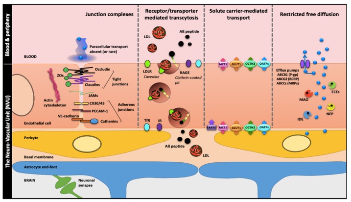
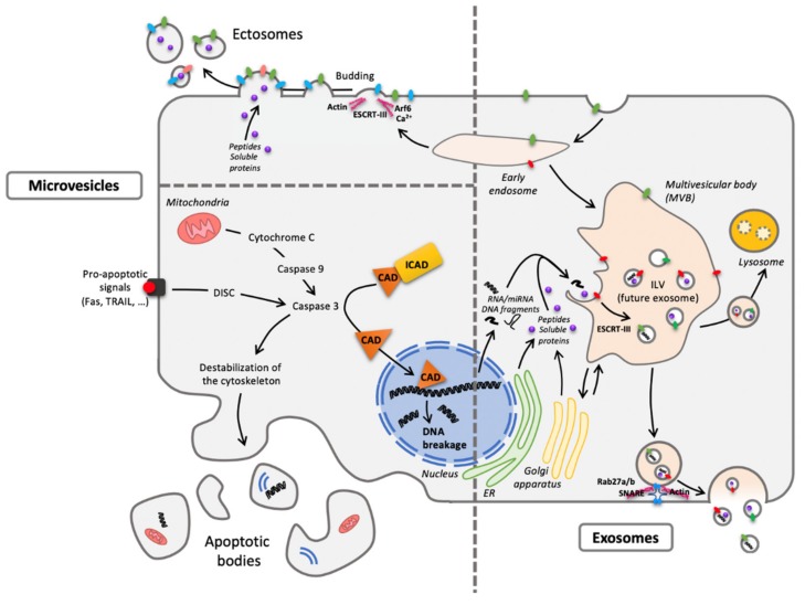
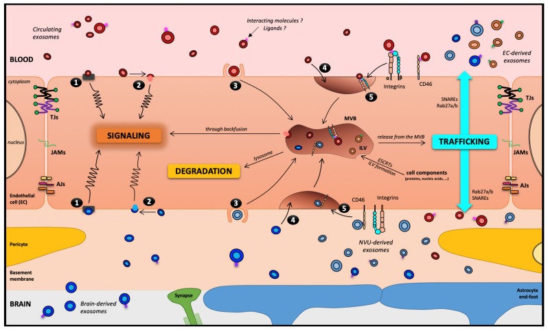
The blood-brain barrier (BBB) is one of the most complex and selective barriers in the human organism. Its role is to protect the brain and preserve the homeostasis of the central nervous system (CNS). The central elements of this physical and physiological barrier are the endothelial cells that form a monolayer of tightly joined cells covering the brain capillaries. However, as endothelial cells regulate nutrient delivery and waste product elimination, they are very sensitive to signals sent by surrounding cells and their environment. Indeed, the neuro-vascular unit (NVU) that corresponds to the assembly of extracellular matrix, pericytes, astrocytes, oligodendrocytes, microglia and neurons have the ability to influence BBB physiology. Extracellular vesicles (EVs) play a central role in terms of communication between cells. The NVU is no exception, as each cell can produce EVs that could help in the communication between cells in short or long distances. Studies have shown that EVs are able to cross the BBB from the brain to the bloodstream as well as from the blood to the CNS. Furthermore, peripheral EVs can interact with the BBB leading to changes in the barrier's properties. This review focuses on current knowledge and potential applications regarding EVs associated with the BBB.

Keywords: blood–brain barrier; brain diseases; exosomes; extracellular vesicles; microvesicles.


Conflict of interest statement

The authors declare no conflicts of interest to disclose.

Figures

Figure 1
Figure 1
The blood-brain barrier (BBB), a solid wall within the brain microvasculature. Brain microvessels endothelial cells (ECs) are the bricks supporting the BBB phenotype with two main components: (i) a physical barrier which restricts transcytosis and seals the paracellular spaces between ECs through apical tight junctions (claudins, tricellulin and occludin linked to actin cytoskeleton by zonula occludens proteins), median junctional adhesion molecules (JAMs) and medio-basolateral Ca2+-dependent adherens junctions; (ii) a metabolic barrier supported by enzymes and efflux pumps restricting nonspecific transport and favor receptor or transporter-specific routes. These features which cement the BBB ‘wall’ are induced, organized and maintained by cell–cell communications between the ECs and the close neighboring cells: brain pericytes, astrocytes (through their end-feet surrounding the brain microvessels) and neurons. These four cell types together with the basal membrane form the neurovascular unit (NVU). Abbreviations: ABCB1: ATP-Binding Cassette sub-family B member 1; ABCCs: ATP-Binding Cassette sub-family C members; ABCG2: ATP-Binding Cassette sub-family G member 2; Aβ: amyloid-β; BCRP: Breast Cancer Resistance Protein; CX30/43: Connexin 30/43; ECEs: Endothelin-Converting Enzymes; EEATs: Excitatory Amino acid Transporter 2; GLUT1: Glucose Transporter 1; IDE: Insulin Degrading Enzyme; IR: Insulin Receptor; LDL: Low-Density Lipoproteins; LDLR: Low-Density Lipoproteins Receptor; MAO: MonoAmine Oxidase; MCT1: Monocarboxylate Transporter 1, MRPs: Multidrug-Resistance Proteins; NEP: Neprilysin; OATPs: Organic Anion Transporting Polypeptides; OCTN2: Organic Cation/Carnitine Transporter 2; PECAM-1 (also CD31): Platelet Endothelial Cell Adhesion Molecule-1; P-gp: P-glycoprotein; RAGE: Receptor for Advanced Glycation End-products; TfR: Transferrin Receptor; ZOs: Zonula Occludens.
Figure 2
Figure 2
One cell, different extracellular vesicles (EVs). The two main forms are microvesicles (MVs) and EXOs differing from their origin. Concerning MVs, ectosomes result from membrane budding (left pane, top) and apoptotic bodies from cellular vesiculation following apoptosis (left panel, bottom). As to exosomes (EXOs), they have an endosomal origin, generated by the formation of intraluminal vesicles (ILV) within the multivesicular body (MVB) (right panel) and released through exocytosis routes. Despite a quite homogenous diameter from 30 to 150 nm, EXOs are difficult to distinguish (size exclusion chromatography or differential ultracentrifugation) from the smallest MVs. Abbreviations: CAD: Caspase-Activated DNAse; DISC: Death-Inducing Signaling Complex; ESCRT: Endosomal Sorting Complexes Required for Transport; ER: Endoplasmic Reticulum; ICAD: Inhibitor of Caspase-Activated DNAse; Rab: RAS-related protein; SNARE: Soluble N-ethylmaleimide-sensitive-factor Attachment protein REceptor; TRAIL: Tumor-necrosis-factor Related Apoptosis Inducing Ligand.
Figure 3
Figure 3
The blood–brain barrier, theoretically a motorway for a melting-pot of EXOs. Five routes have been described for EXOs interacting with a receiving cell: (1) association with a protein G-coupled receptor on the cell surface, inducing a signaling cascade; (2) adhesion to the cell surface and fusion, releasing the EXOs content in the cytoplasm, which can lead to several types of events, including cell signaling; (3) macropinocytosis; (4) nonspecific/lipid raft; or (5) receptor-mediated transcytosis, leading to its entry into the cell through the endocytic pathway and its storage in the MVB. Then, three outcomes remain possible for EXOs: (i) degradation by lysosomes; (ii) signaling induction through a backfusion event in the MVB releasing its content in the cytoplasm; or (iii) trafficking from the MVB to the plasma membrane as neoformed ILVs in the receiving cell. It is worth noting that except in pathological models, under TNF-α treatment [50], EVs and EXOs have not been described to cross the BBB through the paracellular pathway. Abbreviations: AJs: Adherens Junctions; EC: Endothelial Cells; ESCRT: Endosomal Sorting Complexes Required for Transport; ILV: IntraLuminal Vesicles; JAMs: Junctional Adhesion Molecules; NVU: Neuro-Vascular Unit; SNARE: Soluble N-ethylmaleimide-sensitive-factor Attachment protein REceptor; TJs: Tight Junctions.

References

    1. Peluffo H., Unzueta U., Negro-Demontel M.L., Xu Z., Váquez E., Ferrer-Miralles N., Villaverde A. BBB-targeting, protein-based nanomedicines for drug and nucleic acid delivery to the CNS. Biotechnol. Adv. 2015;33:277–287. doi: 10.1016/j.biotechadv.2015.02.004. - DOI - PubMed
    1. Saraiva C., Praça C., Ferreira R., Santos T., Ferreira L., Bernardino L. Nanoparticle-mediated brain drug delivery: Overcoming blood–brain barrier to treat neurodegenerative diseases. J. Control. Release. 2016;235:34–47. doi: 10.1016/j.jconrel.2016.05.044. - DOI - PubMed
    1. Karamanos Y., Pottiez G. Proteomics and the blood–brain barrier: How recent findings help drug development. Expert Rev. Proteomics. 2016;13:251–258. doi: 10.1586/14789450.2016.1143780. - DOI - PubMed
    1. Cecchelli R., Berezowski V., Lundquist S., Culot M., Renftel M., Dehouck M.-P., Fenart L. Modelling of the blood-brain barrier in drug discovery and development. Nat. Rev. Drug Discov. 2007;6:650–661. doi: 10.1038/nrd2368. - DOI - PubMed
    1. Abbott N.J. Astrocyte-endothelial interactions and blood-brain barrier permeability. J. Anat. 2002;200:629–638. doi: 10.1046/j.1469-7580.2002.00064.x. - DOI - PMC - PubMed

LinkOut - more resources