

An Alternatively Translated Connexin 43 Isoform, GJA1-11k, Localizes to the Nucleus and Can Inhibit Cell Cycle Progression
- PMID: 32244859
- PMCID: PMC7175147
- DOI: 10.3390/biom10030473
An Alternatively Translated Connexin 43 Isoform, GJA1-11k, Localizes to the Nucleus and Can Inhibit Cell Cycle Progression
Abstract
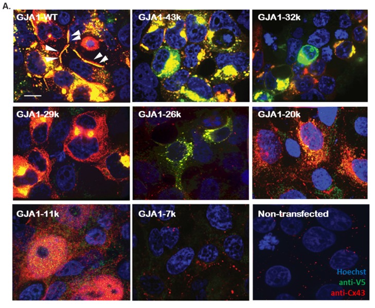
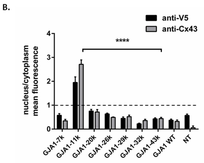
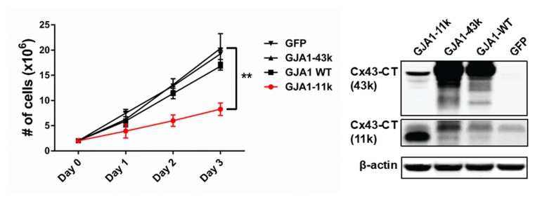
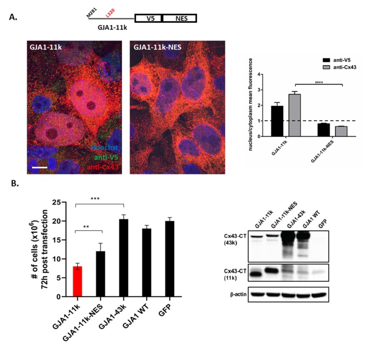
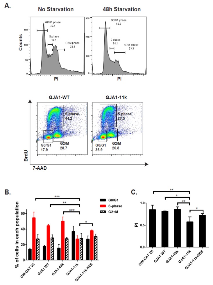
Connexin 43 (Cx43) is a gap junction protein that assembles at the cell border to form intercellular gap junction (GJ) channels which allow for cell-cell communication by facilitating the rapid transmission of ions and other small molecules between adjacent cells. Non-canonical roles of Cx43, and specifically its C-terminal domain, have been identified in the regulation of Cx43 trafficking, mitochondrial preconditioning, cell proliferation, and tumor formation, yet the mechanisms are still being explored. It was recently identified that up to six truncated isoforms of Cx43 are endogenously produced via alternative translation from internal start codons in addition to full length Cx43, all from the same mRNA produced by the gene GJA1. GJA1-11k, the 11kDa alternatively translated isoform of Cx43, does not have a known role in the formation of gap junction channels, and little is known about its function. Here, we report that over expressed GJA1-11k, unlike the other five truncated isoforms, preferentially localizes to the nucleus in HEK293FT cells and suppresses cell growth by limiting cell cycle progression from the G0/G1 phase to the S phase. Furthermore, these functions are independent of the channel-forming full-length Cx43 isoform. Understanding the apparently unique role of GJA1-11k and its generation in cell cycle regulation may uncover a new target for affecting cell growth in multiple disease models.
Keywords: cell cycle; connexin43; internal translation; trafficking.
Conflict of interest statement
We have no conflict of interest to disclose.
Figures
Similar articles
-
Intracellular trafficking pathways of Cx43 gap junction channels.Biochim Biophys Acta Biomembr. 2018 Jan;1860(1):40-47. doi: 10.1016/j.bbamem.2017.05.018. Epub 2017 May 30. Biochim Biophys Acta Biomembr. 2018. PMID: 28576298 Free PMC article. Review.
-
GJA1-20k, a Short Isoform of Connexin43, from Its Discovery to Its Potential Implication in Cancer Progression.Cells. 2025 Jan 24;14(3):180. doi: 10.3390/cells14030180. Cells. 2025. PMID: 39936974 Free PMC article. Review.
-
Autoregulation of connexin43 gap junction formation by internally translated isoforms.Cell Rep. 2013 Nov 14;5(3):611-8. doi: 10.1016/j.celrep.2013.10.009. Epub 2013 Nov 7. Cell Rep. 2013. PMID: 24210816 Free PMC article.
-
GJA1-20k Arranges Actin to Guide Cx43 Delivery to Cardiac Intercalated Discs.Circ Res. 2017 Oct 13;121(9):1069-1080. doi: 10.1161/CIRCRESAHA.117.311955. Epub 2017 Sep 18. Circ Res. 2017. PMID: 28923791 Free PMC article.
-
Dynamic UTR Usage Regulates Alternative Translation to Modulate Gap Junction Formation during Stress and Aging.Cell Rep. 2019 May 28;27(9):2737-2747.e5. doi: 10.1016/j.celrep.2019.04.114. Cell Rep. 2019. PMID: 31141695 Free PMC article.
Cited by
-
Identification of Cx43 variants predisposing to ventricular fibrillation in the acute phase of ST-elevation myocardial infarction.Europace. 2023 Feb 8;25(1):101-111. doi: 10.1093/europace/euac128. Europace. 2023. PMID: 35942675 Free PMC article.
-
Using human induced pluripotent stem cell-derived cardiomyocytes to understand the mechanisms driving cardiomyocyte maturation.Front Cardiovasc Med. 2022 Aug 12;9:967659. doi: 10.3389/fcvm.2022.967659. eCollection 2022. Front Cardiovasc Med. 2022. PMID: 36061558 Free PMC article.
-
Connexins in the Heart: Regulation, Function and Involvement in Cardiac Disease.Int J Mol Sci. 2021 Apr 23;22(9):4413. doi: 10.3390/ijms22094413. Int J Mol Sci. 2021. PMID: 33922534 Free PMC article. Review.
-
High Glucose-Induced Apoptosis Is Linked to Mitochondrial Connexin 43 Level in RRECs: Implications for Diabetic Retinopathy.Cells. 2021 Nov 10;10(11):3102. doi: 10.3390/cells10113102. Cells. 2021. PMID: 34831325 Free PMC article.
-
Connexin46 in the nucleus of cancer cells: a possible role as transcription modulator.Cell Commun Signal. 2025 Mar 27;23(1):153. doi: 10.1186/s12964-025-02151-w. Cell Commun Signal. 2025. PMID: 40148950 Free PMC article.
References
-
- Beardslee M.A., Lerner D.L., Tadros P.N., Laing J.G., Beyer E., Yamada K.A., Kléber A.G., Schuessler R.B., Saffitz J.E. Dephosphorylation and intracellular redistribution of ventricular connexin43 during electrical uncoupling induced by ischemia. Circ. Res. 2000;87:656–662. doi: 10.1161/01.RES.87.8.656. - DOI - PubMed
Publication types
MeSH terms
Substances
Grants and funding
LinkOut - more resources
Full Text Sources
Miscellaneous
