. 2020 Mar 20;21(6):2130.
doi: 10.3390/ijms21062130.

Use of STAT6 Phosphorylation Inhibitor and Trimethylglycine as New Adjuvant Therapies for 5-Fluorouracil in Colitis-Associated Tumorigenesis

Affiliations

Use of STAT6 Phosphorylation Inhibitor and Trimethylglycine as New Adjuvant Therapies for 5-Fluorouracil in Colitis-Associated Tumorigenesis

Mónica G Mendoza-Rodríguez et al. Int J Mol Sci. .

Abstract

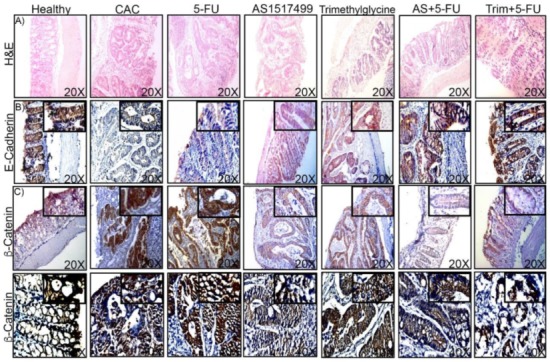
Colorectal cancer (CRC) is one of the most widespread and deadly types of neoplasia around the world, where the inflammatory microenvironment has critical importance in the process of tumor growth, metastasis, and drug resistance. Despite its limited effectiveness, 5-fluorouracil (5-FU) is the main drug utilized for CRC treatment. The combination of 5-FU with other agents modestly increases its effectiveness in patients. Here, we evaluated the anti-inflammatory Trimethylglycine and the Signal transducer and activator of transcription (STAT6) inhibitor AS1517499, as possible adjuvants to 5-FU in already established cancers, using a model of colitis-associated colon cancer (CAC). We found that these adjuvant therapies induced a remarkable reduction of tumor growth when administrated together with 5-FU, correlating with a reduction in STAT6-phosphorylation. This reduction upgraded the effect of 5-FU by increasing both levels of apoptosis and markers of cell adhesion such as E-cadherin, whereas decreased epithelial-mesenchymal transition markers were associated with aggressive phenotypes and drug resistance, such as β-catenin nuclear translocation and Zinc finger protein SNAI1 (SNAI1). Additionally, Il-10, Tgf-β, and Il-17a, critical pro-tumorigenic cytokines, were downmodulated in the colon by these adjuvant therapies. In vitro assays on human colon cancer cells showed that Trimethylglycine also reduced STAT6-phosphorylation. Our study is relatively unique in focusing on the effects of the combined administration of AS1517499 and Trimethylglycine together with 5-FU on already established CAC which synergizes to markedly reduce the colon tumor load. Together, these data point to STAT6 as a valuable target for adjuvant therapy in colon cancer.

Keywords: 5-FU; STAT6; Trimethylglycine; adjuvant therapies; colorectal cancer; drug resistance.


Conflict of interest statement

The authors declare no conflict of interest.

Figures

Figure 1
Figure 1
Adjuvant effect of AS1517499 and Trimethylglycine on 5-fluorouracil (5-FU) treatment in colitis-associated colon cancer (CAC). (A) Experimental design of the in vivo model to induce CAC. The scheme indicates the days of exposure to Azoxymethane (AOM) and dextran sodium sulfate (DSS) and treatments with AS1517499 and Trimethylglycine in combination with 5-FU and as single therapies. (B,C) Average number and size of tumors in mice with CAC without treatment (CAC), 5-FU, AS1517499, Trimethylglycine, and combination of AS1517499+5-FU (AS+5-FU) and Trimethylglycine+5-FU (Trim+5-FU). (D) Images of colons opened longitudinally showing macroscopic morphology after CAC induction and treated and non-treated (CAC) with 5-FU, AS1517499, and Trimethylglycine on day 90. (E,F). Representative Western blot and densitometry analysis of total extracts from colon tissues of mice treated with 5-FU and STAT6 inhibitor, Trimethylglycine, and their combination. In all of them, p-STAT6 levels were normalized relative to the total STAT6 protein levels in CAC without treatments. p value lower than 0.001 (***); p = 0.01 (**) and p = 0.05 (*). Data are representative of two independent experiments. n = 4–6 mice per group.
Figure 2
Figure 2
STAT6 inhibition favors recovering of cell adhesion markers in mice treated with 5-FU and decreases β-catenin nuclear localization. (A) Representative H&E-stained colonic sections from Healthy tissue (Healthy), CAC without treatment, CAC+5-FU (5-FU), CAC+ AS1517499 (AS), and Trimethylglycine (Trim) as singles therapies and the adjuvant effect with 5-FU, AS+5-FU, and Trim+5-FU. (B) Immunohistochemical staining of E-Cadherin and, β-catenin staining (C,D) in colon tissue obtained from mice with the different treatments previously mentioned. The arrows show the localization of β-catenin in the epithelial cells. The sections of tissue were analyzed with SP8 confocal microscope at 20× and 40×. The magnification was obtained of 40×. Data are representative of two independent experiments with 4–6 mice per group.
Figure 3
Figure 3
The inhibition of STAT6 increased apoptosis in colitis-associated colon cancer. (A) Representative stains of colon tissue in TUNEL assay in CAC treated with STAT6 inhibitors and 5-FU and in combinatory therapies. Green color indicates apoptosis in representative tissue and color blue was DAPI stained for nuclei. (B) Fluorescence quantification in colon tissue of TUNEL+ cells. The data are presented as the percentage of mean fluorescence, which was expressed as arbitrary units. Scale bars = 20 μm. The data are expressed as the mean ± SE from 4–6 mice per group representative of two independent experiments. **, p < 0.01; ***, p < 0.001.
Figure 4
Figure 4
STAT6 inhibition modulates the expression of proteins involved in inhibition apoptosis and cell survival. (A,B) Representative Western blot and densitometry analysis of ERK1/2 phosphorylation in CAC without treatments, 5-FU, AS1517499, and Trimethylglycine as single therapies and the combination as adjuvants AS+5-FU and Trim+5-FU. (C,D). Representative Western blot and densitometry analysis of ERCC1 expression under the condition previously mentioned. (E,F) Representative Western blot and densitometry analysis of SNAI1 expression under the condition previously mentioned. In all of them, the levels of the protein analyzed were normalized relative to the protein levels in CAC without treatments. Densitometry analyses of Western blots show the results of at least two independent experiments with four mice per group. p value lower than 0.001 (***); p = 0.01 (**) and p = 0.05 (*).
Figure 5
Figure 5
Adjuvant therapy for 5-FU modulates the percentages of circulating inflammatory monocytes and granulocytes in CAC. Flow cytometric analysis was performed with Ly6G and Ly6C markers expressed in the cell surface of circulating cells. (A) A representative graph displaying the proportion of CD11b+Ly6Chi monocytes gated on CD11b+ living cells population isolated from the circulation of mice with CAC without treatment, 5-FU, AS1517499, and Trimethylglycine, and a combination of these treatments on day 90 after AOM administration. (B) A representative graph displaying CD11b+Ly6ClowLy6G+ cells from the previously mentioned treatments and combinations in animals on day 90 after AOM administration. (C) Percentage of CD4+ population cells. (D) Percentage of CD8+ cells isolated from the circulation of the mice on day 90 after AOM/DSS administration and treatment with 5-FU and STAT6 inhibitors. The data are representative of two independent experiments that included n = 4–6 mice/group. Values are expressed as the mean ± SE. p value lower than 0.001 (***); p = 0.01 (**) and p = 0.05 (*).
Figure 6
Figure 6
Pro-tumorigenic cytokines are regulated by the adjuvants AS1517499 and Trimethylglycine in CAC. (AD). Relative expression of Il-10, Tgf-β, Il-17a, and Cxcr2 genes determined by qPCR in CAC without treatment (black bars), AS1517499 (red bars), Trimethylglycine (green bars), as single therapies and AS1517499+5-FU (AS+5-FU) (brown bars), and Trimethyglycine+5-FU (Trim+5-FU) (orange bars); all relative expressions were compared with the expression levels in healthy tissues (white bars). Data are presented as individual values with median and range from two independent experiments performed in triplicate. p value lower than 0.001 (***); p = 0.01 (**) and p = 0.05 (*).
Figure 7
Figure 7
Trimethylglycine decreases STAT6 phosphorylation in a human epithelial colon cancer cell line. (AC) Representative Western blots showing the protein levels of phosphorylated STAT6 in HCT-116 colonic human cells. (A). Epithelial HCT-116 cells were stimulated with recombinant IL-4 for 20 min and 24 h with and without Trimethylglycine (IL-4-20′+Trim and IL-4-24 h+Trim). (B) Representative Western blot showing the levels STAT6 phosphorylation with the stimulus of Trimethylglycine, IL-4, and Trimethylglycine plus IL-4 at 24h. (C). Representative Western blot showing the levels of STAT6 phosphorylation using the STAT6 inhibitor AS1517499 (300mM). (D). Representative immunofluorescence assay for P53 protein on HCT-116 cells exposed to IL-4 and Trimethylglycine. Alexa 488 (green) color indicates the expression of P53 in these cells and DAPI (blue) was used to visualize nuclei; scale bars = 20 μm. Confocal analyses were performed using the Leica TCS SP8 confocal microscopy system.

Similar articles

Cited by

References

    1. Ferlay J., Colombet M., Soerjomataram I., Mathers C., Parkin D.M., Pineros M., Znaor A., Bray F. Estimating the global cancer incidence and mortality in 2018: GLOBOCAN sources and methods. Int. J. Cancer. 2019;144:1941–1953. doi: 10.1002/ijc.31937. - DOI - PubMed
    1. Ullman T.A., Itzkowitz S.H. Intestinal inflammation and cancer. Gastroenterology. 2011;140:1807–1816. doi: 10.1053/j.gastro.2011.01.057. - DOI - PubMed
    1. Coussens L.M., Werb Z. Inflammation and cancer. Nature. 2002;420:860–867. doi: 10.1038/nature01322. - DOI - PMC - PubMed
    1. Rawla P., Sunkara T., Barsouk A. Epidemiology of colorectal cancer: Incidence, mortality, survival, and risk factors. Prz. Gastroenterol. 2019;14:89–103. doi: 10.5114/pg.2018.81072. - DOI - PMC - PubMed
    1. Jonker D., Rumble R.B., Maroun J., Gastrointestinal Cancer Disease Site Groupof Cancer Care Ontario’s Program in Evidence-Based Care Role of oxaliplatin combined with 5-fluorouracil and folinic acid in the first- and second-line treatment of advanced colorectal cancer. Curr. Oncol. 2006;13:173–184. - PMC - PubMed

MeSH terms