Review
. 2020 Mar 20;21(6):2135.
doi: 10.3390/ijms21062135.

Intervertebral Disc Diseases PART 2: A Review of the Current Diagnostic and Treatment Strategies for Intervertebral Disc Disease

Affiliations
Review

Intervertebral Disc Diseases PART 2: A Review of the Current Diagnostic and Treatment Strategies for Intervertebral Disc Disease

Pang Hung Wu et al. Int J Mol Sci. .

Abstract

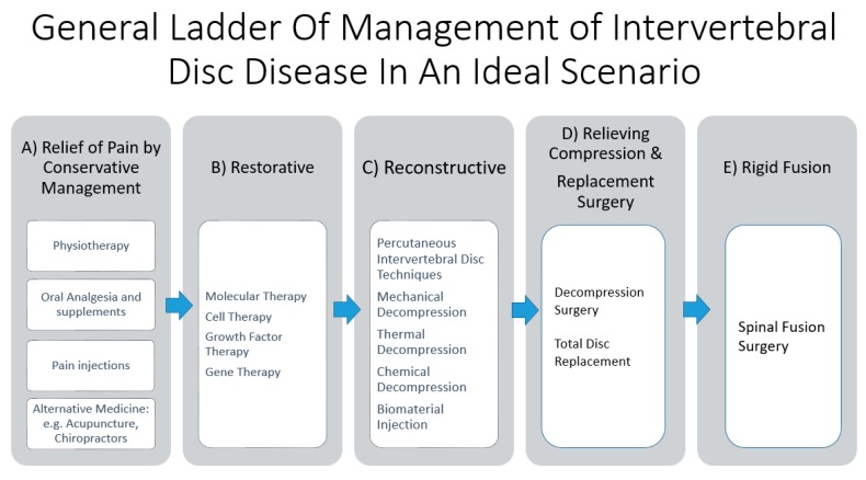
With an aging population, there is a proportional increase in the prevalence of intervertebral disc diseases. Intervertebral disc diseases are the leading cause of lower back pain and disability. With a high prevalence of asymptomatic intervertebral disc diseases, there is a need for accurate diagnosis, which is key to management. A thorough understanding of the pathophysiology and clinical manifestation aids in understanding the natural history of these conditions. Recent developments in radiological and biomarker investigations have potential to provide noninvasive alternatives to the gold standard, invasive discogram. There is a large volume of literature on the management of intervertebral disc diseases, which we categorized into five headings: (a) Relief of pain by conservative management, (b) restorative treatment by molecular therapy, (c) reconstructive treatment by percutaneous intervertebral disc techniques, (d) relieving compression and replacement surgery, and (e) rigid fusion surgery. This review article aims to provide an overview on various current diagnostic and treatment options and discuss the interplay between each arms of these scientific and treatment advancements, hence providing an outlook of their potential future developments and collaborations in the management of intervertebral disc diseases.

Keywords: degenerative disc disease; gene therapy; intervertebral disc; spinal decompression; spinal endoscopy; spinal fusion; spinal injection; spinal molecular therapy; spine pain management; stem cells.


Conflict of interest statement

The authors declare no conflict of interest.

Figures

Figure 1
Figure 1
General ladder of management of intervertebral disc disease in an ideal scenario. (A) Relief of pain by conservative management comprises of physiotherapy, oral analgesia, and nutrition supplements supplemented with or without pain injections. Alternative medicine, such as acupuncture and chiropractor practice, has been described but it is beyond the scope of this review. (B) Restorative therapy involves molecular therapy, such as gene, growth factor, and cell therapy, which aim to restore the anabolic function and decrease the catabolic function of the disc in an attempt to repair the disc damage. (C) Reconstructive therapy involves reshaping the disc through decompression, such as mechanical, thermal, and chemical decompression, and biomaterial injection, such as polyvinyl alcohol cryogel, which has biomechanical properties similar to an intervertebral disc. (D) Relief surgery, such as decompression surgery, relieves compression of the neural elements from the associated pathological thickening of the ligamentum flavum or bulging disc from degenerative disc disease. Replacement surgery, such as total disc replacement, removes the affected diseased disc segment and replacing it with an artificial disc, hence preserving the motion of the lumbar spinal segment. (E) Rigid fusion surgery is the most invasive and definitive management, where the motion of the spinal segment is sacrificed, with the intervertebral disc removed and replaced with bone grafts to facilitate fusion of the lumbar segment. It is of note that many of the restorative and reconstructive therapies are under experimental and clinical trial stages; widespread practice on the general population is not recommended unless more evidence is gathered. Hence, only in the “ideal” situation when all the treatment options are available, can we fully utilize this ladder of management.
Figure 2
Figure 2
A: Degenerative disc disease with Pfirrmann grade III disc showing an inhomogeneous structure, and an unclear distinction of nucleus and annulus and type 1 Modic changes with intermediate signal intensity in the T2 image with slightly decreased disc height and disc bulge. B: After radiofrequency ablation of the disc, sinuvertebral nerve, and basivertebral nerve, there is shrinkage of the degenerative disc and there is an increase in the signal of Modic changes in the adjacent vertebra body. However, with the current imaging technique, there are limitations in quantifying the effects of these end plate changes objectively. Further development in this area of assessment would be beneficial to assess treatment effects on the end plate and disc in early DDD (figure reproduced with permission courtesy of Kim et al. [12]).
Figure 3
Figure 3
Decision flow chart in clinical practice for patients with stable early degenerative disc disease. Patients with early DDD manifested as Pfirmann grade 1–3 disc on MRI are assessed for symptoms. Asymptomatic patients and symptomatic patients who have not tried conservative management (*) should undergo a trial of conservative management first. Patients with persistent discogenic pain who failed conservative treatments can be considered for discography. A negative discography makes disc-related symptoms less likely. One should consider ruling out other causes of back pain related to the spinal column, such as facet joint pain, tumor, infection, and fracture, etc., or referred pain from other visceral organs, or musculoskeletal back pain. A positive discography is suggestive of disc-related symptoms. This group of patients are consulted for options of invasive or noninvasive treatment strategies. A noninvasive conservative management option for pain relief consists of physiotherapy, oral medications, exercises, and alternative medicine. Invasive options include pain relief by pain generator injections. Injection of steroid and local anesthesia can be applied to pain generators, such as facet block to the facet joint, epidural steroid injection to the epidural space, and peri radicular injections for neuropathy or percutaneous epidural neurolysis when adhesion between the dura and intervertebral disc diseases is observed. Reconstruction of disc morphology, with percutaneous intervertebral disc techniques, such as mechanical, chemical, and thermal decompression and biomaterial injection, is another option. Many of these therapies are still in experimental stages. Restorative treatment, such as molecular treatment, such as gene, growth factor, and cell therapy, is another option, but most of these therapies are still at the clinical trial stages. Surgery is often the last definitive treatment option if other conservative treatment strategies fail.
Figure 4
Figure 4
Decision flow chart in clinical practice for patients with late degenerative disc disease, Pfirrmann grade 4–5. We assess for any related pure axial back pain, neural compression, and instability. If there is no neural compression or instability and only pure axial back pain, the patient should be offered a trial of conservative noninvasive treatment as a first line of treatment (#). It is controversial regarding total disc replacement and fusion surgery as a first line of definitive treatment for back pain. Both are invasive and irreversible procedures; hence, they should be considered as a last option in pure axial back pain even in advanced degenerative disc disease (*). If conservative management fails, discography should be considered. In a negative discography, we need to rule out other causes of back pain and continue conservative management. In a positive discography with pure axial back pain in advanced disc degeneration, the less invasive option of injection to pain generators, such as epidural steroid injection, percutaneous epidural neurolysis, peri-radicular injection, and facet joint blocks, can be considered for temporal relief of symptoms. Clinicians can consider other more definitive palliative management, such as radiofrequency ablation, which can be applied by minimally invasive techniques, such as endoscopic radiofrequency ablation of the sinuvertebral, basivertebral nerve, and disc, or fluoroscopic-guided percutaneous intervertebral disc treatment, such as radiofrequency ablation of the disc. Neural compression without instability or disc collapse can be treated with decompression only for patients without discogenic back pain. While for patients with discogenic back pain and neural compression, decompression surgery with additional endoscopic or other methods of treatment of the sinuvertebral and basivertebral nerve can be done [12]. Total disc replacement is done when there is disc collapse with the intention to preserve motion; fusion is done when there is disc collapse with the intension of rigid fusion. If there is any symptomatic gross instability, fusion surgery should be considered.
Figure 5
Figure 5
Transforaminal endoscopic lumbar discectomy (TELD). Using a beveled-type working cannula with an 8 mm outer diameter. The endoscope is introduced into the surgical field, with the entire procedure performed using constant saline irrigation. A shows the exiting nerve root in view with its surrounding epidural fat tissue. B shows the drilling of the superior articular facet under direct endoscopic vision. C shows the dissection of the foraminal ligament. D showes the exposure of the disc after manipulation of the working channel and endoscope to bring the disc in view. E shows the removal of the disc with endoscopic forceps. F shows the decompressed disc underneath floating neural elements. The blue stain on the disc was caused by indigo carmine disc injection, which served as a colored marker for ease of identification of disc herniation. G shows the herniation of the left L3/4 disc compressing on the neural element in sagittal MRI, which was removed and demonstrated in post-operative MRI of the same level sagittal cut in H. I shows the herniation of the left L3/4 disc compressing on the neural element in the central, paracentral, and foraminal region axial MRI, which was removed and demonstrated in post-operative axial MRI of the same level axial cut in J.
Figure 6
Figure 6
Posterior lumbar endoscopic unilateral laminotomy with bilateral decompression and discectomy over left lumbar five and sacral one. A shows the ligamentum flavum was lifted off from the caudal attachment in left L5/S1. B shows detachment of the remnant ligamentum flavum from the superior articular process of left side of S1. C shows the bulging disc, which is stained with indigo carmine. D shows discectomy performed using endoscopic forceps. E shows a prolapsed disc with neural element compression in the sagittal view MRI, which is decompressed in the same cut sagittal MRI in F. G shows a prolapsed disc with paracentral traversing nerve root compression with background thickening of the ligamentum flavum and spinal stenosis in the axial view MRI, which is decompressed with the ligamentum flavum removed and discectomy performed in H of the same cut axial MRI.
Figure 7
Figure 7
Right-sided L4/5 recurrent disc herniation after previous open laminotomy and discectomy 2 years ago, the patient underwent uniportal endoscopic transforaminal lumbar interbody fusion of right L4/5(ETLIF). A shows the recurrence of disc herniation with facet arthritis in the MRI axial cut of L4/5. B shows the same axial cut with left transforaminal lumbar interbody fusion showing resection of the facet joint and cage in an optimal position. C and D show the pre and post-operative status of the right facet. Note the previous right L4 laminotomy in C, the facet was resected, and the cage introduced in an optimal position as shown in D. E and F show the sagittal view of the pre and pos-operative status. Note the increase in the foraminal height and intervertebral height as a result of the right L4/5 ETLIF. G and H show the increase in the coronal disc height pre and post-operatively in right L4/5 ETLIF. I and J show the pre and post-operative standing neutral XR with the L4/5 interbody cage and standard posterior inserted pedicle screws in L4 and L5. K and L show the 3D reconstruction of pre and post-operative right L4/5 ETLIF.
Figure 8
Figure 8
A: Uniportal interlaminar endoscopic approach to the disc showing the relationship of the lumbar five (L5) traversing nerve root, which is retracted away by the working channel, exposing the disc of L4/5, the basivertebral nerve is located above the pedicle of left L5, and there is grade 3 neovascularization and inflammatory granulation tissue with adhesion around the basivertebral nerve region. In most circumstances, the basivertebral nerve is too fine to be seen by endoscopic vision. B: The same region in the same patient after radio frequency was applied to shrink the degenerative disc, and to ablate the pathological neovascularization with underlying inflammatory tissues and the basivertebral nerve. The typical response is twitching of the buttock when the correct location of the basivertebral nerve is ablated. C: Another patient with similar steps in retraction of the traversing nerve root and exposing neovascularized tissue and the location of the basivertebral nerve. D: Radiofrequency ablation applied to neovascularized tissue, disc, and basivertebral nerve (figure reproduced with permission from Kim et al. [12]).

Similar articles

Cited by

References

    1. Vos T.P., Flaxman A.D.P., Naghavi M.P., Lozano R.P., Michaud C.M.D., Ezzati M.P., Shibuya K.P., Salomon J.A.P., Abdalla S.M., Aboyans V.P., et al. Years lived with disability (YLDs) for 1160 sequelae of 289 diseases and injuries 1990–2010: A systematic analysis for the Global Burden of Disease Study 2010. Lancet. 2012;380:2163–2196. doi: 10.1016/S0140-6736(12)61729-2. - DOI - PMC - PubMed
    1. Walker B.F. The prevalence of low back pain: A systematic review of the literature from 1966 to 1998. J. Spinal Disord. 2000;13:205–217. doi: 10.1097/00002517-200006000-00003. - DOI - PubMed
    1. Sharma A., Sargar K. Temporal evolution of disc in young patients with low back pain and stress reaction in lumbar vertebrae. Am. J. Neuroradiol. 2017;38:1647–1652. doi: 10.3174/ajnr.A5237. - DOI - PMC - PubMed
    1. Gerhardt J., Bette S., Janssen I., Gempt J., Meyer B., Ryang Y.M. Is eighty the new sixty? Outcomes and complications after lumbar decompression surgery in elderly patients over 80 years of age. World Neurosurg. 2018;112:e555–e560. doi: 10.1016/j.wneu.2018.01.082. - DOI - PubMed
    1. Boden S.D., Davis D.O., Dina T.S., Patronas N.J., Wiesel S.W. Abnormal magnetic-resonance scans of the lumbar spine in asymptomatic subjects. A prospective investigation. J. Bone Jt. Surg. Am. 1990;72:403–408. doi: 10.2106/00004623-199072030-00013. - DOI - PubMed

MeSH terms

Supplementary concepts