. 2020 Mar 20;21(6):2136.
doi: 10.3390/ijms21062136.

A Compensatory U1snRNA Partially Rescues FAH Splicing and Protein Expression in a Splicing-Defective Mouse Model of Tyrosinemia Type I

Affiliations

A Compensatory U1snRNA Partially Rescues FAH Splicing and Protein Expression in a Splicing-Defective Mouse Model of Tyrosinemia Type I

Dario Balestra et al. Int J Mol Sci. .

Abstract

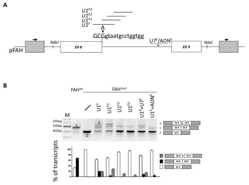
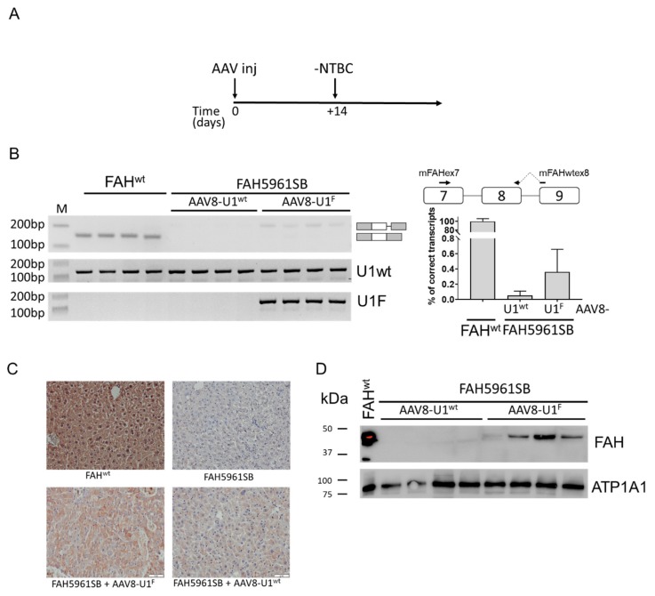
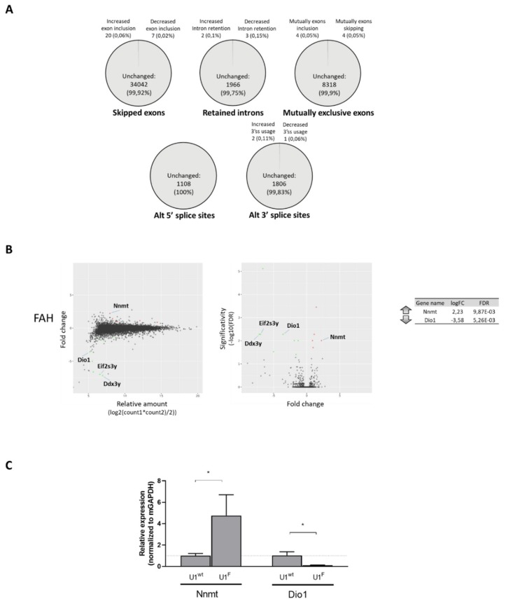
The elucidation of aberrant splicing mechanisms, frequently associated with disease has led to the development of RNA therapeutics based on the U1snRNA, which is involved in 5' splice site (5'ss) recognition. Studies in cellular models have demonstrated that engineered U1snRNAs can rescue different splicing mutation types. However, the assessment of their correction potential in vivo is limited by the scarcity of animal models with the targetable splicing defects. Here, we challenged the U1snRNA in the FAH5961SB mouse model of hepatic fumarylacetoacetate hydrolase (FAH) deficiency (Hereditary Tyrosinemia type I, HT1) due to the FAH c.706G>A splicing mutation. Through minigene expression studies we selected a compensatory U1snRNA (U1F) that was able to rescue this mutation. Intriguingly, adeno-associated virus-mediated delivery of U1F (AAV8-U1F), but not of U1wt, partially rescued FAH splicing in mouse hepatocytes. Consistently, FAH protein was detectable only in the liver of AAV8-U1F treated mice, which displayed a slightly prolonged survival. Moreover, RNA sequencing revealed the negligible impact of the U1F on the splicing profile and overall gene expression, thus pointing toward gene specificity. These data provide early in vivo proof-of-principle of the correction potential of compensatory U1snRNAs in HTI and encourage further optimization on a therapeutic perspective, and translation to other splicing-defective forms of metabolic diseases.

Keywords: FAH; RNA therapeutics; Tyrosinemia type I; U1snRNA; aberrant splicing; fumarylacetoacetate hydrolase deficiency; mouse models.


Conflict of interest statement

D.B., D.S., M.F., S.L., N.Z., C.C., N.P., P.B. and S.F.J.v.d.G. have no competing interests to declare. M.P. is inventor of a patent (PCT/IB2011/054573) on modified U1snRNAs. M.P.had role in the design of the study; in the interpretation of data; in the writing of the manuscript.

Figures

Figure 1
Figure 1
The mouse FAH c.706G>A mutation can be rescued by engineered U1snRNAs. (A) Schematic representation of the mouse FAH minigene with exonic and intronic sequences represented by boxes and lines, respectively (not in scale). Exonic and intronic nucleotides are indicated in upper and lower cases respectively. The nucleotide change (G>A) leading to the FAH deficiency in FAH5961SB mice is indicated (arrow). The targeting regions of the engineered U1snRNAs or the U7snRNA/AONF are reported as continuous and dotted lines, respectively. The cryptic 5′ss at position +63 in intron 8 is indicated by an asterisk. (B) FAH splicing patterns in Hepa1-6 cells transiently transfected with the wild-type (FAHwt) or mutated (FAHmut) minigenes, alone or in combination with U1/U7snRNA or the AONF. The schematic representation of the transcripts (numbers 1 to 3, with exons not in scale) is reported on the right. Amplified products were separated on 2% agarose gel. M, 100 bp molecular weight marker. The lower panel reports the evaluation of amplicons by denaturing capillary electrophoresis (see Figure S1A). Histograms report the relative percentage of each transcript expressed as mean ± standard deviation (SD).
Figure 2
Figure 2
The compensatory U1F partially rescues FAH expression in vivo. (A) Schematic representation of the protocol designed to perform the experiments in mice and exploiting the AAV8-mediated delivery of the U1snRNAs. Mice, kept on NTBC in their drinking water, were injected with 1*1013 vg/kg of body weight of AAV8-U1F or AAV8-U1wt and transferred to normal drinking water without NTBC (-NTBC) 14 days later (+14). (B) FAH splicing pattern profiles in mouse livers, together with the schematic representation of transcripts, reported on the right side. The quantification of correctly spliced FAH transcript by qPCR is indicated on the right, with the schematic organization of FAH gene region and the exploited primers (on top). Results are reported as percentage of correctly spliced transcripts (mean ± SD). (C) Immunohistochemical analysis of FAH expression in liver sections of wild type (FAHwt) and FAH5961SB mice, either untreated or treated with AAV8-U1wt or AAV8-U1F. Representative examples of liver sections stained with a specific anti-FAH antibody (brown). Images are taken at 20× magnification. Scale bar, 50 µm. (D) Western blotting analysis through a specific anti-FAH antibody in liver homogenates of wild type (FAHwt) and FAH5961SB mice treated with AAV8-U1wt or AAV8-U1F. The mouse ATPase Na+/K+ Transporting Subunit Alpha 1 (ATP1A1) was exploited as load control. The protein marker, reporting the molecular size of bands, is reported on the left.
Figure 3
Figure 3
The compensatory U1F does not have a widespread effect on splicing and overall gene expression. (A) Fraction of each category (SE, RI, MXE, A5SS and A3SS) of Gencode-annotated splicing events showing increased or decreased alternative isoform use in hepatocytes from FAH5961SB mice treated with AAV8-U1F as compared with those treated with AAV8-U1wt. For each category, the number and the percentage (%) of events is indicated in the graph (FDR ≤0.05; Inclusion Level Difference ≤−0.2 or ≥0.2). (B) Comparison of the gene expression profile in hepatocytes from FAH5961SB mice injected with AAV8-U1F as compared with those injected with the control AAV8-U1wt. Only genes coding for proteins and having a fold change higher than two are shown. MA plot showing the relationship between the average expression value (on the X-axis) and the fold change (Y-axis) for each gene in the genome (left panel). Volcano plot showing the relationship between the fold-change (on the X-axis) and the significance of the differential expression test (Y-axis) for each gene (middle panel). Black dots represent the genes that are not significantly differentially expressed, while red and green dots represent the genes significantly UP- and DOWN-regulated, respectively. List of coding genes UP and Down regulated and mapping in autosomes (right panel). Only genes having a fold-change (FC) >2 were reported. Adjusted p-values for multiple tests (by the Benjamini–Hochberg procedure) are reported as “false discovery rate” (FDR) value. (C) Validation of RNAseq data by qPCR. Histograms report the relative gene expression ± standard deviation (SD) from three independent experiments, normalized for the house keeping mouse GAPDH.

Similar articles

Cited by

References

    1. Sterne-Weiler T., Howard J., Mort M., Cooper D.N., Sanford J.R. Loss of exon identity is a common mechanism of human inherited disease. Genome Res. 2011;21:1563–1571. doi: 10.1101/gr.118638.110. - DOI - PMC - PubMed
    1. Baralle M., Baralle F.E. The splicing code. BioSystems. 2018;164:39–48. doi: 10.1016/j.biosystems.2017.11.002. - DOI - PubMed
    1. De Conti L., Baralle M., Buratti E. Exon and intron definition in pre-mRNA splicing. Wiley Interdiscip. Rev. RNA. 2013;4:49–60. doi: 10.1002/wrna.1140. - DOI - PubMed
    1. Baralle M., Baralle D., De Conti L., Mattocks C., Whittaker J., Knezevich A., Ffrench-Constant C., Baralle F.E. Identification of a mutation that perturbs NF1 gene splicing using genomic DNA samples and a minigene assay. J. Med. Genet. 2003;40:220–222. doi: 10.1136/jmg.40.3.220. - DOI - PMC - PubMed
    1. Tanner G., Glaus E., Barthelmes D., Ader M., Fleischhauer J., Pagani F., Berger W., Neidhardt J. Therapeutic strategy to rescue mutation-induced exon skipping in rhodopsin by adaptation of U1 snRNA. Hum. Mutat. 2009;30:255–263. doi: 10.1002/humu.20861. - DOI - PubMed

LinkOut - more resources