Review
. 2020 Mar 20;12(3):739.
doi: 10.3390/cancers12030739.

The Multifactorial Role of PARP-1 in Tumor Microenvironment

Affiliations
Review

The Multifactorial Role of PARP-1 in Tumor Microenvironment

Juan Manuel Martí et al. Cancers (Basel). .

Abstract

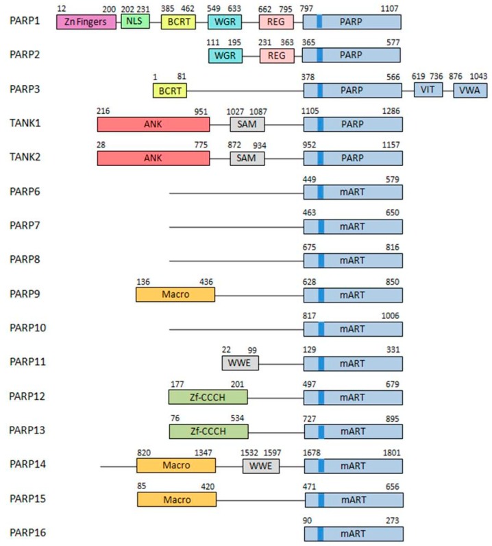
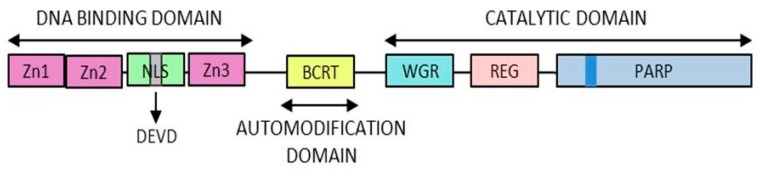
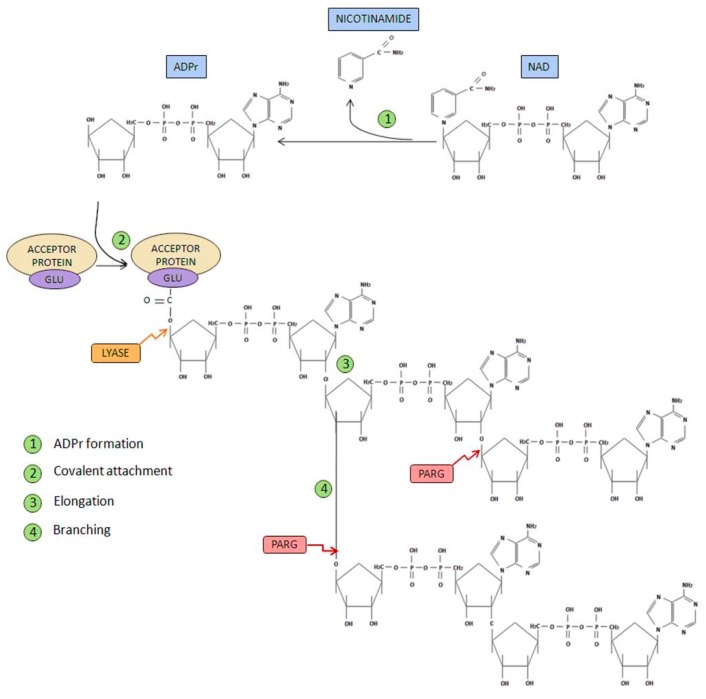
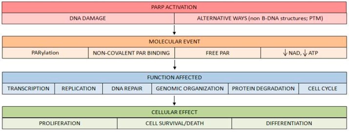
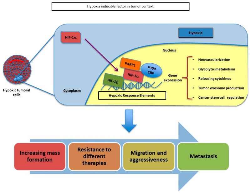
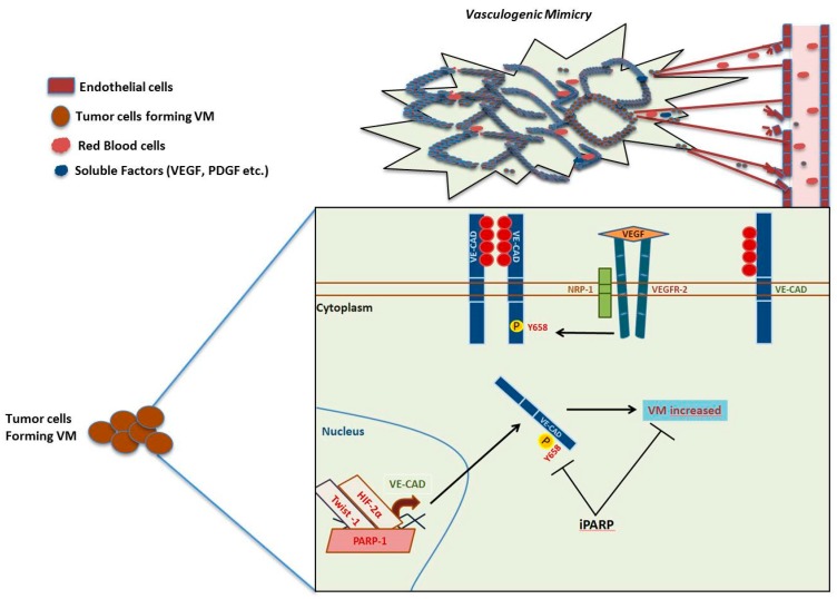
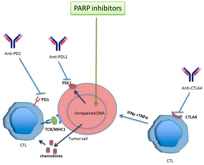
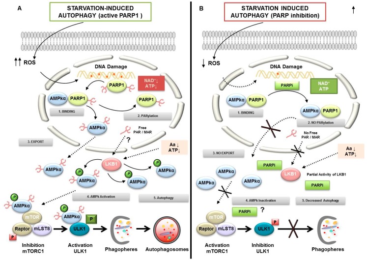
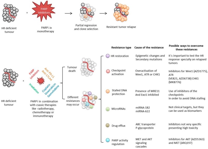
Poly(ADP-ribose) polymerases (PARPs), represent a family of 17 proteins implicated in a variety of cell functions; some of them possess the enzymatic ability to synthesize and attach poly (ADP-ribose) (also known as PAR) to different protein substrates by a post-translational modification; PARPs are key components in the cellular response to stress with consequences for different physiological and pathological events, especially during neoplasia. In recent years, using PARP inhibitors as antitumor agents has raised new challenges in understanding their role in tumor biology. Notably, the function of PARPs and PAR in the dynamic of tumor microenvironment is only starting to be understood. In this review, we summarized the conclusions arising from recent studies on the interaction between PARPs, PAR and key features of tumor microenvironment such as hypoxia, autophagy, tumor initiating cells, angiogenesis and cancer-associated immune response.

Keywords: PARP inhibitors; PARPs; PARylation; Tumor microenvironment; autophagy; hypoxia.


Conflict of interest statement

The authors declare “no conflict of interests”.

Figures

Figure 1
Figure 1
Poly(ADP-ribose) polymerases (PARP) family. The structure of the different members of the PARP family is described. Different domains are detailed in different colors. Brighter blue shows the PARP signature sequence, common throughout all the members of the family [5].
Figure 2
Figure 2
PARP1 structure. The different domains of PARP1 are presented here. The zinc fingers responsible for the DNA binding capacity, the automodification domain thanks to which PARP1 is modified by polymer and the catalytic domain containing the “PARP signature”.
Figure 3
Figure 3
PARP metabolism. Different steps in polymer formation are shown in green. In contrast, the proteins involved in polymer degradation are shown in red and yellow [14,15].
Figure 4
Figure 4
Molecular events following PARP activation. Once PARP is activated, downstream events of PARP signaling take place, involving either covalent PARylation of substrates, non-covalent binding of PAR polymer to proteins bearing a PAR-binding motif, release of free PAR to the cell or lowering cellular NAD+/ATP levels. Via these pathways, PARP/PARylation regulate functions such as transcription, replication, DNA repair, protein degradation and cell cycle, mediating various cellular phenomena such as proliferation, cell survival and cell death or differentiation.
Figure 5
Figure 5
Schematic representation of HIF-α and PARP1 association throughout the adaptation of tumoral cells during hypoxia. The union of PARP1 to HIF-a helps to its stabilization and allows the transcription of several genes involved in tumor growth, resistance, migration and metastasis.
Figure 6
Figure 6
Vasculogenic mimicry pathway and PARP. VE-cadherin can be phosphorylated by stimuli of VEGF by VEGFR2 activity and the co-receptor (NRP-1). The soluble factor (VEGF) increases the phosphorylation of Y658 of VE-cadherin and the consequent internalization. Inhibition of PARP decreases the phosphorylation Y658 of VE-cadherin and finally the possibility to form VM in aggressive melanoma cells (B16F10 cells).
Figure 7
Figure 7
PARP inhibitors together with immune checkpoint inhibitors potentiate antitumor immune-mediated response. Activated T lymphocytes react against tumor antigens. The inhibition of PARP increases the number of lymphocytes infiltrating the tumor after the upregulation of chemokines, promoting an immune response mediated by CTLs. In spite PARP inhibitors modulate positively the upregulation of PDL-1 (favouring tumor scape from immune control) the anti-CTLA4 activates T cells to promote an antitumor response. Anti PD1/PDL-1 reverses CTL inhibition provoked by PARP inhibitor –induced PDL-1 expression. In this way, anti-PD1/PDL-1 can synergize with PARP inhibitors to ammeliorate antitumor immune response.
Figure 8
Figure 8
Regulation of autophagy by PARylation. (1) PARP1 forms a complex with AMPKα in nucleus. (A) PARP activation conditions: During starvation-induced autophagy, ROS release leads DNA damage and PARP1 overactivation. Self-PARylated PARP1 interact with AMPKα1 subunit (2). The complex is disrupted and PAR-AMPKα is exported to cytosol (3). The presence of PAR-AMPK and the continuous absence of amino acids and ATP depletion favor total activation of AMPKα population by LKB1, inhibition of mTORC1, interaction PAR-phospho-AMPK/ULK1, and autophagosome formation (4). LKB1 activity is presumably modified in a PARylation-dependent manner. (B) PARP inhibited: Starvation-induced ROS production was abrogated after PARP inhibition. Following AMPKα1/PARP1 interaction (1) the AMPKα1 subunit is not PARylated and AMPK nuclear export is inhibited (2 and 3). In spite of nutrient and energy depletion, AMPKα is inactive; mTORC1 is partially activated and interacts with ULK1, favoring its inhibition (4). Finally, the autophagosomes production will be delayed.
Figure 9
Figure 9
Summary of PARP inhibition approaches during cancer treatments, different possible outcomes and ways designed to overcome the possible appearance of resistances.

References

    1. Hottiger M.O., Hassa P.O., Luscher B., Schuler H., Koch-Nolte F. Toward a unified nomenclature for mammalian ADP-ribosyltransferases. Trends Biochem. Sci. 2010;35:208–219. doi: 10.1016/j.tibs.2009.12.003. - DOI - PubMed
    1. Peralta-Leal A., Rodriguez-Vargas J.M., Aguilar-Quesada R., Rodriguez M.I., Linares J.L., de Almodovar M.R., Oliver F.J. PARP inhibitors: New partners in the therapy of cancer and inflammatory diseases. Free Radic. Biol. Med. 2009;47:13–26. doi: 10.1016/j.freeradbiomed.2009.04.008. - DOI - PubMed
    1. Chambon P., Weill J.D., Mandel P. Nicotinamide mononucleotide activation of new DNA-dependent polyadenylic acid synthesizing nuclear enzyme. Biochem. Biophys. Res. Commun. 1963;11:39–43. doi: 10.1016/0006-291X(63)90024-X. - DOI - PubMed
    1. Meyer-Ficca M.L., Meyer R.G., Jacobson E.L., Jacobson M.K. Poly (ADP-ribose) polymerases: Managing genome stability. Int. J. Biochem. Cell Biol. 2005;37:920–926. doi: 10.1016/j.biocel.2004.09.011. - DOI - PubMed
    1. Meyer-Ficca M.L., Meyer R.G., Coyle D.L., Jacobson E.L., Jacobson M.K. Human poly (ADP-ribose) glycohydrolase is expressed in alternative splice variants yielding isoforms that localize to different cell compartments. Exp. Cell Res. 2004;297:521–532. doi: 10.1016/j.yexcr.2004.03.050. - DOI - PubMed